1559
Improved visualization of lumbar nerve roots diffusion tensor imaging using iZoom-IRIS without fat-suppression pre-pulse1Radiology, Eastern Chiba Medical Center, Chiba, Japan, 2Faculty of Health Sciences, Institute of Medical, Pharmaceutical and Health Sciences, Kanazawa University, Ishikawa, Japan, 3Philips Japan, Tokyo, Japan, 4Philips Healthcare (Shanghai) Ltd., Shanghai, China, 5Philips Australia & New Zealand, North Ryde, Australia
Synopsis
Keywords: Neurography, MSK
Motivation: DTI based on single-shot EPI is established method to evaluate lumbar nerve roots compression, but EPI-DTI has a high geometric distortion.
Goal(s): To improve the image quality of the lumbar nerve roots, thereby improving the accuracy of tractography visualization and quantitative measurements.
Approach: To reduce distortion, we developed a method to reduce the number of phase encodes per acquisition by combining iZoom 2D RF excitation, IRIS and LIPO (SSGR) only fat suppression.
Results: Compared to the conventional EPI-DTI, LION iZoom IRIS-DTI improves the tractography image quality. LION iZoom IRIS-DTI was superior to the EPI-DTI, in accuracy of FA values measurement and ICC.
Impact: LION iZoom IRIS-DTI is effective in reducing image distortion and might improve the accuracy and reproducibility of FA measurements of lumbar nerve roots.
PURPOSE
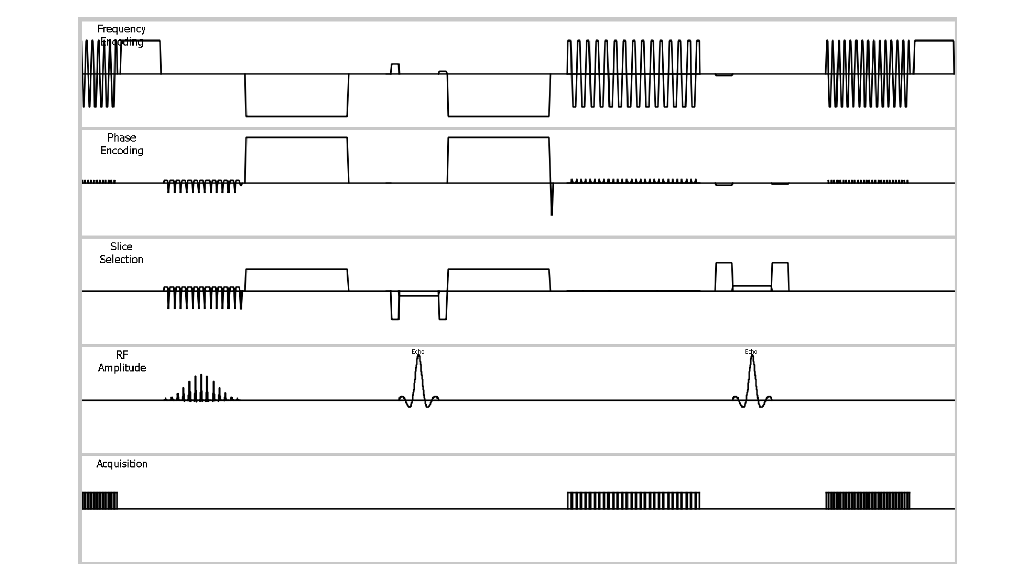
Diffusion tensor imaging (DTI) based on single-shot echo planar imaging sequence (EPI-DTI) is established method to evaluate lumbar nerve roots compression, because several studies have shown that DTI and tractography of human lumbar nerves can visualize and quantitatively evaluate lumbar nerves by fractional anisotropy (FA)1,2. However, EPI-DTI has a severe problem such as high geometric distortion. To solve these problems, we previously proposed a DTI based on single-shot turbo spin echo sequence (TSE-DTI)3,4, but is sometimes suffers from its low SNR, resulting in long scan time.To reduce distortion while maintaining clinically acceptable SNR, we developed a combination of 2-dimensional spatially selective excitation technique (iZoom) and multi-shot EPI-DWI with 2D image navigator (Image Reconstruction using Image-space Sampling function: IRIS). Moreover, the slice selection gradient reversal (SSGR, LIPO)5 technique was optimized (LIPO-Only: LION-DWI6,7) for maximizing the fat suppression without fat suppression pre-pulse to prevent SNR drop by applying pre-pulses. We combined altogether, called LION iZoom IRIS-DTI [Fig.1]. The purpose of this study is to evaluate the effect of accuracy and reproducibility of the quantitative values in lumbar nerve roots using LION iZoom IRIS-DTI.
METHODS
All subjects were examined with 3.0T whole-body clinical system (Ingenia CX, Philips Healthcare). The study was approved by the local IRB, and written informed consent was obtained from all subjects. Conventional EPI-DTI, LION iZoom IRIS-DTI, and 3D T2-TSE were performed on lumbar nerve roots in 6 healthy volunteers. The ability of tractography (length and number of fibers, misalignment with actual position) and FA values of L4-S1 levels were compared between conventional EPI-DTI and LION iZoom IRIS-DTI. In addition, intraclass correlation coefficients (ICC) were calculated based on the results of two measurements made by two radiological technologists for all FA values.Imaging parameters for EPI-DTI were; axial, voxel size = 3.0 × 3.0 × 4.0mm3, FOV = 350 × 275mm2, b-value = 800s/mm2, MPG = 15 directions, EPI factor = 55, fat suppression = SPIR and LIPO, TR = 5466ms, TE = 61ms, and total acquisition time = 4m20s.
Imaging parameters for LION iZoom IRIS-DTI were; axial, voxel size = 3.0 × 3.0 × 4.0mm3, FOV = 350 × 120mm2, b-value = 600s/mm2, MPG = 15 directions, EPI factor = 19, fat suppression = LIPO only, TR = 5336ms, TE = 61ms, and total acquisition time = 5m52s.
RESULTS AND DISCUSSION
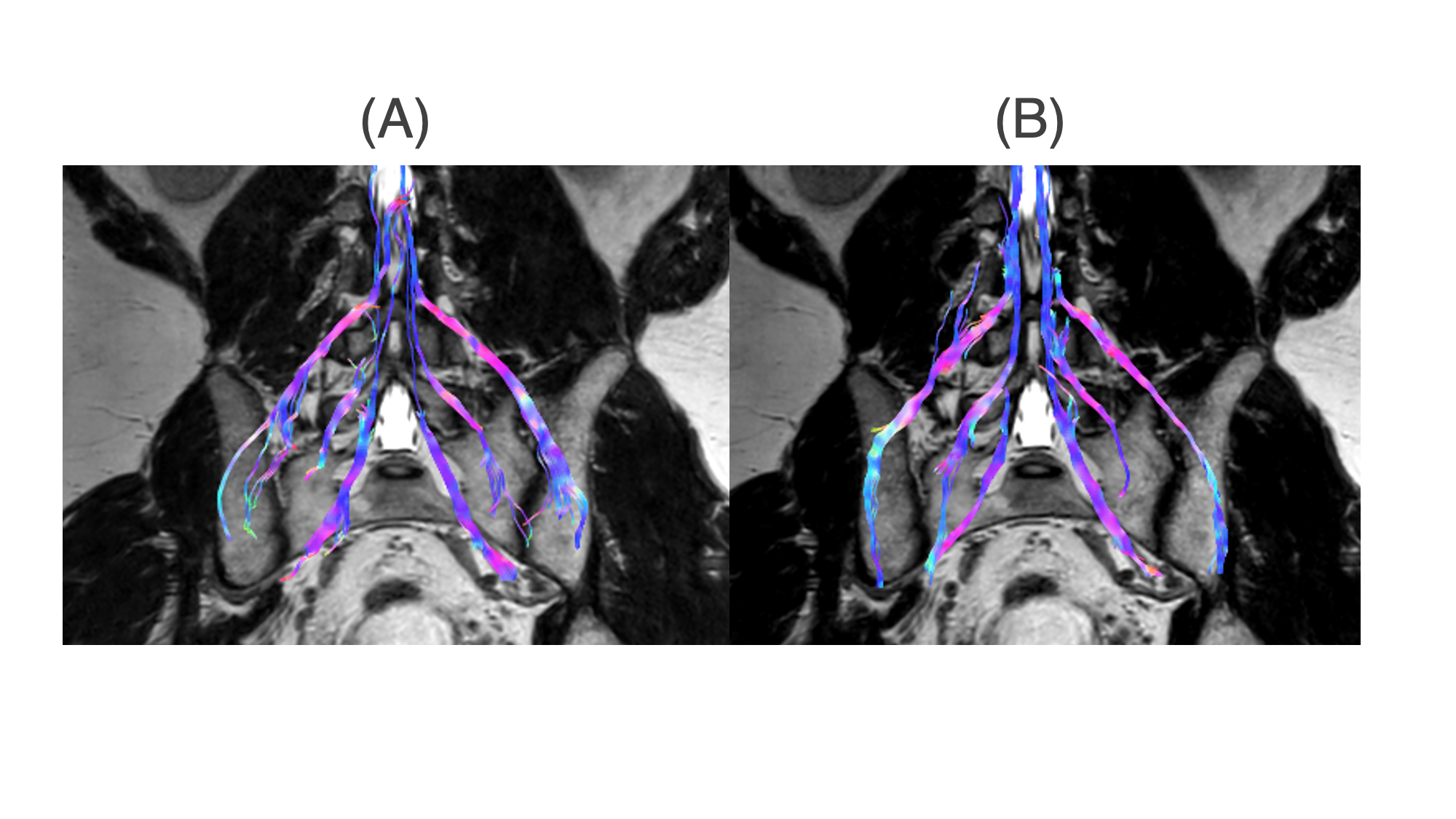
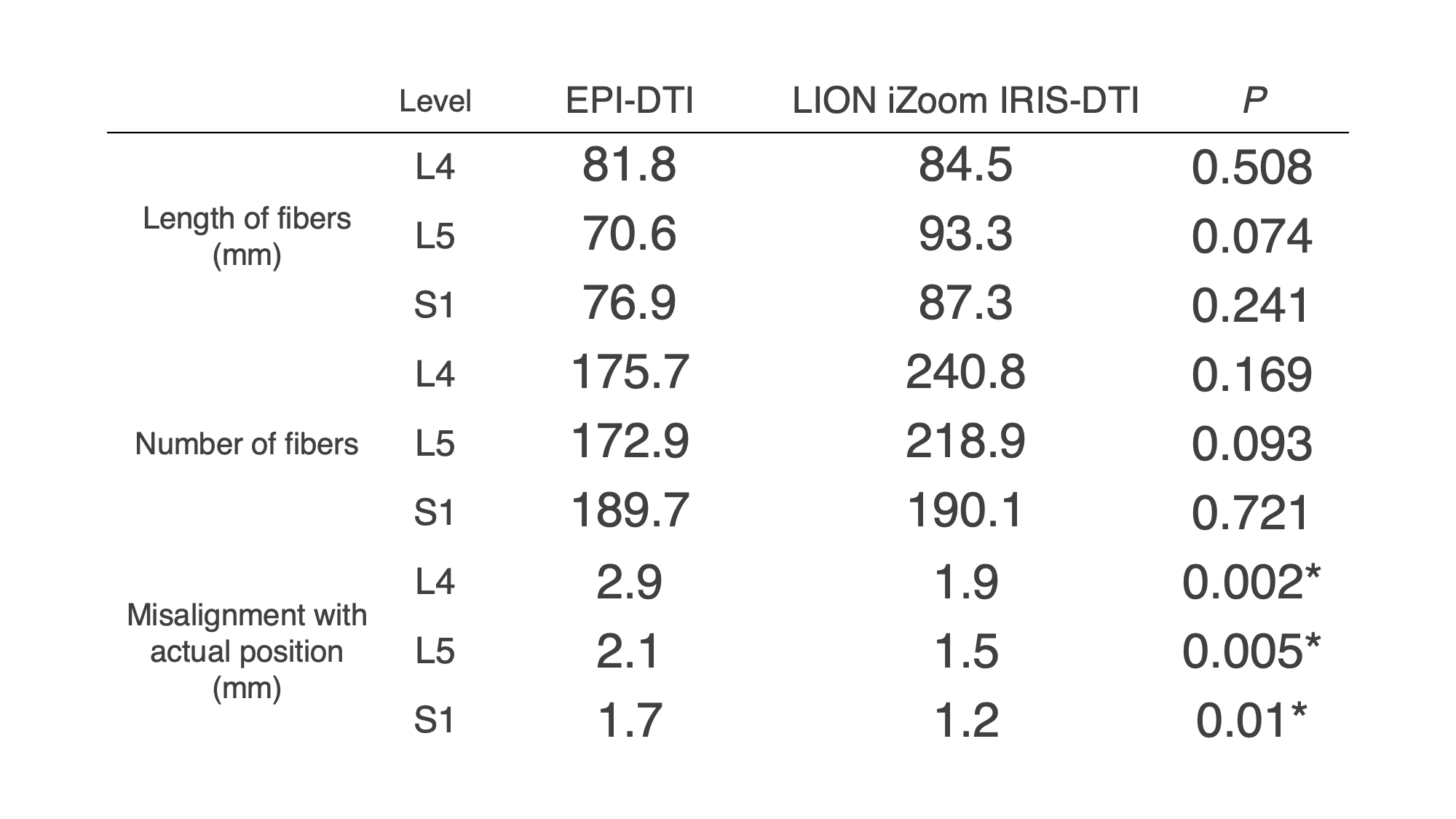
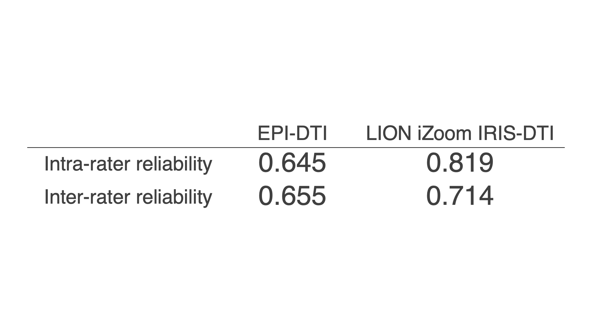
Representative tractography of the two imaging methods is shown in Fig. 2. The length and number of fibers were better for LION iZoom IRIS-DTI than for EPI-DTI, but not significantly. The misalignment with the actual position was larger for EPI-DTI, and the degree of misalignment tended to be more pronounced at higher levels in particularly [Fig. 3]. Although there was no significant difference in FA between the two imaging methods, the standard deviation was smaller in LION iZoom IRIS-DTI (EPI-DTI = 0.325 ± 0.042, LION iZoom IRIS-DTI = 0.338 ± 0.035). ICC (intra-rater reliability and inter-rater reliability) in LION iZoom IRIS-DTI (0.819 and 0.714) were higher than those of EPI-DTI (0.645 and 0.655) [Fig.4]. These results suggest that LION iZoom IRIS-DTI is effective in reducing image distortion and contributes to improve both image quality of tractography and reproducibility of FA value measurements.CONCLUSION
LION iZoom IRIS-DTI might be promising in reducing image distortion and improving the accuracy and reproducibility of FA measurements of lumbar nerve roots.Acknowledgements
No acknowledgement found.References
1. BalbiV, et al. Tractography of lumbar nerve roots: initial results. Eur Radiol 2011;21(6):1153–9.
2. Eguchi Y, et al. Quantitative evaluation and visualization of lumbar foraminal nerve root entrapment by using diffusion tensor imaging: preliminary results. Am J Neuroradiol 2011;32(10):1824–9.
3. Sakai T, et al. Distortion-free diffusion tensor imaging for evaluation of lumbar nerve roots: Utility of direct coronal single-shot turbo spin-echo diffusion sequence. Magnetic Resonance Imaging 49 (2018) 78–85.
4. Sakai T, et al. Improvement of distortion-free diffusion tensor imaging for evaluation of lumbar nerve roots: Utility of direct coronal single-shot turbo spin-echo diffusion sequence with a split acquisition. ISMRM2019 #2887
5. Nagy Z, et al. Efficient fat suppression by slice-selection gradient reversal in twice-refocused diffusion encoding. Magn Reson Med. 2008 Nov;60(5):1256-60.
6. Murayama D, et al. Reduced acquisition time of female pelvis diffusion weighted imaging (DWI) using LIPO-only (LION) DWI. ISMRM2023 #2960
7. Sakai T, et al. Volume isotropic thin-slice high-quality brain DWI with no fat-suppression pre-pulse. ISMRM2023 #3970
Figures