1549
Assessment of Myofiber-type Transformation in Rats with Discogenic Low Back Pain Using Diffusion Tensor Image of Paraspinal Muscles1The First Affiliated Hospital of Kunming Medical University, Kunming, China, 2The Second Affiliated Hospital of Kunming Medical University, Kunming, China, 3Kunming Medical University, Kunming, China, 4GE HealthCare MR Research, Beijing, China
Synopsis
Keywords: Muscle, Diffusion Tensor Imaging, Low back pain , Muscle fiber
Motivation: The development of discogenic low back pain (DLBP) and myofiber-type transformation in paraspinal muscles are strongly related. However, the impact of diffusion tensor imaging (DTI) in identifying myofiber-type transformation in paraspinal muscles remains uncertain.
Goal(s): Exploring the value of DTI in evaluating myofiber-type transformation in paraspinal muscles, and contributing to improved clinical diagnosis and intervention strategies for DLBP.
Approach: Prospective case-controlled animal experiments and establish the DLBP model in rats. DTI scanning and immunofluorescence of myofibers were performed to assess myofiber-type transformation.
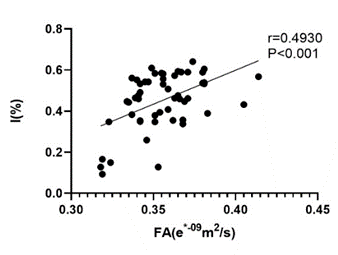
Results: The fractional anisotropy (FA) was identified as a noninvasive imaging marker for detecting myofiber-type transformation in DLBP rats (r=0.4930, P<0.05).
Impact: Detection of myofiber-type in muscle biopsies is challenging. By utilizing DTI, clinicians can diagnose pathological alterations in paraspinal muscles early on, intervene quickly, and alleviate the burden on patients before the pain worsens.
Introduction
Discogenic low back pain (DLBP) is a clinical disorder caused by disc pathology and has a large socioeconomic impact and burden. The composition of paravertebral muscles plays crucial role in DLBP, but muscle biopsies in patients were challenging. Diffusion tensor imaging (DTI) has shown promise as an imaging biomarker for skeletal muscles, but its ability to characterize myofiber-type in DLBP patients remains unknown. This study aims to investigate the value of DTI in assessing myofiber-type transformation in the paraspinal muscles of DLBP patients using a DLBP rat model.Methods
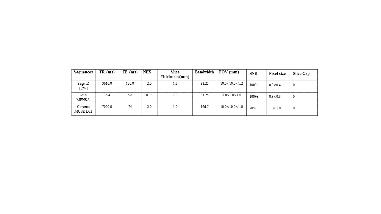
Objectives:The study was approved by the ethics committee for animal research (Kmmu20220762). Ninety healthy female Sprague Dawley rats were randomly divided into DLBP group, sham-operated group, and normal group, then follow the time from modelling procedure, each group was subdivided into 1-month group,3 months group, and 6 months group, nine groups total(N=10/group), and each group of rats was matched for age, weight, and size, etc. The DLBP group was established by disrupting L4/5 and L5/6 intervertebral discs in rats under X-ray fluoroscopy, successful modeling was confirmed by nucleus pulposus signal reduction on T2WI and behavioral experiments indicating DLBP manifestations.MRI Acquisition :All MR examinations were performed on 3.0T scanner (Architect, GE Healthcare Systems, USA) equipped with a 3.0T 16-channel rat-specific coil (CG-MUC49-H300-AG, Shanghai Chenguang Medical Technologies Co., LTD, China). DTI was obtained using a SpinEcho-EPI sequence (Table 1). Image and data measurement using post-processing workstations (AW4.7, GE, USA) were conducted. Subsequently, immunofluorescence of myofibers was performed on the rats.
Data Analysis: Statistical analyses were conducted using SPSS software (version 21.0; SPSS Inc, Chicago, IL, USA); One-way ANOVA was used to examine the means of different groups, while the Bonferroni procedure was employed to conduct repeated comparisons between multiple groups. Spearman analysis assessed the correlation between DTI parameters and the percentage of I/II myofiber in the rat paravertebral muscles. P<0.05 was considered significant.
Results
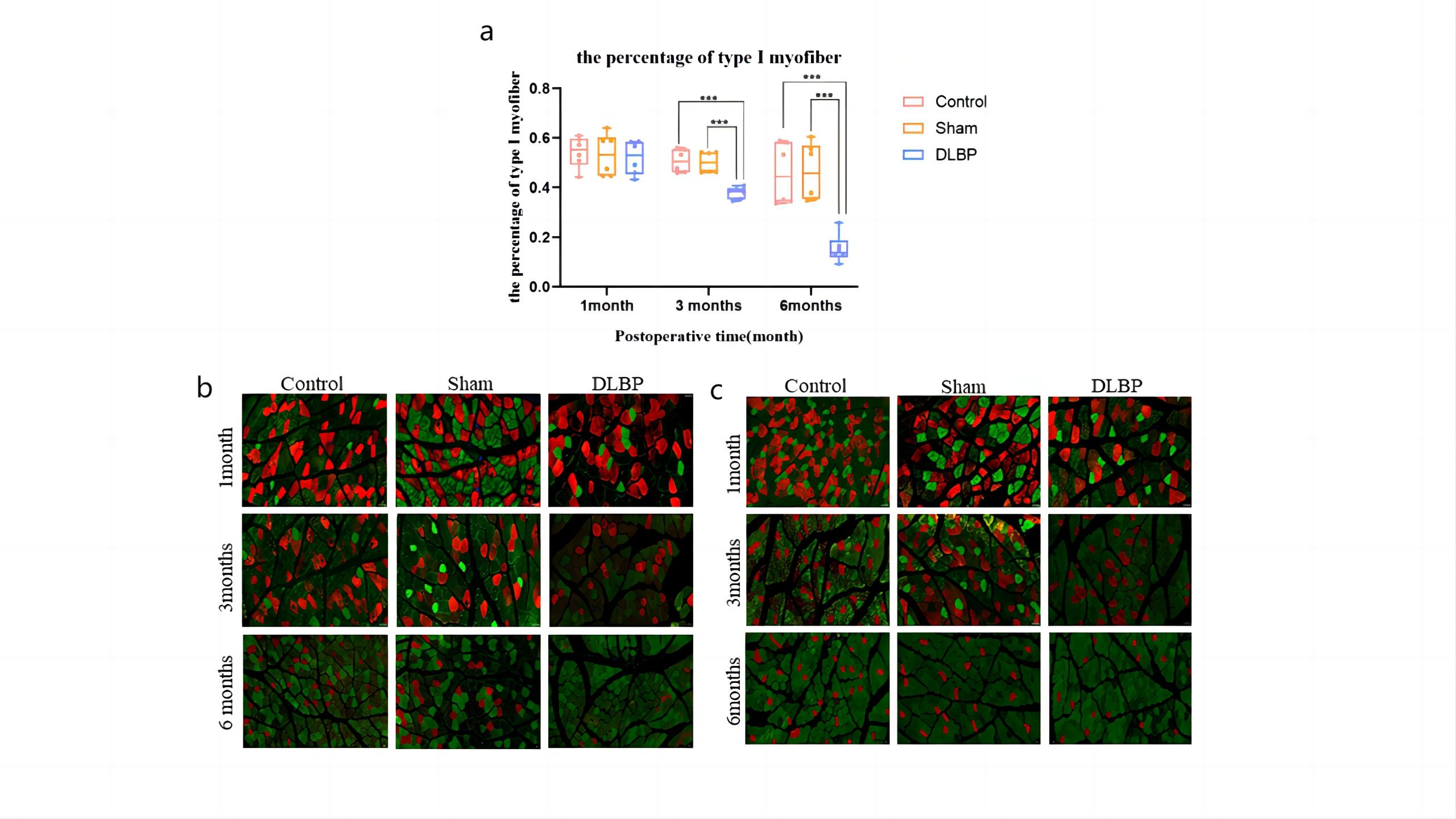
Compared to the sham-operated and normal groups, the DLBP groups exhibited a gradual decrease in FA values in the paraspinal muscles(P<0.05) (Fig.1,2). Additionally, the DLBP groups showed a significant decrease in the percentage of type I myofiber and an increase in the percentage of type II myofiber (P<0.05) (Fig.3). Notably, the percentage of type I myofiber demonstrated a moderate positive correlation with FA values (r=0.4930, P<0.05) (Fig.4).Discussion
In our study, we observed a decrease in FA value, a significant decrease in the percentage of type I myofiber, and an increase in the percentage of type II myofiber, which indicated alterations in the myofiber composition. The transformation of myofiber types can be influenced by various factors. Such as, regarding the relationship between the Two-Compartmental Diffusion MR Signal Model proposed by Karampinos et al and DTI, and it is consistent with the concept that microstructural changes within the muscle tissue can affect the diffusion properties observed in DTI 1. Meanwhile, We summarized that DTI parameters were influenced by the microstructure and pathological changes of tissues:(1) The type of myofiber. Type II is thicker than type I, water molecules were more easily diffused, which leads to a decrease in FA 2; (2) The fat fraction in muscle. Increased fat content limits the diffusion of water molecules within tissues, leading to FA increased 3;(3) Mechanical overload and chronic inflammation, these resulted in myofiber remodeling and adaptations to the altered mechanical demands 4;(4) The neural and hormonal factors 5. Thus, combined with the above theoretical analysis:(1) The transformation of type I fibers with high mitochondrial density to type II fibers with low mitochondrial density leads to a decrease in volume of the interstitial space, and the FA value decreases. (2) The previous study found that fat infiltration and CSA changes in the paravertebral muscle of DLBP rats were not obvious. However clinical studies have confirmed that the paravertebral muscle of DLBP patients presents fat infiltration, muscle atrophy, and myofiber transformation, therefore, the paravertebral muscles of DLBP patients were subjected to the combined effects of muscle atrophy and fat infiltration, an increase in the FA may occur, the change was greater than the decrease of FA caused by the transformation of type I to type II fibers alone, Finally, the change of DTI parameters of paravertebral muscle in DLBP patients may present the opposite result to that in DLBP rats.Conclusion
our study provides evidence that DTI can serve as a valuable tool for investigating the myofiber-type transformation associated with DLBP rats. However further research is warranted to explore the underlying mechanisms and to determine the clinical relevance of these findings in DLBP patients.Acknowledgements
Grant Support:Our research was supported by the National Natural Science Foundation of China (No. 82260338), Yunnan Fundamental Research Projects (No.202201AC070669,202201AU070051, 202301AS070016) and Yunnan Provincial Basic Research Program-Provincial and University Joint Special Project(202301AY070001-114).
References
1. Karampinos DC, King KF, Sutton BP, Georgiadis JG. Myofiber Ellipticity as an Explanation for Transverse Asymmetry of Skeletal Muscle Diffusion MRI In Vivo Signal. Annals of Biomedical Engineering 2009;37:2532-46.
2. Lee E, Xing F, Ahn S, et al. Magnetic resonance imaging based anatomical assessment of tongue impairment due to amyotrophic lateral sclerosis: A preliminary study. J Acoust Soc Am 2018;143:EL248.
3. Tadano K, Okamoto Y, Isobe T, et al. Changes in skeletal muscle diffusion parameters owing to intramyocellular lipid. Magnetic Resonance Imaging 2020;73:70-5.
4. Tan ET, Zochowski KC, Sneag DB. Diffusion MRI fiber diameter for muscle denervation assessment. Quant Imaging Med Surg 2022;12:80-94.
5. Galinowski, A, Miranda, M, Lemaitre, H, Martinot, M, Vulser, H, et al. Resilience and brain connectivity.EUR PSYCHIAT. 2013; 28 (S2): 59-59.
Figures
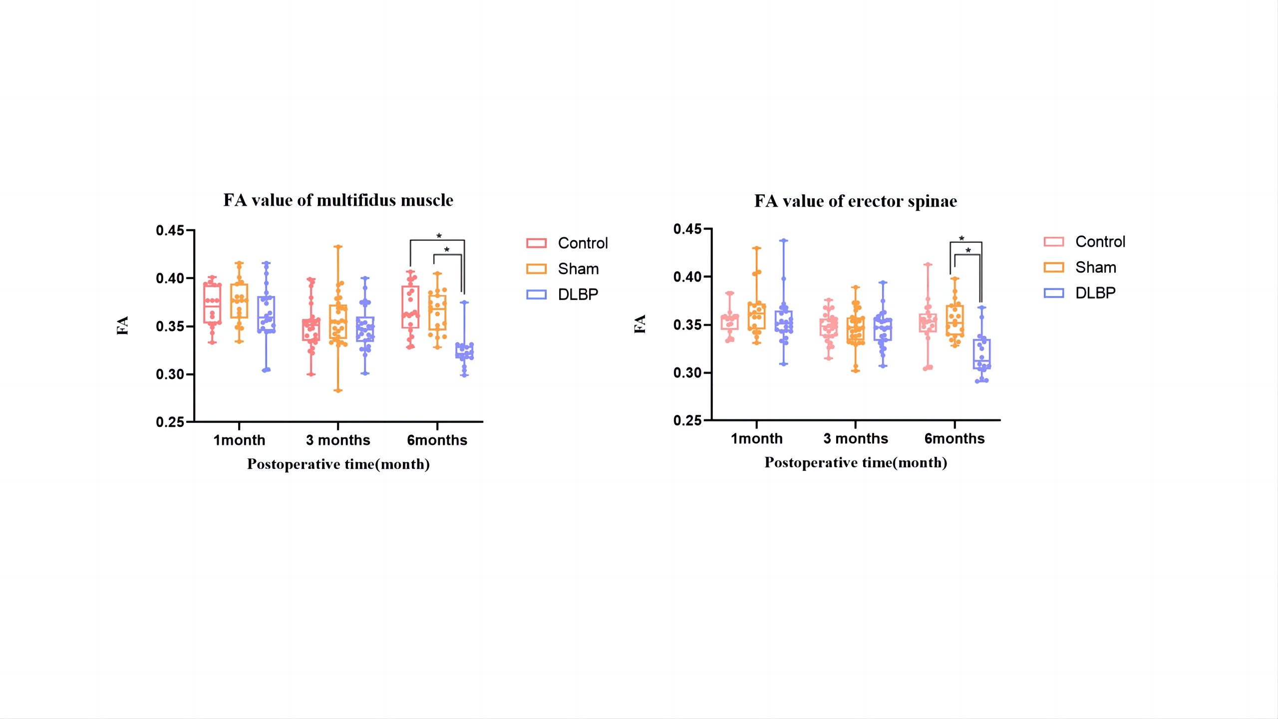
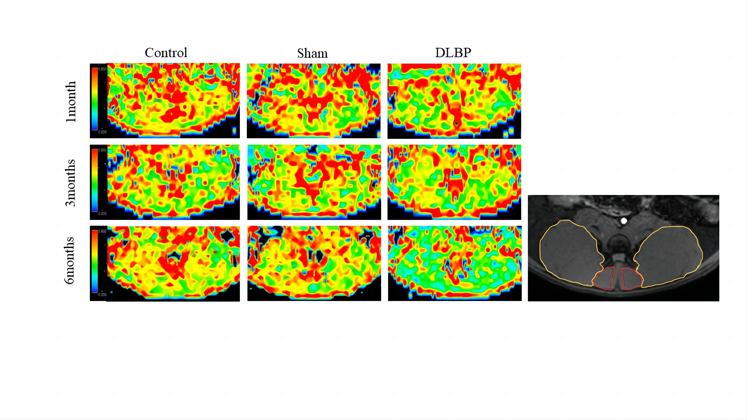
Figure 2:On the left is the FA parameters maps of multifidus and erector spinae muscles at L4/5 and L5/6 levels in three groups of rats at different time points(Blue-green-yellow-red in muscle represents a change from low to high FA values).
On the right is an anatomical image (The red box shows the area of interest for the multifidus muscle,and the orange box shows the area of interest for the erector spinae) for reference.