1546
MRI changes of lumbar vertebrae and paraspinal muscles predict vertebral fractures in patients with type 2 diabetes1The First Affiliated Hospital of Kunming Medical University, YunNan KunMing, China, 2GE HealthCare MR Research, Beijing, China
Synopsis
Keywords: Muscle, Diabetes, Vertebral Bone Quality score, paravertebral muscles; magnetic resonance imaging
Motivation: Patients with T2DM are at an increased risk of vertebral fractures, but the specific risk factors remain unclear.
Goal(s): Investigate the correlation between changes in lumbar vertebrae and paravertebral muscles observed through MRI and the occurrence and severity of vertebral fractures in T2DM patients.
Approach: Retrospectively collect general information and lumbar spine MRI images of patients diagnosed with T2DM. Develop a diagnostic model based on MRI vertebral body and paravertebral muscle parameters and evaluate the model's diagnostic performance.
Results: Higher VBQ scores, decreased relative CSA of paravertebral muscles, and increased fat infiltration of the paravertebral muscles indicate an elevated risk of fractures.
Impact: The findings of this study could potentially have a significant impact on the management and prevention of vertebral fractures in T2DM patients. By identifying specific MRI-based risk factors, clinicians can improve fracture risk assessment and implement targeted interventions.
Introduction
Fracture risk in patients with type 2 diabetes mellitus (T2DM) is influenced by various factors. While the prediction of fracture risk in T2DM patients primarily focuses on bone-related aspects1. However, paravertebral muscles have been found to play a crucial role in providing spinal stability and protecting against vertebral fractures2. The relationship between MRI-based changes in lumbar vertebrae, paravertebral muscles, and the occurrence and extent of vertebral fractures in T2DM patients remains poorly understood. Therefore, this study aims to explore the relationship between MRI-based changes in lumbar vertebrae and paravertebral muscles and their influence on the occurrence and extent of vertebral fractures in patients with T2DM. By investigating these associations, we can gain valuable insights into the comprehensive factors contributing to fracture risk in T2DM patients.Methods
This study obtained ethical approval from the First Affiliated Hospital of Kunming Medical University. Informed consent was obtained from all participants. We collected general information and lumbar spine MRI images of patients diagnosed with T2DM from January 1, 2021, to January 31, 2022. A total of 168 patients with T2DM were included in this study, including 81 patients in the fracture group and 87 patients in the non-fracture group. All patients underwent standard preoperative MRI of their lumbar spine, including T2-weighted imaging (T2WI) sagittal, T1-weighted imaging (T1WI) sagittal, T2WI fat-suppressed sagittal, and T2WI axial sequences. The scan covered segments T11-L5. The average scanning time for each participant was seven minutes. The VBQ score was calculated using the sagittal T1WI sequence. Additionally, the axial T2WI sequence was used to outline the paravertebral muscle area and assess fat infiltration using Image J software, as shown in Figure 1. Pearson's correlation analysis was conducted to assess the correlation between VBQ scores and paravertebral muscle changes in patients with T2DM. Univariate and multivariate logistic regression analyses were conducted to identify the risk factors for vertebral fracture in T2DM. Additionally, the model's performance was evaluated using the receiver operating curve (ROC).Results
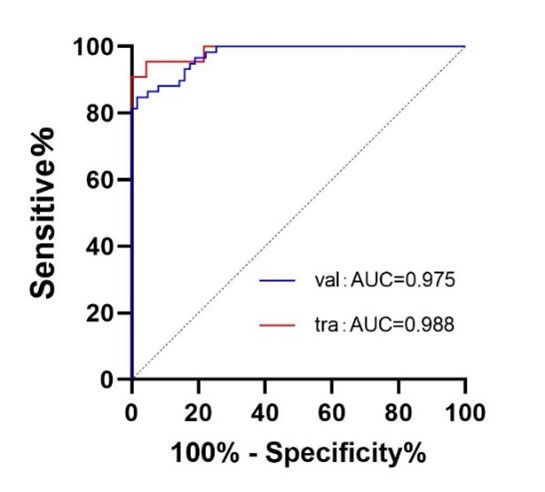
The fracture group patients had significantly higher age and BMI and longer disease duration compared to those in the non-fracture group(P=0.03, P=0.000, P=0.013). The fracture group also exhibited higher VBQ scores and paravertebral muscle fat infiltration (FI) and smaller relative cross-sectional area (r-CSA) of the paravertebral muscles compared to the non-fracture group (P<0.001), these results are shown in Table 1. Regarding fracture severity, just as Table 2 shows, the severe fracture group had smaller paravertebral muscle CSA (pmCSA) compared to the mild fracture group, and the moderate and severe fracture groups had higher FI compared to the mild fracture group (P<0.05). Correlation analysis revealed a slight negative correlation between FI and r-CSA(r=-0.300, P<0.05). Multivariate logistic regression analysis showed that higher BMI, a duration of DM exceeding 5 years, higher VBQ scores, lower r-CSA, and greater FI were associated with a higher risk of fracture in T2DM patients. The ROC curve analysis demonstrated good predictive performance, with an AUC of 0.973 for the training set and 0.979 for the validation set (P=0.011, P<0.001), as shown in Figure 2.Discussion
Our study revealed that T2DM patients with vertebral fractures had a higher VBQ scores compared to those without fractures3, indicating an association between increased VBQ scores and elevated fracture risk. This is consistent with prior research that has also highlighted the impact of VBQ on fracture susceptibility in T2DM patients4,5. The increased VBQ score in T2DM patients suggests an enlargement of the vertebral body volume, which contributes to an elevated risk of fractures6. Consequently, the VBQ score, based on conventional MRI, can serve as a valuable risk factor for identifying vertebral fractures in T2DM patients. Additionally, our study revealed a negative correlation between pmCSA and r-CSA with VBQ scores. Conversely, the FI showed a positive correlation with VBQ scores. This indicates that muscles and bones are an interconnected system, and during the development of T2DM, bone mass decline and muscle degeneration occur simultaneously7. Therefore, in assessing fracture risk in T2DM patients, it is crucial to consider changes in both bone quality and paravertebral muscles. Besides, our study highlights the significant impact of paravertebral muscle characteristics on fracture risk in patients with T2DM. Higher BMI, longer disease duration, lower r-CSA , and greater FI will increase the risk of fractures. These findings emphasize the importance of considering paravertebral muscle status in fracture risk assessment and management strategies for T2DM patients.Conclusion
Increased VBQ score assessed decreased r-CSA of paravertebral muscles, and increased FI by MRI are risk factors for lumbar vertebral fractures in patients with T2DM, and FI is related to the degree of fracture.Acknowledgements
References
1 Williams, R. et al. Global and regional estimates and projections of diabetes-related health expenditure: Results from the International Diabetes Federation Diabetes Atlas, 9th edition. Diabetes Res Clin Pract 162, 108072, doi:10.1016/j.diabres.2020.108072 (2020).
2 Ortiz-Martinez, M. et al. Recent Developments in Biomarkers for Diagnosis and Screening of Type 2 Diabetes Mellitus. Curr Diab Rep 22, 95-115, doi:10.1007/s11892-022-01453-4 (2022).
3 Asokan, A. G. et al. Evaluation of bone mineral density among type 2 diabetes mellitus patients in South Karnataka. J Nat Sci Biol Med 8, 94-98, doi:10.4103/0976-9668.198363 (2017).
4 Hofbauer, L. C. et al. Bone fragility in diabetes: novel concepts and clinical implications. Lancet Diabetes Endocrinol 10, 207-220, doi:10.1016/S2213-8587(21)00347-8 (2022).
5 Ehresman, J. et al. Vertebral bone quality score predicts fragility fractures independently of bone mineral density. Spine J 21, 20-27, doi:10.1016/j.spinee.2020.05.540 (2021).
6 Haffer, H. et al. Bone quality in patients with osteoporosis undergoing lumbar fusion surgery: analysis of the MRI-based vertebral bone quality score and the bone microstructure derived from microcomputed tomography. Spine J 22, 1642-1650, doi:10.1016/j.spinee.2022.05.008 (2022).
7 Wiedmer, P. et al. Sarcopenia - Molecular mechanisms and open questions. Ageing Res Rev 65, 101200, doi:10.1016/j.arr.2020.101200 (2021).
Figures
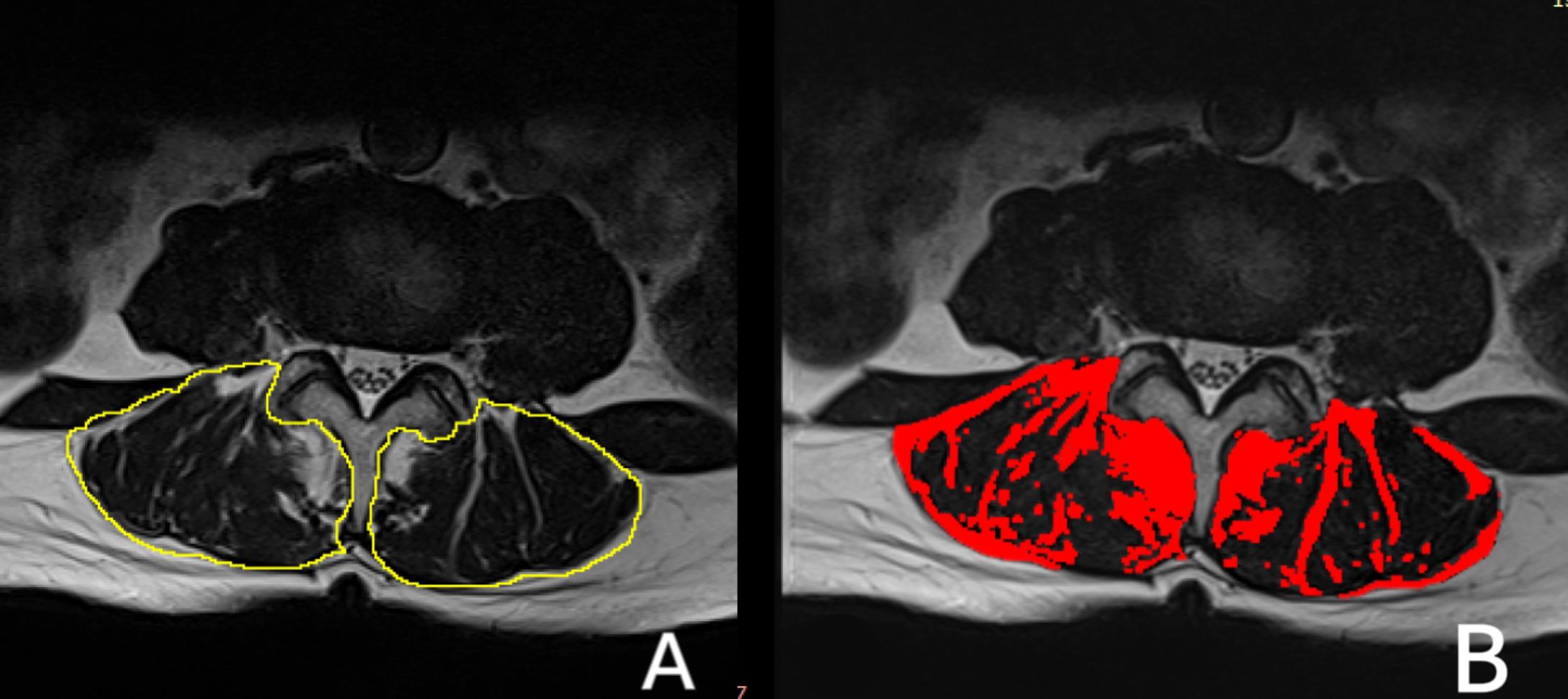
Figure 1 Paravertebral muscle CSA and fat infiltration rate ROI measurement schematic diagram
A: The total CSA of bilateral paraspinal muscles (yellow) is marked below the L3 vertebral body. B: The CSA in the fat area of the paravertebral muscle (red).
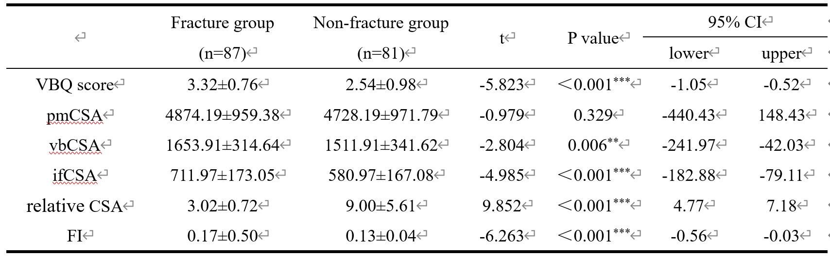
Table 1 Differences in imaging indicators of vertebral body and paravertebral muscle changes between two groups
VBQ score: Vertebral bone quality score; pmCSA:Paravertebral muscles Cross-sectional area; vbCSA: Vertebral body Cross-sectional area; ifCSA: Intermuscular fat Cross-sectional area; All CSA are in mm²; FI: Paravertebral muscles fat infiltration rate;*indicates P<0.05; **indicates P<0.01; ***indicates P<0.001
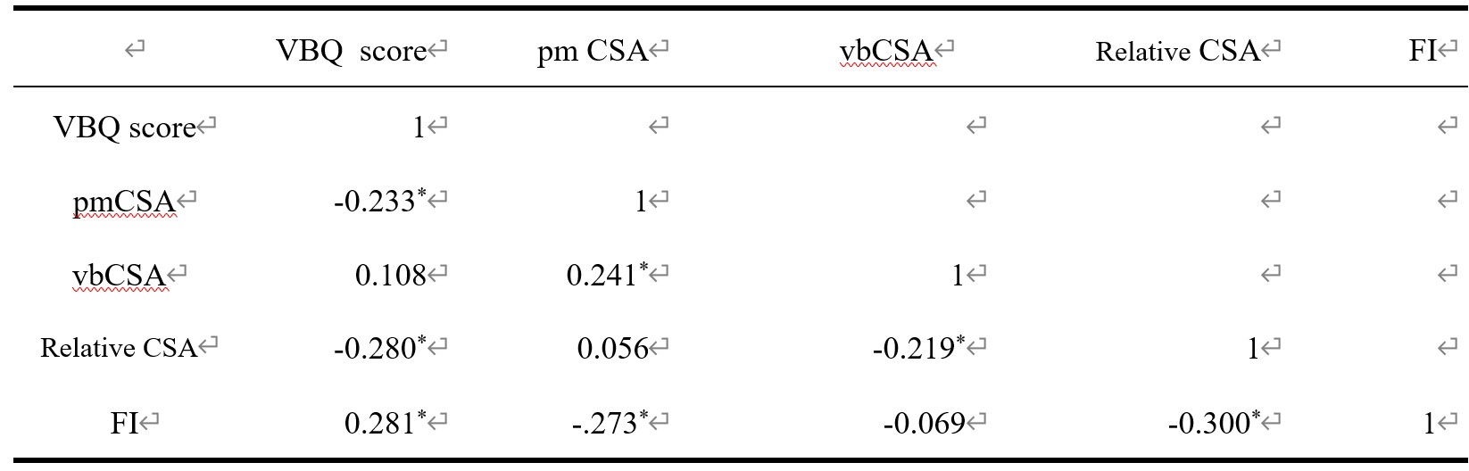
Table 2 Correlation analysis between VBQ score and paravertebral muscle indicators
VBQ score: Vertebral bone quality score; pmCSA:Paravertebral muscles Cross-sectional area; vbCSA: Vertebral body Cross-sectional area; ifCSA: Intermuscular fat Cross-sectional area; All CSA are in mm²; FI: Paravertebral muscles fat infiltration rate; *indicates P<0.05