1538
Quantitative MRI Evaluation of the Supraspinatus-Deltoid Muscle Force Couple with T2* Mapping1Radiology and Biomedical Imaging, University of California San Francisco, San Francisco, CA, United States, 2Department of Orthopaedic Surgery, University of California San Francisco, San Francisco, CA, United States
Synopsis
Keywords: Muscle, MSK, UTE, T2* mapping, shoulder, muscle, supraspinatus, deltoid
Motivation: No previous studies have evaluated relative supraspinatus-deltoid force couple (Supra-Delt-FC) muscle T2*-metrics or considered their potential as biomarkers for Supra-Delt-FC function
Goal(s): Evaluate/compare Supra-Delt-FC T2*-values within 6 supraspinatus injured-case, and 6 (age/sex matched) uninjured-control subjects
Approach: Unilateral, 3T, 3D-Cones, 15-echo-UTE MRI acquisitions were utilized for calculation and evaluation of Supra-Delt-FC muscle T2*-values
Results: Independent evaluations of deltoid and supraspinatus muscle T2*-values revealed no significant differences between injured-case and uninjured-control subjects. However, evaluation of relative T2*-metrics (mean difference & FC-ratios) revealed significant case-to-control differences – suggesting that relative supraspinatus-to-deltoid T2* metrics may be more sensitive to alterations in Supra-Delt-FC and shoulder function following injury
Impact: While independent comparisons of respective supraspinatus and deltoid muscle T2*-values revealed no significant case-to-control differences, relative Supra-Delt-FC muscle T2*-metrics were significantly different between groups. Results suggest that relative Supra-Delt-FC muscle T2*-metrics may be more sensitive to alterations in Supra-Delt-FC function.
Introduction
Within the shoulder, the force couple (FC) generated by the deltoid and supraspinatus muscles plays a pivotal role in joint stability and movement. Shoulder abduction is initiated by the contraction of the supraspinatus which overcomes the resistance of the deltoid as it works to keep the humeral head centered within the glenoid socket. In the setting of supraspinatus tears, the deltoid attempts to compensate for loss of supraspinatus function by exerting more force during abduction – this can result in abnormal translation of the humeral head on the glenoid and subsequently serve to exacerbate pain, impingement and degeneration.1-3 Previous studies have utilized both T2 and T2* qMRI mapping techniques to evaluate rotator cuff health, muscle denervation, and fatty infiltration.4-7 However, no previous studies have utilized T2* mapping of muscle to jointly evaluate synergistic muscles within the supraspinatus-deltoid force couple (Supra-Delt-FC) of the shoulder. As such, the objective of this study was to evaluate Supra-Delt-FC muscle T2* values with case subjects who have sustained a supraspinatus injury and compare to the same metrics within healthy control subjects. We hypothesized that deltoid and supraspinatus muscle T2* values T2* values would be significantly different between injured case subjects and healthy controls.Methods
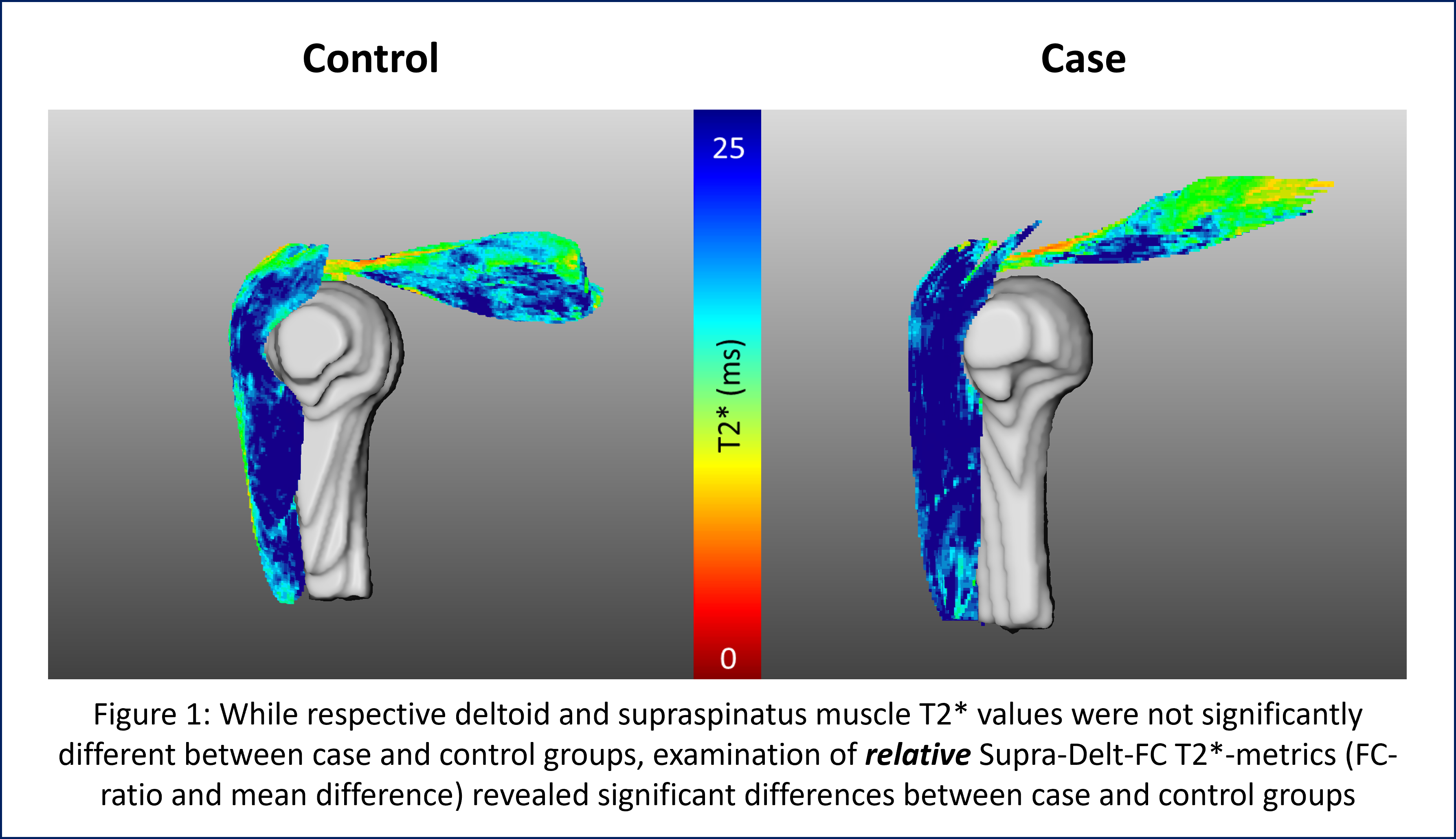
These data were collected as a part of a larger overall IRB-approved study evaluating rotator cuff tendons. Unilateral clinical 3T MRIs were acquired on 12 total subjects (6 supraspinatus injured case subjects/6 age and sex matched healthy controls, 6 females) using a 16-channel shoulder coil. Deltoid and supraspinatus muscles were manually segmented (MeVisLab) from three-dimensional, Cones UTE sequences (TEs: 15 echoes between 0.17-25ms, TR: 36ms, voxel size: 0.8 x 0.8 x 3mm3, RBW: ±83.3kHz, Flip-Angle: 17°). Mean and median meniscal T2* metrics (Figure 1) were calculated via a mono-exponential fit of signal intensity to corresponding echo time (Matlab, Natick, MA). Mean differences between deltoid and supraspinatus T2* values were calculated and T2* FC-ratio values were obtained by dividing deltoid T2* values by the corresponding supraspinatus T2* values. Statistical Analysis: Mann-Whitney U tests were used to determine whether a significant differences in Supra-Delt-FC T2* values existed between case and control groups. Significance was set at p<0.05.Results
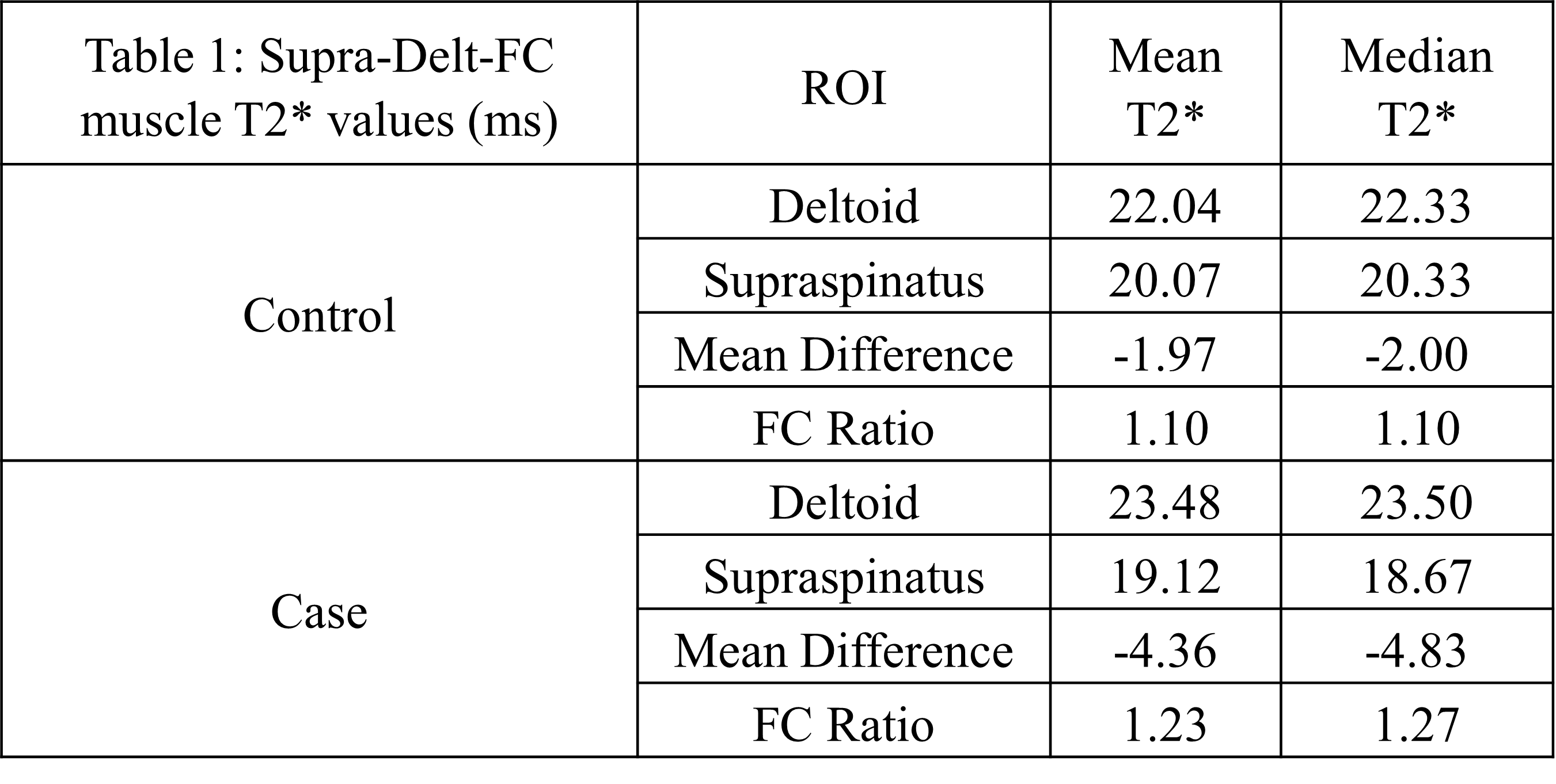
Though not statistically significant, deltoid T2* was prolonged in case subjects (mean: 23.48ms; median: 22.50ms) relative to controls (mean: 22.04ms; median: 22.33ms), while supraspinatus T2* values were relatively shorter within case subjects (mean: 19.12ms; median: 18.67ms) compared to controls (mean: 20.07ms; median: 20.33ms; Table 1, Figure 1). However, significant differences in relative deltoid-to-supraspinatus T2* metrics were found between case and control subjects. Specifically, both the Supra-Delt-FC mean difference and Supra-Delt FC-ratio of T2* values were significantly (p ≤0.02) different between case (mean difference: -4.4ms; FC-ratio: 1.23) and control subjects (mean difference: -1.97; FC-ratio: 1.10). Median T2* metrics were also significantly different (p ≤0.036) between case (mean difference: -4.36; FC-ratio: 1.27) and control (mean difference: -2.00; FC-ratio: 1.10) subjects.Discussion
While respective supraspinatus and deltoid T2* values were not statistically significantly different between case and control subjects on their own, both the deltoid-to-supraspinatus T2* FC-ratios and mean difference T2* values were significantly different between case and control groups – suggesting that relative supraspinatus-to-deltoid muscle T2* metrics are more sensitive to alterations in supraspinatus-deltoid-FC function after supraspinatus injury. Though these findings are bases on a limited number of subjects, results of the current study also suggest that muscles within the FC should be jointly considered, as an evaluation focused on individual muscles alone would have missed the significant differences in FC-ratio with rotator cuff injury. Previous studies have demonstrated that patients with a functionally intact Supra-Delt-FC tend to have better longitudinal outcomes, reduced pain, and improved quality of life compared to those that do not.2,3 While arthroscopic repair has been shown result in increased supraspinatus muscle volume 2 years post-operatively8, its potential to successfully restore shoulder function hinges on the pre-op status of the Supra-Delt-FC muscles – once present, fatty infiltration cannot be reversed and full function cannot be restored. As such, evaluation of supraspinatus and deltoid T2* values may represent quantitative imaging-based biomarkers by which to monitor pre- and post-op Supra-Delt-FC function in the shoulder.Conclusion
Results of the current study suggest that (1) relative supraspinatus-to-deltoid T2*- metrics are sensitive to the loading environment shifts experienced by the deltoids of patients who have sustained a supraspinatus injury and, (2) relative T2*-metrics may have utility as imaging-based biomarkers for Supra-Delt-FC and overall shoulder function – with the potential to serve as quantitative metrics by which to inform individual treatment plans, prognosis, and treatment efficacy in patients with rotator cuff injuries.Acknowledgements
The authors would like to thank GE Healthcare, our funding sources (NIH/NIAMS K01AR075895, UCSF-CTSI UL1 TR001872, and NIH/NIAMS P30AR075055), our MRI-technologists, and support staff for their assistance with this studyReferences
- de Witte P.B. Int J Sports Phys Ther. 2016, PMID: 24289984
- Edwards P.E., Int J Sports Phys Ther. 2016, PMID: 27104061
- Akhtar A., J Clin Orthop Trauma. 2021, PMID: 34012769
- Krepkin K., Skeletal Radiol. 2017, PMID: 27896400
- Argentieri E.C., Muscle Nerve. 2021, PMID: 33501678
- Pownder S.L., Vet Comp Orthop Traumatol. 2023, PMID: 36588292
- Anz A.W., Eur J Radiol. 2014, PMID: 24613548
- Park Y.B., Am J Sports Med. 2016, PMID: 26865396
Figures