1529
Validity of Ultrashort Echo Time MRI-based Finite Element Analysis of Human Femur Compared to Mechanical Testing1Radiology, University of Pennsylvania, Philadelphia, PA, United States, 2Bioengineering, University of Pennsylvania, Philadelphia, PA, United States, 3Orthopaedic Surgery, University of Pennsylvania, Philadelphia, PA, United States, 4Bioengineering, Temple University, Philadelphia, PA, United States
Synopsis
Keywords: Bone, Bone
Motivation: Hip fractures pose a substantial health concern commonly associated with osteoporosis. Bone mineral density from Dual-energy X-ray absorptiometry (DXA) is the standard-of-care test for assessment of osteoporosis and fracture risk, yet many who sustain fragility fractures are above the diagnosis threshold.
Goal(s): The objective of this study was to conduct a preliminary investigation into the validity of MRI Finite Element Analysis (FEA) for predicting bone strength.
Approach: Ultrashort Echo Time MRI was used on cadaveric human femur sections and FEA was performed.
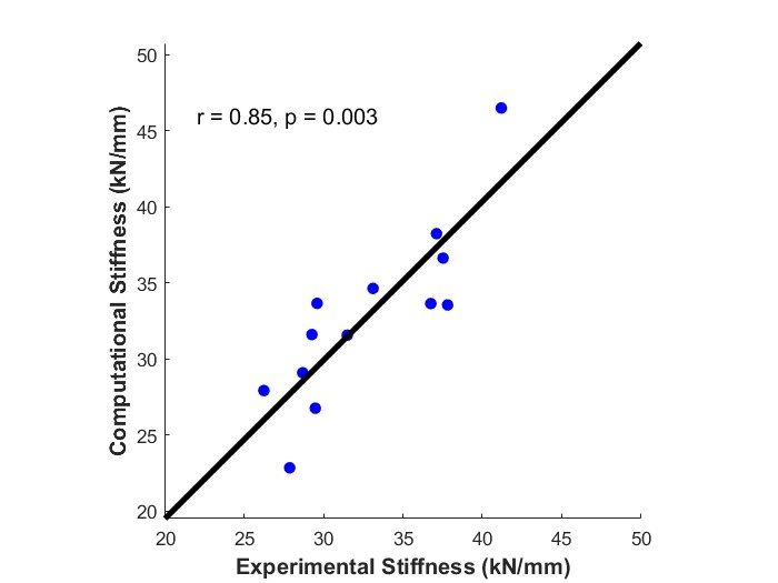
Results: The stiffness derived from UTE FEA showed a strong correlation with stiffness derived from mechanical testing.
Impact: This study compared UTE-based Finite Element Analysis to mechanical testing and found that there was a significant correlation between computational and experimental values. This indicates the potential for UTE MRI to be used in the clinical evaluation of bone strength.
Introduction
Hip fractures are a serious health problem that often affects older patients. These injuries have a high rate of morbidity and mortality and repeatedly impact patients with osteoporosis. However, in vivo evaluation of fracture risk continues to be challenging. DXA is the clinical standard for assessment of osteoporosis and fracture risk, yet many patients who sustain fragility fractures are above the diagnosis threshold3. Conventional MRI-based Finite Element Analysis (FEA) has been shown to provide a more accurate evaluation of bone mechanical competence1. Recently, ultrashort echo time (UTE) MRI sequences have been shown to directly image signal from cortical bone water that is invisible with conventional MRI due to its short T2* decay. UTE-derived cortical bone imaging biomarkers have been shown to predict porosity, collagen density, and whole-bone mechanics2,4. Therefore, the objective of the present study was to conduct a preliminary investigation into the efficacy of UTE MRI FEA for predicting bone strength as a clinically viable, non-invasive method for evaluating fracture risk.Methods
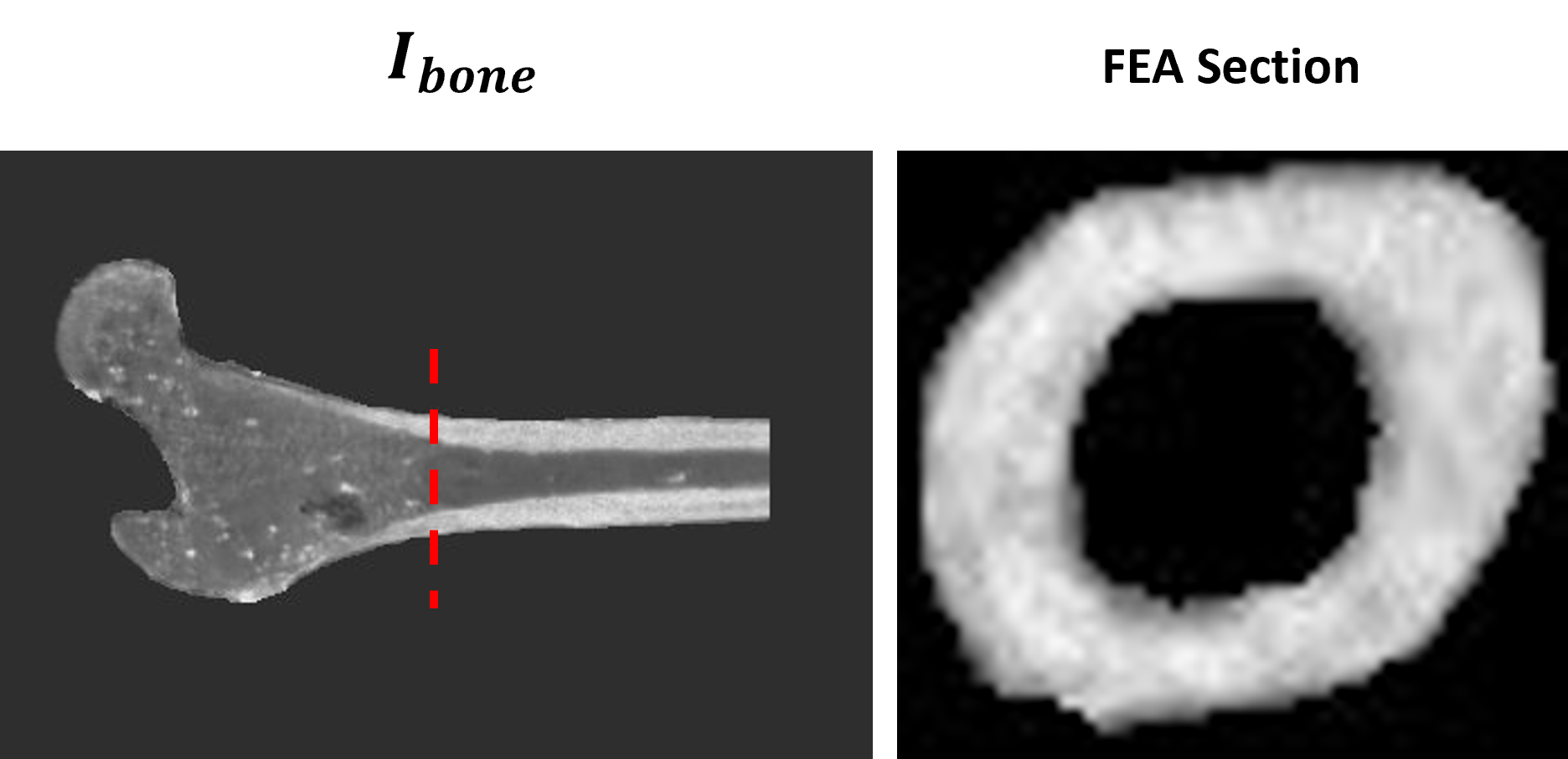
Human cadaveric femur specimens (n=13, age 72.1 ± 14.9 years) were stored at -30° C and thawed for 12-16 hours prior to experimentation. Specimens were imaged with a Prisma 3T MRI scanner (Siemens, Erlangen, Germany) with an 18-channel flexible body coil array wrapped tightly around the femurs. A custom dual-echo UTE sequence was acquired with TE1=50 µs, TE2=2400 µs, TR=7 ms, field-of-view 280 mm3, flip angle 12 degrees, 120k center-out spokes, dwell time 2 µs, and off-line reconstruction resulting in a reconstructed matrix size of 480x480x480. The TEs were chosen such that the first echo acquires signal from the long-T2 components of marrow and soft tissue, as well as the short-T2 species in bone, whereas the short-T2 signal has entirely decayed by the second echo. Therefore, to generate a bone-specific image where cortical bone is the brightest component, Eq 1 was used:$$I_{bone}=\frac{S_{TE_1}-S_{TE_2}}{S_{TE_1}+S_{TE_2}}$$
Mechanical competence of the specimen was determined by compression testing using 20-mm thick bone samples from the region immediately distal to the lesser trochanter. Samples were cut using an IsoMet 1000 Precision Cutter and carefully washed with phosphate-buffered saline (PBS) to remove marrow tissue. Specimens underwent uniaxial compression tests using a servo-hydraulic material testing machine (Instron 8874, Instron, Norwood, MA) equipped with a 100 kN load cell. The 20-mm proximal femur segments were placed loosely between two parallel steel platens and compressed under displacement control at a rate of 0.06 mm/s between -50 and 2000N. Samples were cycled 3 times to a load of -24,000 N or until catastrophic failure. The linear portion of the load-displacement curves from the 24 kN ramp were used to determine stiffness (N/mm).
To replicate the mechanical testing parameters for the FEA, a 20-mm slice of the proximal femur shaft directly below the lesser trochanter was selected. Each femur section was rescaled between 0 and 100 with the maximum value being set to the maximum intensity within the cortex. Masks were used on the background as well as the marrow, so the final axial image included only the cortical bone (Figure 1). Once preprocessed, the virtual bone sections were run through a custom linear FEA (kN/mm) to simulate stiffness under uniaxial compression in the superior-inferior direction.
Results
Once stiffness measurements were obtained both computationally and experimentally, a linear correlation model was created, and the correlation coefficient and p-value were calculated using MATLAB. There was a significant correlation between FEA-derived mechanical stiffness and ground-truth mechanical testing with r=0.85 and p=0.003 (Figure 2).Discussion
The stiffness results from the FEA performed on MRI UTE scans show a significant and strong correlation with mechanical testing measurements. Our results indicate the potential for MRI to be used in the clinical evaluation of fracture risk. Ongoing work is exploring additional ways to leverage UTE measurement of cortical bone composition to further improve FEA models. However, the present study does have limitations. First, our relatively small sample size provides challenge when drawing conclusions, and further investigation should be done with more specimen. In addition, we performed a linear FEA, which simulates a fracture in the superior inferior direction, yet many hip fractures do not occur in just one plane. Simulation of a sideways fall should also be done to analyze its correlation with mechanical measurements as this may be a more rigorous representation of osteoporotic fall-fractures. The clinical significance of this work is the ability of our UTE-based analysis method to provide a more comprehensive and accurate assessment of fracture risk compared to current clinical methods such as DXA.Acknowledgements
No acknowledgement found.References
1. Jerban, Saeed, et al. “MRI-based mechanical competence assessment of bone using Micro Finite Element Analysis (micro-FEA): Review.” Magnetic Resonance Imaging, vol. 88, 2022, pp. 9–19, https://doi.org/10.1016/j.mri.2022.01.009.
2. Jones, Brandon C., et al. “Automated, calibration-free quantification of cortical bone porosity and geometry in postmenopausal osteoporosis from ultrashort echo time MRI and deep learning.” Bone, vol. 171, 2023, p. 116743, https://doi.org/10.1016/j.bone.2023.116743.
3. Kanis, J.A, et al. “Ten-year risk of osteoporotic fracture and the effect of risk factors on screening strategies.” Bone, vol. 30, no. 1, 2002, pp. 251–258, https://doi.org/10.1016/s8756-3282(01)00653-6.
4. Rajapakse, Chamith S., et al. “MRI-based assessment of proximal femur strength compared to mechanical testing.” Bone, vol. 133, 2020, p. 115227, https://doi.org/10.1016/j.bone.2020.115227.
Figures