1521
Toward efficient arterial spin labeling imaging of knee bone marrow at 7T by utilizing a localized parallel transmit water excitation pulse design1University of Minnesota, Minneapolis, MN, United States
Synopsis
Keywords: Bone, Bone, Ultra High Field, 7T, knee, bone marrow, pTx RF pulse, spatial-spectral RF pulse design, water excitation, fat supression
Motivation: To make knee bone marrow arterial spin labeling (ASL) imaging a viable and clinically practical approach.
Goal(s): To increase knee bone marrow ASL imaging signal-to-noise efficiency and quality by using parallel transmit spatial-spectral localized water excitation pulses for ASL image readouts.
Approach: Spatial-spectral water excitation pulses inside a 2D arbitrarily-shaped region of interest were designed and validated in a knee phantom and healthy volunteers using an 8-channel transceiver knee coil.
Results: The designed spatial-spectral pulses could produce localized water excitation inside targeted regions with effective fat suppression.
Impact: The validated pulse design provides an effective way for localized water excitation and eliminate the need for additional fat saturation with reduced RF power deposition, having a great potential to improve knee ASL imaging efficiency at ultrahigh field.
Introduction
Given its demonstrated clinical potential (1), there is an increasing interest to perform arterial spin labeling (ASL) imaging of knee bone marrow (called knee ASL imaging in the following to be brief) at ultrahigh field (UHF), owing to gains in perfusion signal-to-noise ratio (SNR) and parallel imaging performance (2). However, to fully realize the benefits of UHF, technical challenges related to UHF in general and those related to knee ASL imaging in particular need to be overcome (3,4). Here we implemented a parallel transmit (pTx) spatial-spectral (spsp) pulse design for image readouts of knee ASL imaging to produce uniform water excitation inside a targeted region and suppress fat in the presence of RF and main field inhomogeneities was demonstrated with both phantom and human studies at 7 Tesla (7T).Methods
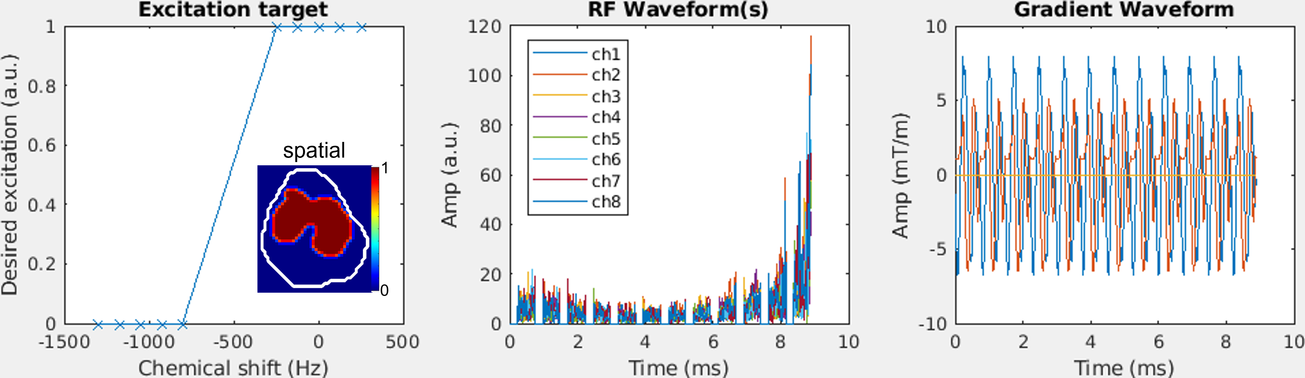
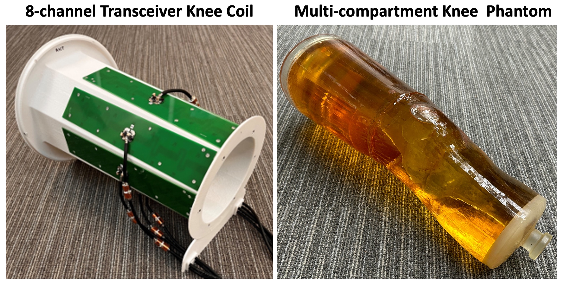
Studies were performed on a Siemens Terra (Siemens, Erlangen, Germany) equipped with a body gradient (200 T/m/s slew rate, 80 mT/m Gmax) using a prototype 8-channel transceiver knee coil (Figure 1), with each element being a fractionated dipole antenna (5). A 3D-printed knee phantom used for our validation study has two compartments: the outside-bone compartment filled with polyvinylpyrrolidone (PVP) solution with matched dielectric properties to the human leg tissue at 7T and the bone compartment with peanut oil to mimic bone marrow fat (Figure 1).The pulse design was implemented in Matlab (MathWorks, Natick, MA, USA), formulated as a least squares minimization and solved using the conjugate gradient algorithm as in our previous study (6). The designed pulses (Figure 2) consisted of a train of twelve 0.74 ms sub-pulses with each corresponding to a 2D spiral pulse for an excitation target having both spatial and spectral components. The spatial component defined a uniform excitation (of 10-degree flip angles) inside a 2D ROI and the spectral component a water passband plus a fat stopband (centered at water and fat resonance frequencies, respectively).
The validation was achieved via both phantom and human studies using 3 mm isotropic resolution ∆B0 and B1+ maps acquired with vendor-provided pulse sequences. The validation studies with the phantom used a single ROI covering the entire slice of interest for our pulse design while the validation studies with healthy volunteers also used a ROI only covering the bone marrow. A pTx-capable gradient recalled echo (GRE) sequence was developed for 3D GRE image acquisition at 2-mm isotropic resolutions with sagittal orient, 2-fold in-plane acceleration, 2.63-ms TE and 80-ms TR. For comparison purpose, images with matched parameters were also obtained using the coil operated in its circularly polarized (CP) mode for excitation (mimicking traditional single transmission).
Results
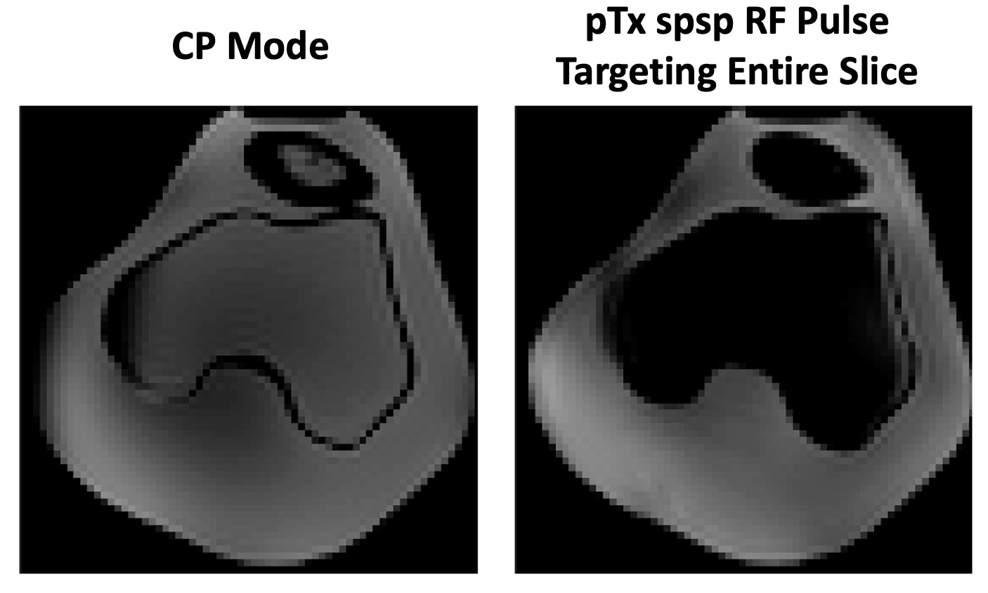
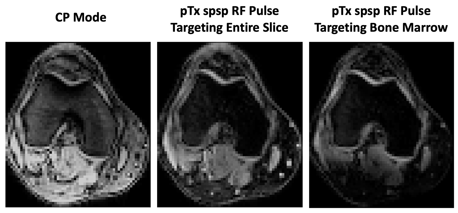
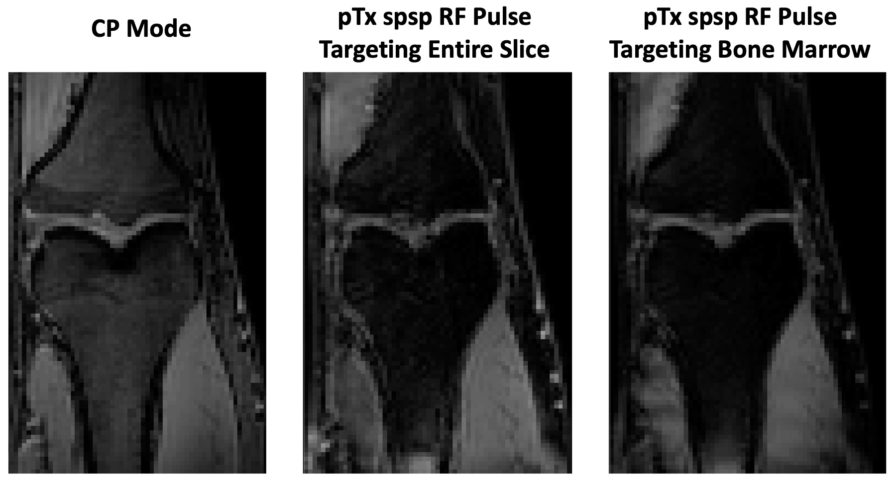
The designed pTx spsp pulses achieved effective fat suppression in the bone compartment of the knee phantom and produced desired excitation in the compartment outside of the bone with PVP solution (Figure 3). Human studies further confirmed that the designed pulses could provide localized water excitation and effectively suppressed bone marrow fat with a target covering the entire slice, as well as a target only covering the bone marrow, which substantially reduced water signal from surrounding tissues (Figure 4). The effective bone marrow fat suppression across a large field of view in the head-foot direction further indicated that the designed pTx spsp pulses were robust against spatial variations in RF fields and B0 off-resonances (Figure 5).We have implemented and validated a pTx spsp pulse design, for localized water excitation within an arbitrarily-shaped 2D ROI with robust bone marrow fat suppression, in both a phantom and healthy volunteers. The designed pulses will help increase knee ASL imaging efficiency by eliminating the need for additional fat saturation due to reduced RF power deposition.
We will further investigate the design of universal pulses for plug-and-play pTx (7) and its integration with the image readouts of knee ASL imaging sequences, such as those used for our previous knee bone perfusion studies (1,3,8): single-shot fast spin echo (9) and readout-segmented echo planar imaging (10) readouts.
Conclusion
The pTx spatial-spectral design, as validated at 7T, could provide localized water excitation with robust bone marrow fat suppression, having a great potential to improve knee ASL imaging efficiency at ultrahigh field.Acknowledgements
The authors would like to acknowledge Patrick Liebig and Yulin Chang from Siemens Healthineers for their assistance with the calculation of multichannel B1+ mapping using the vendor sequence and with sequence development, respectively. This study was supported in part by National Institute of Health grants including R56EB033365, 1R01EB033365, P41 EB027061, and S10 OD025256.References
1. Li X, Johnson CP, Ellermann J. Measuring Knee Bone Marrow Perfusion Using Arterial Spin Labeling at 3 T. Sci Rep 2020;10(1):5260.
2. Li X, Auerbach EJ, Van de Moortele PF, Ugurbil K, Metzger GJ. Quantitative single breath-hold renal arterial spin labeling imaging at 7T. Magnetic resonance in medicine : official journal of the Society of Magnetic Resonance in Medicine / Society of Magnetic Resonance in Medicine 2018;79(2):815-825.
3. Li X, Johnson CP, Ellermann J. 7T bone perfusion imaging of the knee using arterial spin labeling MRI. Magnetic resonance in medicine : official journal of the Society of Magnetic Resonance in Medicine / Society of Magnetic Resonance in Medicine 2020;83(5):1577-1586.
4. Li X, Ellermann J, Metzger GJ. Evaluation of Potential Benefits of 7T for Knee Epiphyseal Bone Marrow ASL Imaging. Proc. Intl. Soc. Mag. Reson. Med. 2023: 0135.
5. Raaijmakers AJ, Ipek O, Klomp DW, Possanzini C, Harvey PR, Lagendijk JJ, van den Berg CA. Design of a radiative surface coil array element at 7 T: the single-side adapted dipole antenna. Magnetic resonance in medicine : official journal of the Society of Magnetic Resonance in Medicine / Society of Magnetic Resonance in Medicine 2011;66(5):1488-1497.
6. Wu X, Vaughan JT, Ugurbil K, Van de Moortele PF. Parallel excitation in the human brain at 9.4 T counteracting k-space errors with RF pulse design. Magnetic resonance in medicine : official journal of the Society of Magnetic Resonance in Medicine / Society of Magnetic Resonance in Medicine 2010;63(2):524-529.
7. Gras V, Vignaud A, Amadon A, Le Bihan D, Boulant N. Universal pulses: A new concept for calibration-free parallel transmission. Magnetic resonance in medicine : official journal of the Society of Magnetic Resonance in Medicine / Society of Magnetic Resonance in Medicine 2017;77(2):635-643.
8. Li X, Johnson CP, and Ellermann J. Knee Epiphyseal Bone Marrow Perfusion Imaging Using FAIR RESOLVE. In: Proceedings of the 28th Annual Meeting of ISMRM, ISMRM 2020: Abstract 1142.
9. Hennig J, Nauerth A, Friedburg H. RARE imaging: a fast imaging method for clinical MR. Magnetic resonance in medicine : official journal of the Society of Magnetic Resonance in Medicine / Society of Magnetic Resonance in Medicine 1986;3(6):823-833.
10. Porter DA, Heidemann RM. High resolution diffusion-weighted imaging using readout-segmented echo-planar imaging, parallel imaging and a two-dimensional navigator-based reacquisition. Magnetic resonance in medicine : official journal of the Society of Magnetic Resonance in Medicine / Society of Magnetic Resonance in Medicine 2009;62(2):468-475.
Figures
-
Figure 1. The 8-channel transceiver (8TxRx) fractionated dipole
array (left) for knee MRI and multi-compartment knee phantom (right) used in
phantom validation study. The knee phantom
was 3D-printed to have two
compartments: the outside-bone compartment filled with polyvinylpyrrolidone
(PVP) solution with matched dielectric properties to the human leg tissue at 7T
and the bone compartment with peanut oil to mimic bone marrow fat.