1519
Long-T2- and Fat-Suppressed Cortical Bones Imaging by Using Quiet MRI PETRA Sequence1Faculty of Science and Engineering, University of Nottingham Ningbo China, Ningbo, China, 2Xingaoyi Medical Equipment Co. Ltd, Ningbo, China, 3Nottingham Ningbo China Beacons of Excellence Research and Innovation Institute, Ningbo, China
Synopsis
Keywords: Skeletal, Bone
Motivation: Visualizing cortical bone with short T2 value is challenging since long-T2 tissues and fat usually obscure it.
Goal(s): This study develops a new long-T2- and lipidic tissue-suppressed sequence with silent acoustic noise to improve patient scanning comfort.
Approach: We use an AFP inversion RF with controlled timing to maintain the long-T2 components at a low level and optimize the sequence parameters to null the fat signal.
Results: We apply the PETRA to reduce the SPL to only 1.9 dBA higher than the background. The head cortical bone structure can be successfully visualized with positive contrast.
Impact: The designed silent long-T2 tissue-suppressed PETRA sequence can be potentially useful for investigating the newborns bone development, which can decrease neonatal hearing injury risks and might be helpful for early-age skeletal maturation assessment.
Introduction
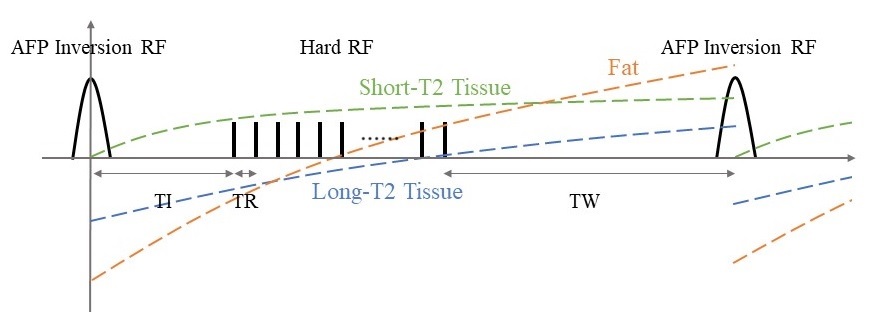
Cortical bones have an ultrashort T2 value of less than 1ms that can hardly be seen in MRI images.1 The development of ultrashort echo time (UTE) and zero echo time (ZTE) techniques allow MRI to provide the noninvasive and nonradiative approach for short-T2 tissue imaging.1-3 However, the visualization of cortical bone might be concealed by long-T2 tissues like fat and water in many soft tissues. Previous studies use dual-echo UTE subtraction, relaxation-matched excitation, etc. for soft tissue suppression.4, 5 In this study, we designed a new long adiabatic inversion prepared 3D isotropic pointwise encoding time reduction with radial acquisition (PETRA) sequence, improving the cortical bone imaging with lower acoustic noise for patient comfort enhancement and a wider range of T1 values nulling for these soft tissues than standard inversion recovery methods.6-8 As shown in Figure 1, the long-T2 tissues experience an inversion recovery after a long-duration adiabatic fast passage (AFP) RF pulse, whereas the short-T2 tissues go through the saturation recovery process.9 The time between adjacent inversion pulses is carefully controlled so the long-T2 tissues are not fully recovered and remain at a low level. The acquisition is located when the lipidic signal is weak to achieve fat suppression simultaneously.Methods
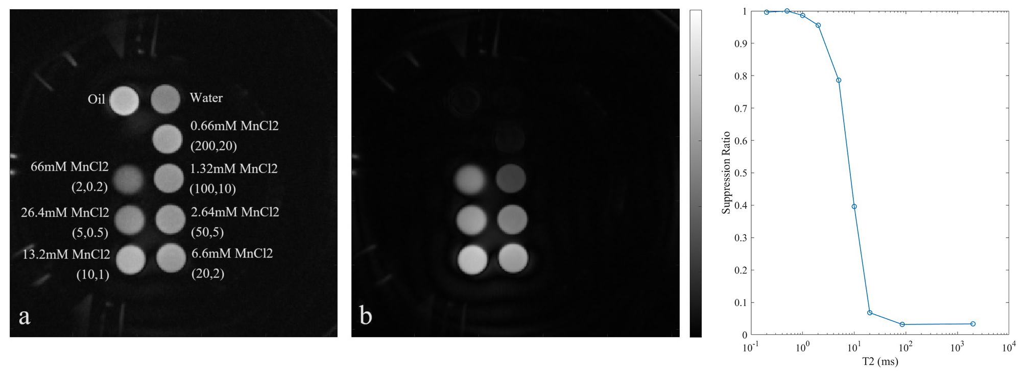
Bloch simulation is performed to optimize the sequence parameters, including hard RF flip angle (FA), inversion time (TI) and waiting time (TW) for larger SNR of cortical bone and its CNR with fat and water in different soft tissues. The sequence is designed with the following parameters: AFP inversion pulse duration=20 ms, FOV=280 mm, matrix=256*256*256, FA=12 degrees, sampling frequency=10 kHz, TR=4 ms, TE=100 us, TI=40 ms and TW=150 ms. The phantom study is conducted on an XGY SuperScan-1.5T MR scanner (Xingaoyi Medical Equipment Company, China) for MnCl2 solutions with different concentrations of 0.66, 1.32, 2.64, 6.6, 13.2, 26.4, and 66 mM, forming (T1 T2) values of (200 20), (100 10), (50 5), (20 2), (10 1), (5 0.5), (2 0.2) ms. It is also applied for in vivo brain and knee imaging. The images are reconstructed using the 3D non-uniform Fourier transform (NUFFT) in Matlab (MathWorks, USA). The acoustic noise is measured by a 4955 microphone (Bruel Kjaer, Denmark) and a 2250 sound level meter (Bruel Kjaer, Denmark) in the isocenter of the scanner bore and compared with the 3D radial UTE sequence.Results
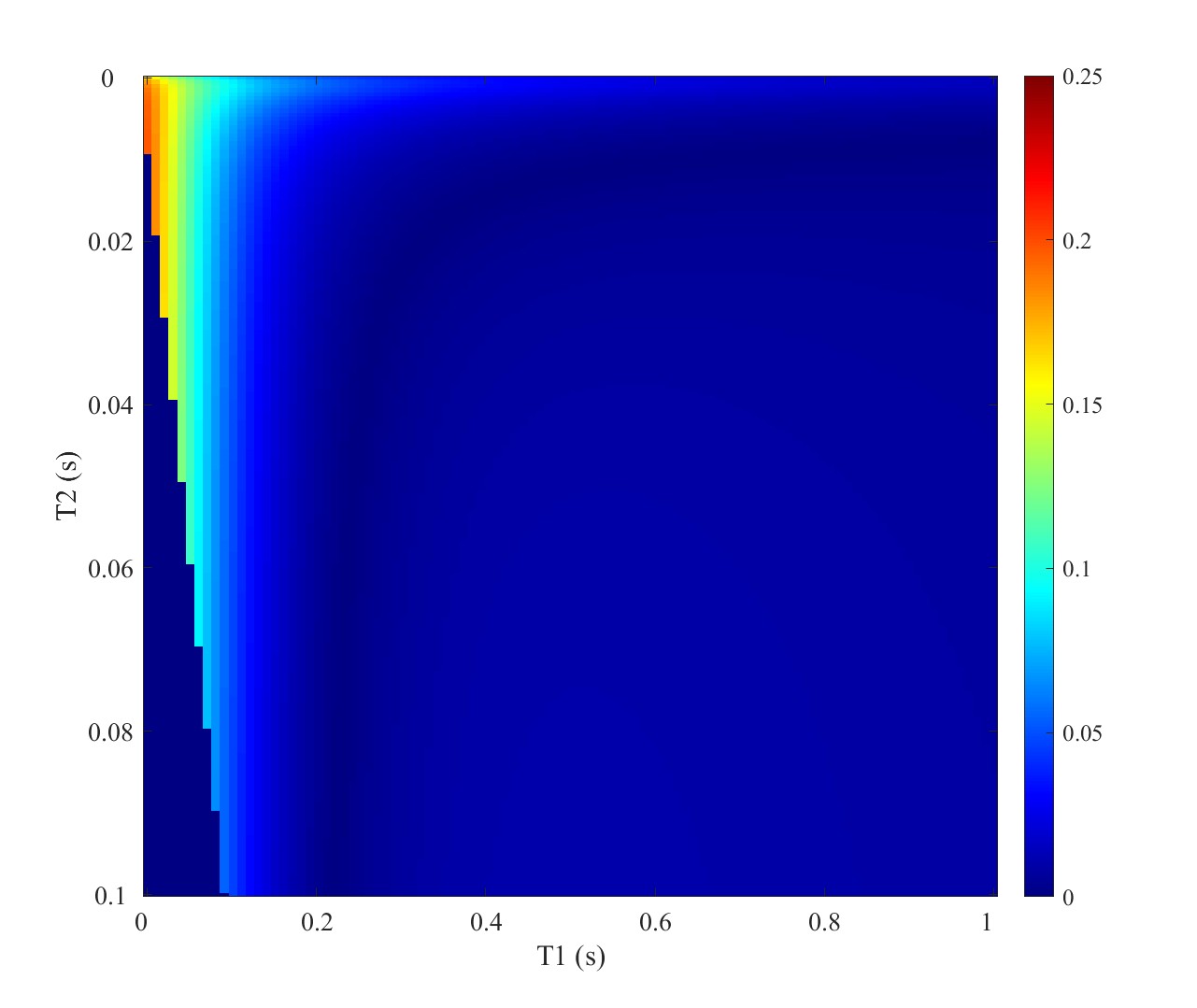
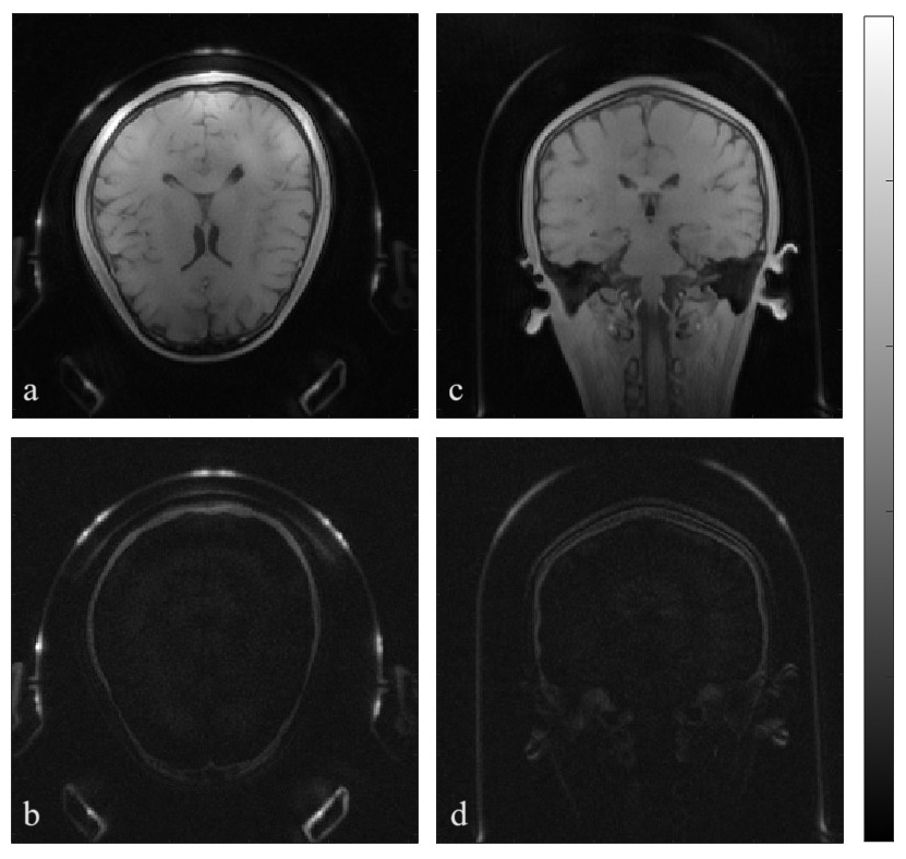
Figure 2 shows the signal intensity of tissues of different T1 and T2 values in Matlab simulation to show the range of excited short T2 values. The images of MnCl2 solutions together with water and oil are shown in Figure 3. Compared with the image without the magnetization preparation, low-concentration solutions and fat signals are suppressed significantly, remaining with only 3.28% oil and 3.46% water signals. The imaging using normal PETRA and long-T2 tissue-suppressed PETRA sequences of head cortical bones are demonstrated in Figure 4. Our PETRA sequence's measured average sound pressure level (SPL) is 66.8 dBA, which is decreased by 20.3 dBA than the 3D radial UTE sequence and is only 1.9 dBA higher than the background noise.Discussion
The results show that long T2 tissues containing different T1 values like gray matter, white matter and muscle, etc. can be significantly reduced and the lipidic tissues are also suppressed that can avoid obscuring short-T2 cortical bones like in non-fat-suppressed images, whereas 0.2-1 ms short-T2 components are remained. The SPL is also much lower than the commonly used radial UTE sequences for short-T2 imaging due to the small gradient switching between each TR in the PETRA sequence.Conclusion
We designed and demonstrated a PETRA sequence potentially useful for cortical bone visualization, successfully suppressing long-T2 soft tissues, including fat on a 1.5 T MRI scanner. Its near background acoustic noise can mitigate patient restlessness and improve hearing safety during scanning, which might be helpful for the neonate bone development assessment.Acknowledgements
No acknowledgement found.References
1. Eric C, Du J, and Chung C. UTE Imaging in the Musculoskeletal System. J. Magn. Reson. Imaging. 2015;41(4):870-83.
2. Weiger M, Pruessmann P. MRI with zero echo time. eMagRes. 2012;1:311–322.
3. Gatehouse D, Bydder M. Magnetic resonance imaging of short T2 components in tissue. Clin. Radiol. 2003;58(1):1-19.
4. Ethan J, Urvi V, Pejman G, et al. Improved cortical bone specificity in UTE MR imaging. Magn. Reson. Med. 2017;77(2):684-695.
5. Du J, Bydder M. Qualitative and quantitative ultrashort-TE MRI of cortical bone. NMR Biomed. 2013;26(5):489-506.
6. Grodzki M, Peter J, and Bjoern H. Ultrashort echo time imaging using pointwise encoding time reduction with radial acquisition (PETRA). Magn. Reson. Med. 2012;67(2): 510-518.
7. McJury M. Acoustic noise and magnetic resonance imaging: a narrative/descriptive review. J. Magn. Reson. Imaging. 2022;55(2):337-346.
8. Mark Q, Andrew J, Letendr A, et al. Anxiety in patients undergoing Mr Imaging. Radiology. 1989;170(2):463-466.
9. Ya-Jun M, Hyungseok J, Zhao W, et al. Myelin imaging in human brain using a short repetition time adiabatic inversion recovery prepared ultrashort echo time (STAIR-UTE) MRI sequence in multiple sclerosis. Radiology. 2020;297:392-404.
Figures