1512
Evaluation of Therapeutic Effect of Myocardial Infarction Patch Using Cardiac Magnetic Resonance Imaging1Department of Radiology, Sir Run Run Shaw Hospital, Zhejiang University School of Medicine, Hangzhou, China, 2MR Research Collaboration Team, Siemens Healthineers Ltd, Shanghai, China
Synopsis
Keywords: Myocardium, Heart, Myocardial infarction,Patch,Cardiac magnetic resonance
Motivation: Evaluation of therapeutic effect of myocardial infarction(MI) patch using cardiac magnetic resonance (CMR) imaging
Goal(s): Cooperation between medicine and polymer science
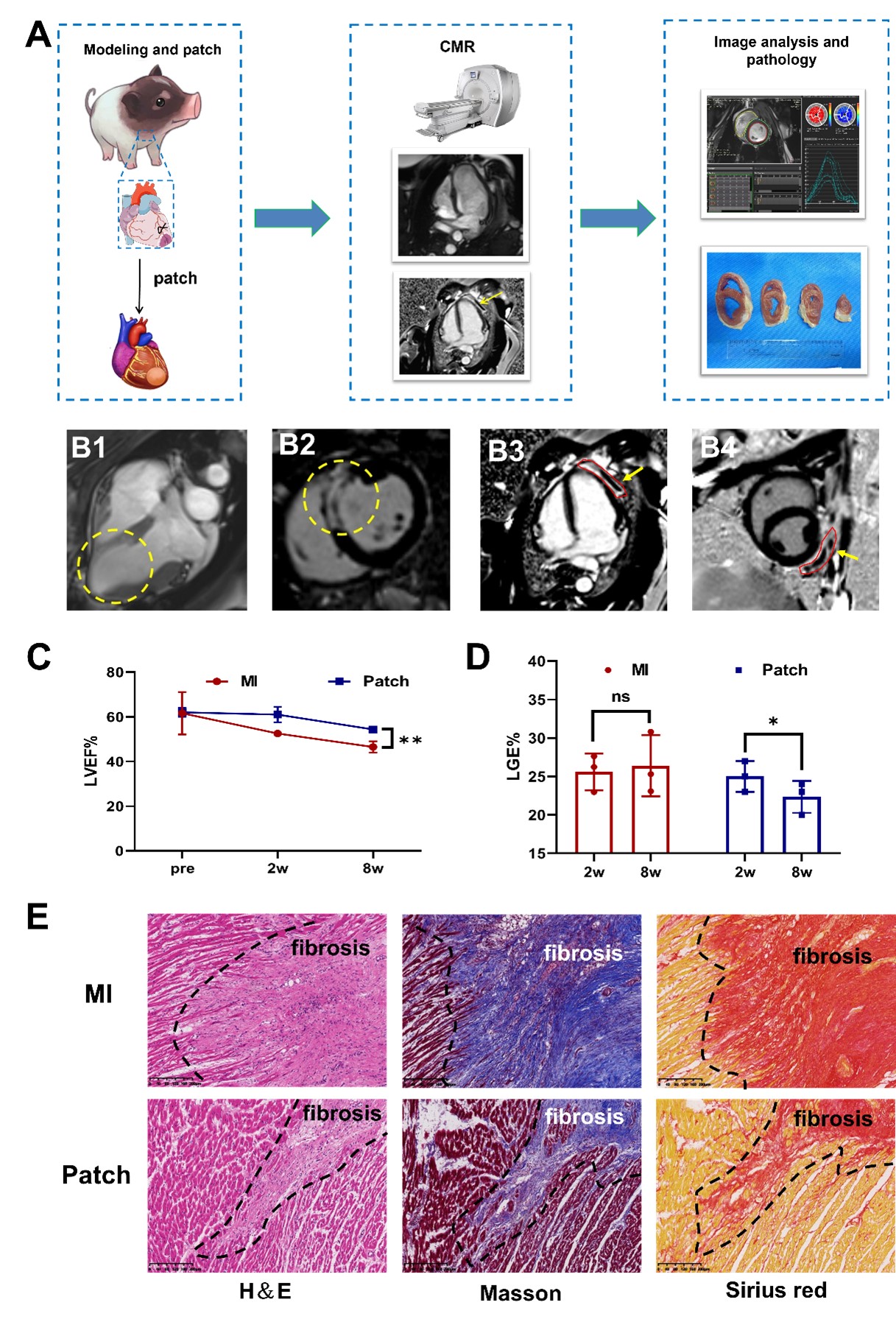
Approach: We made a pig model of MI. The MI pigs in the patch group attached the patch to the epicardial side, while the MI pigs in the control group were not treated. We performed CMR imaging before operation, 2 weeks and 8 weeks after modeling. Finally, the heart was taken for pathological verification.
Results: Compared with the control group, the patch group pig had thicker ventricular wall, better cardiac function and smaller LGE%. Histology and imaging correspond well.
Impact: By non-invasively analyzing the heart's structure and function, CMR helps us evaluate the potential of using patches to treat MI and offers valuable insights for preventing ventricular aneurysms after heart attacks in clinical practice.
Background
After a heart attack, changes in the heart's structure can lead to ventricular aneurysms, and effective treatments are currently limited. Lately, the idea of using "cardiac patches" has gained popularity. These patches are made of biopolymer materials, which are thin and elastic, and are firmly attached to the outer surface of the damaged heart tissue through minimally invasive surgery to provide support and prevent ventricular aneurysms[1-2]. The retention stability and long-term curative effect of the patch can be monitored by imaging means. Cardiac magnetic resonance (CMR) imaging allows us to measure fibrosis in the heart and assess its function. In this study, we used CMR to analyze the heart's shape and function in pigs with heart attacks before and after applying the patch, aiming to objectively measure the patch's therapeutic effects.Materials and Methods
We conducted experiments on female Bama miniature pigs (n=6). We induced heart attacks by ligating the left anterior descending coronary artery and confirmed the success of the procedure with ECG. In the control group, we closed the chest after the artery ligation, while in the patch group, we implanted the patch and then closed the chest. We conducted CMR scans before surgery and at 2 and 8 weeks post-surgery using a 3Tscanner (MAGNETOM Skyra, Siemens Healthcare, , Erlangen, Germany). The main sequences include Cine (short axis, 2-chamber, and 4-chamber were acquired via steady-state free proceeding (SSFP) sequence (slice thickness= 8 mm, slice space = 2 mm, TR = 3.5 , TE =1.5 , temporal resolution 47.5ms, flip angle = 45°- 56°) and LGE(obtained 10 min after the injection of 0.15 mmol·Kg-1 gadolinium). We analyzed the data using CVI 42 software, version 5.13.5 (Circle Cardiovascular Imaging Inc., Calgary, Canada), which included assessing left ventricular ejection fraction, LGE percentage, and left ventricular strain. After CMR, we euthanized the animals and examined their hearts with histological staining, including TTC, HE, Masson, and Sirius red.Results
The CMR results showed that in the control group, the local ventricular wall became thinner and formed ventricular aneurysms. In contrast, the ventricular wall in the patch treatment group remained thicker, and we observed epicardial patches with low signal intensity (p<0.05). Compared to the control group, the patch group had a higher left ventricular ejection fraction, higher segmental strain index, and lower LGE percentage (p < 0.05). Pathological staining revealed that the patch group had less myocardial fibrosis compared to the control group (p < 0.05).Discussion
Cardiac patch, a novel treatment strategy, brings new hope for the treatment of ventricular aneurysm. Although the current research is still in the preclinical stage, its prospect is worth looking forward to[2]. CMR imaging has become an important means of heart disease examination, so we are very confident that it can fully reflect the efficacy of biomaterials.In this study, the ventricular wall in the patch group could still maintain a certain thickness, and there was no ventricular aneurysm bleeding, which showed that the heart shape of this group was closer to a healthy state than that of the control group. Similarly, compared with the control group, the ejection fraction of the pigs in the patch group was higher and the LGE% was lower, which also proved that the patch helped to maintain the heart function. Histologically, we can clearly see that the area of myocardial fibrosis in the patch group is smaller than that in the control group.We are still in the process of this project, but from the existing results, the image quality and measurement accuracy of CMR are satisfactory, and the therapeutic effect of patch is gratifying.Conclusion
Cardiac patch can maintain the thickness of ventricular wall and protect cardiac function, which is a promising treatment strategy to prevent ventricular pathological remodeling. CMR is a non-invasive method for evaluating the effectiveness of cardiac patches and serves as a valuable imaging tool for quantitatively assessing the functional impact of these materials.Clinical Application
Imaging plays a crucial role in assessing the effectiveness of biomaterials. By non-invasively analyzing the heart's structure and function, CMR helps us evaluate the potential of using patches to treat myocardial infarction and offers valuable insights for preventing ventricular aneurysms after heart attacks in clinical practice.Acknowledgements
Special thanks to Professor Yang Zhu from the MOE Key Laboratory of Macromolecular Synthesis and Functionalization, Department of Polymer Science and Engineering, Zhejiang University and MR Research Collaboration Team of Siemens Healthineers Ltd for their support of this study.
This work was supported by the National Natural Science Foundation of China (Grant No. 81873908, for HJH) and the Fundamental Research Funds for the Central Universities (226-2023-00066).
References
[1] Yao Y, Li A, Wang S, et al. Multifunctional elastomer cardiac patches for preventing left ventricle remodeling after myocardial infarction in vivo[J]. Biomaterials, 2022, 282:121382.
[2] Chang Tianqi, Liu Chunxia, Lu Kunyan, et al. Biomaterials based cardiac patches for the treatment of myocardial infarction[J]. Journal of Materials Science & Technology, 2021, 94:77-89.
Figures