1509
Gray-Blood Late Gadolinium Enhancement (GB-LGE) for Improved Myocardial Scar Evaluation1Department of Radiology, Sir Run Run Shaw Hospital, Zhejiang University School of Medicine, Hangzhou, Zhejiang Province, China, Hangzhou, China, 2MR Research Collaboration Team, Siemens-Healthineers Ltd., Shanghai, China, Shanghai, China
Synopsis
Keywords: Myocardium, Cardiovascular
Motivation: Traditional bright-blood late gadolinium enhancement (BB-LGE) technique often obscures the visualization of endocardial LGE. In contrast, black blood technique overcomes this limitation but sacrifices contrast between the myocardium and blood pool.
Goal(s): By selecting specific TI (inversion time) values, gray blood images can be generated to achieve the desired contrast among the blood pool, myocardium, and scar tissue.
Approach: We conducted a comparative analysis of image quality, diagnostic confidence score, detection of delayed enhancement lesions, and the contrast-to-noise ratio (CNR) between BB-LGE and GB-LGE.
Results: The implementation of GB-LGE enhances diagnostic confidence and improves CNR between the blood pool and myocardial scar tissue.
Impact: The proposed GB-LGE sequence in this study enhances scar detection rates and diagnostic confidence, addressing limitations associated with equipment and technology. It optimizes clinical workflow efficiency while offering a universal approach with potential for widespread adoption.
Introduction
Late gadolinium enhancement cardiac magnetic resonance (LGE-CMR) imaging is a well-established medical imaging technique for assessing myocardial scar[1,2]. Over the past two decades, LGE-CMR has emerged as the gold standard for noninvasive myocardial scar assessment[3]. However, traditional bright-blood LGE (BB-LGE) is susceptible to missing subendocardial myocardial scar located at the blood pool's edge due to the high signal intensity of blood[4]. To address this limitation, black-blood LGE technology was introduced. While black-blood LGE significantly enhances the detection of subendocardial myocardial scar, it reduces the contrast between the blood pool and myocardium[5-7]. Thus, this study introduces a novel LGE technology termed Gray-blood LGE (GB-LGE) with the goal of improving the contrast between the blood pool, normal myocardium, and myocardial scar. It aims to explore whether this technology can enhance image quality, diagnostic efficiency, and the characterization of myocardial scar through qualitative and quantitative analysis.Methods
This prospective study collected data from 84 patients who underwent CMR examination between May 2021 and June 2022. Patients were randomly divided into two groups, A and B. All patients underwent MRI on a 3T scanner (MAGNETOM Skyra, Siemens Healthineers AG, Erlangen, Germany) or a 1.5T scanner (MAGNETOM Aera, Siemens Healthineers AG, Erlangen, Germany). In Group A, patients underwent BB-LGE followed by GB-LGE, while in Group B, patients underwent GB-LGE followed by BB-LGE. The inversion time (TI) values were set to when the signal intensity of the myocardium and blood pool crossed zero, respectively. Magnitude images of BB-LGE and GB-LGE were reconstructed using phase-sensitive inversion recovery (PSIR) technology. The crucial sequences parameters were as follows: 3T: FOV=360×320; slice thickness=8mm; Slices pacing=2mm; TR=9.24ms; TE=1.24ms; Flip angle=40o; Matrix=166×256; Interlayer resolution=1.85×1.33×8.00mm3; 1.5T:FOV=360×320; slice thickness=8mm;Slice spacing=2mm;TR=24.4ms;TE=1.39ms;Flip angle=45o;Matrix=186×256; Interlayer resolution=2.07×1.50×8.00mm3; Independent observers evaluated and analyzed the images for overall image quality, diagnostic confidence score, identification of delayed enhancement lesions outside the left ventricle, and contrast-to-noise ratio (CNR) in each case.Results
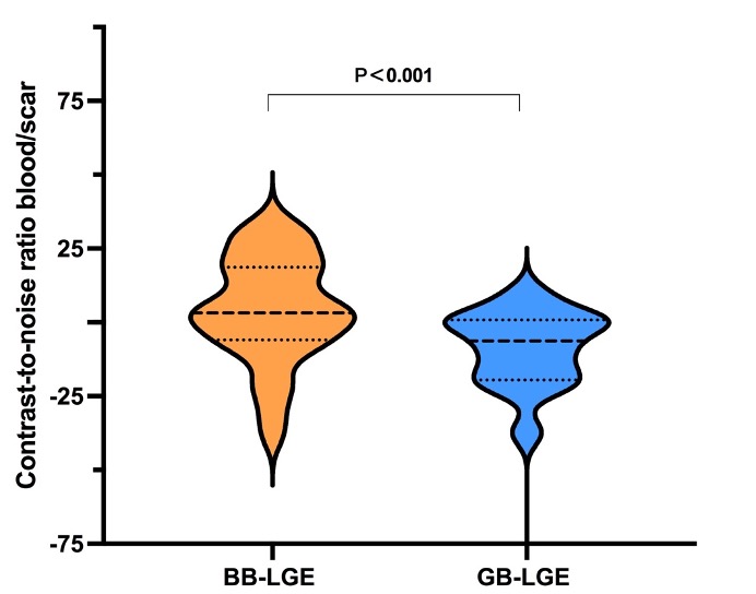
Compared to BB-LGE, GB-LGE significantly improved observer confidence scores (63.3% vs. 36.7%, p=0.039). Quantitative analysis of myocardial scar revealed no significant differences in scar mass [14.6 (8.3, 23.6) g vs. 13.6 (7.7, 24.2) g, p=0.451] and scar extent (LGE %) [14.2 (6.4, 26.5) % vs. 14 (7.9, 21.7) %, p=0.379] between these two imaging methods. However, CNR between the blood pool and myocardial scar significantly improved [-6.3 (-19.5, 0.84) vs. 3.2 (-5.9, 18.7), p<0.001], with no significant difference in CNR between myocardial scar and normal myocardium [30.3 (20.6, 46.7) vs. 30.1 (19.5, 65.7), p=0.271].Discussion
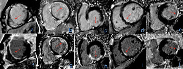
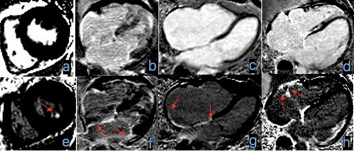
This study aims to assess the novel GB-LGE (Gray Blood Late Gadolinium Enhancement) imaging method for the detection of myocardial scars, all without necessitating additional MT-prep or T2-prep pulses and without modifying scanning protocol parameters. This method has been validated across multiple field strength devices. The results demonstrate that GB-LGE exhibits greater sensitivity compared to BB-LGE in detecting subendocardial delayed enhancement regions that are challenging to visualize using traditional techniques. Additionally, GB-LGE enhances the contrast-to-noise ratio (CNR) between scar and blood pools, improving image contrast between scar tissue and normal myocardium, thereby enhancing diagnostic confidence among observers. Furthermore, it exhibits advantages in the detection of left ventricular extramural delayed enhancement.Conclusions
GB-LGE, as developed in this study, outperforms traditional BB-LGE in myocardial scar detection. It enhances image contrast and reader diagnostic confidence. GB-LGE is not constrained by specific equipment or technology, offering a straightforward operation process and efficient image optimization. These attributes significantly enhance clinical work efficiency and hold substantial clinical application and promotion value.Acknowledgements
This work was supported by the National Natural Science Foundation of China (Grant No. 81873908, for HJH).References
[1] Kim H W, Rehwald W G, Jenista E R, et al. Dark-Blood Delayed Enhancement Cardiac Magnetic Resonance of Myocardial Infarction[J]. JACC Cardiovasc Imaging, 2018, 11(12): 1758-1769.
[2] Francis R, Kellman P, Kotecha T, et al. Prospective comparison of novel dark blood late gadolinium enhancement with conventional bright blood imaging for the detection of scar[J]. J Cardiovasc Magn Reson, 2017, 19(1): 91.
[3] Holtackers R J, Van De Heyning C M, Nazir M S, et al. Clinical value of dark-blood late gadolinium enhancement cardiovascular magnetic resonance without additional magnetization preparation[J]. J Cardiovasc Magn Reson, 2019, 21(1): 44.
[4] Holtackers R J, Van De Heyning C M, Chiribiri A, et al. Dark-blood late gadolinium enhancement cardiovascular magnetic resonance for improved detection of subendocardial scar: a review of current techniques[J]. J Cardiovasc Magn Reson, 2021, 23(1): 96.
[5] Kellman P, Xue H, Olivieri L J, et al. Dark blood late enhancement imaging[J]. J Cardiovasc Magn Reson, 2016, 18(1): 77.
[6] Liu C-Y, Wieben O, Brittain J H, et al. Improved delayed enhanced myocardial imaging with T2-Prep inversion recovery magnetization preparation[J]. Journal of Magnetic Resonance Imaging, 2008, 28(5): 1280-1286.
[7] Foo T K F, Wolff S D, Gupta S N, et al. Enhanced viability imaging: Improved contrast in myocardial delayed enhancement using dual inversion time subtraction[J]. Magnetic Resonance in Medicine, 2005, 53(6): 1484-1489.
Figures