1506
Free Breathing Single-Beat Myocardial Late Gadolinium Enhancement Imaging1Department of Medicine (Cardiovascular Division), Beth Israel Deaconess Medical Center and Harvard Medical School, Boston, MA, United States, 2Siemens Medical Solutions USA, Boston, MA, United States, 3Cardiovascular MR R&D, Siemens Healthcare Ltd., Calgary, AB, Canada, 4Radiology, Beth Israel Deaconess Medical Center and Harvard Medical School, Boston, MA, United States
Synopsis
Keywords: Myocardium, Cardiovascular
Motivation: To overcome the limitations of LGE imaging, including respiratory motion artifacts and lengthy scan times, while enhancing myocardial scar imaging.
Goal(s): To develop a deep learning-based free-breathing single-beat LGE.
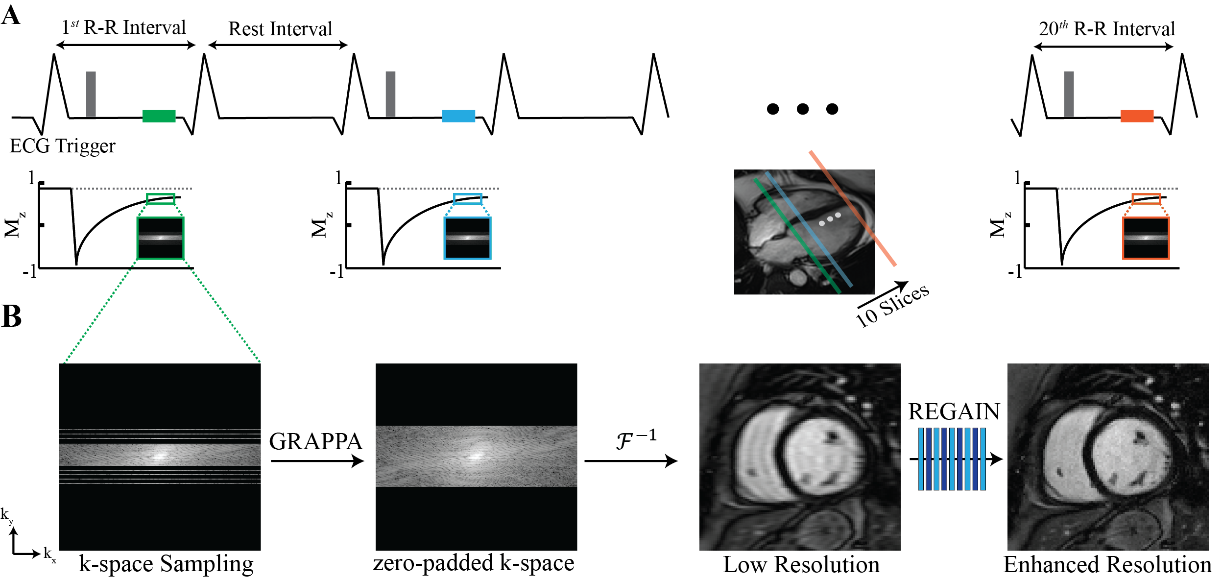
Approach: Free-breathing single-beat low-resolution 2D LGE images are acquired and followed by resolution enhancement generative adversarial inline neural network (REGAIN) to enhance the spatial resolution. Each slice was acquired in a single beat, followed by one beat for signal recovery. The entire left ventricular dataset was acquired in 20 heartbeats.
Results: REGAIN improved image sharpness and quality of single-beat 2D LGE acquired with 4.7-fold acceleration with spatial resolution of 1.5 × 5 mm2.
Impact: A rapid single-beat 2D LGE imaging can reduce CMR scan time, increase patient comfort, and reduce sensitivity to breathing motion.
INTRODUCTION
Late gadolinium enhancement (LGE) CMR is commonly used for imaging scars. In LGE, images are often acquired using an ECG-segmented data acquisition. Typically, 2D LGE imaging is collected within a single breath-hold per slice. The acquisition time usually takes 3-5 minutes for full left ventricular (10 slices) coverage. Alternatively, a single shot with or without multiple averaging can be used; however, this often results in lower spatial resolution or artifacts/blur due to misregistration or errors in registration. 3D LGE has also been proposed using respiratory navigators or self-gating approaches; however, scan time remains long. Parallel imaging can be used in 2D LGE, however, acceleration rate is limited to $$$\leq$$$2. In this study, we sought to develop a free-breathing single-beat 2D LGE imaging technique that allows full left ventricular (LV) coverage within 20 heartbeats. Image resolution was enhanced using a deep-learning model, resolution enhancement generative adversarial inline neural network (REGAIN)1.METHODS
This study aims to acquire a whole-heart LGE in just 20 heartbeats by acquiring a reduced k-space equivalent to a low-resolution image at every other heartbeat, with sufficient intervals for signal recovery. A schematic of the acquisition is depicted in Fig. 1A. Only 30% of the total k-space is acquired at every other heartbeat while keeping 32 lines in the center of k-space and 2-fold GRAPPA acceleration followed by zero-padding to the intended matrix size. Subsequently, REGAIN is used to reconstruct these low-resolution images (Fig.1 B).REGAIN Reconstruction:
REGAIN is a deep learning-based reconstruction technique built on the enhanced super-resolution generative adversarial network (ESRGAN). REGAIN uses densely connected convolutions with residual blocks within its generator while employing a relativistic average GAN for the discriminator to generate realistic details. In addition to ESRGAN loss functions, REGAIN adds constraints on the spatial frequency with an $$$\ell_1$$$ fast-Fourier transform. The acquired reduced k-space with 2-fold in-plane acquisition was first reconstructed with GRAPPA. This reconstructed k-space does not have full phase-encode acquisition and corresponds to a low-resolution image. Subsequently, REGAIN enhances the image resolution, and a schematic of the reconstruction pipeline is given in Fig. 1C. REGAIN was trained on 1616 subjects with ECG-gated segmented cine acquisitions and was not retrained with LGE data; instead, the pre-trained model on cine data was utilized.
Imaging Experiments: We prospectively recruited 23 subjects (60 ± 14 years, 11 males) referred for a clinical CMR exam for LGE imaging. Imaging was performed at 3T (MAGNETOM Vida Siemens Healthineers, Erlangen, Germany) with the following parameters: TR/TE = 2.9/1.5 ms, α = 52°, FOV = 300-400 × 300-400 mm2, acquisition matrix = 256 × 64, in-plane resolution = 1.5 × 5 mm2, acquisition window = 120 ms, slice-thickness = 10mm. 32 fully-sampled center phase encoding lines per cardiac cycle with 2-fold GRAPPA acceleration were acquired. This acquisition was zero-filled to a matrix size = 256 × 224 which represents the low-resolution image. REGAIN was integrated with the Siemens Framework for Image Reconstruction (FIRE)2 prototype and reconstructed the images from voxel size = 1.5 × 5 mm2 to 1.5 × 1.5 mm2.
RESULTS
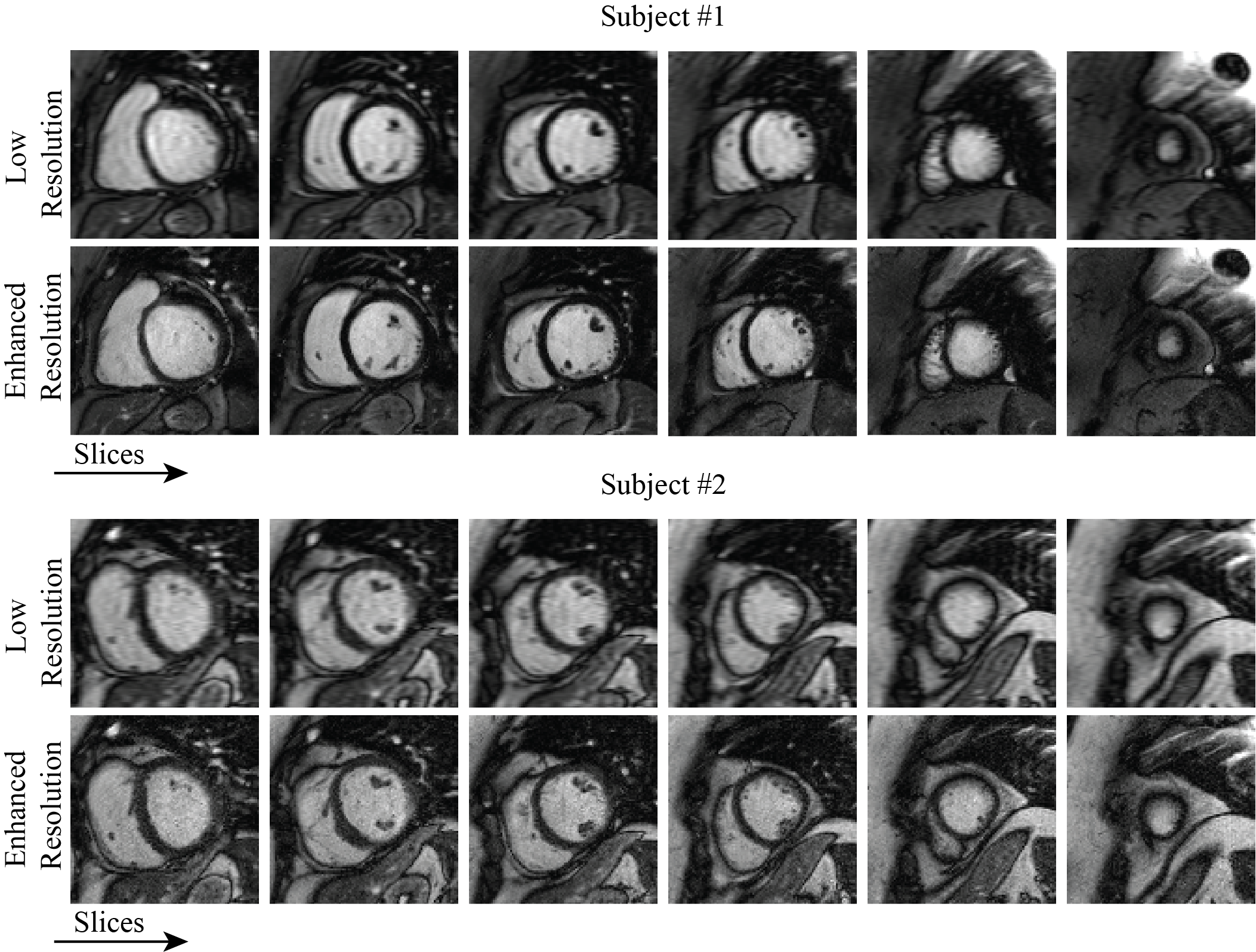
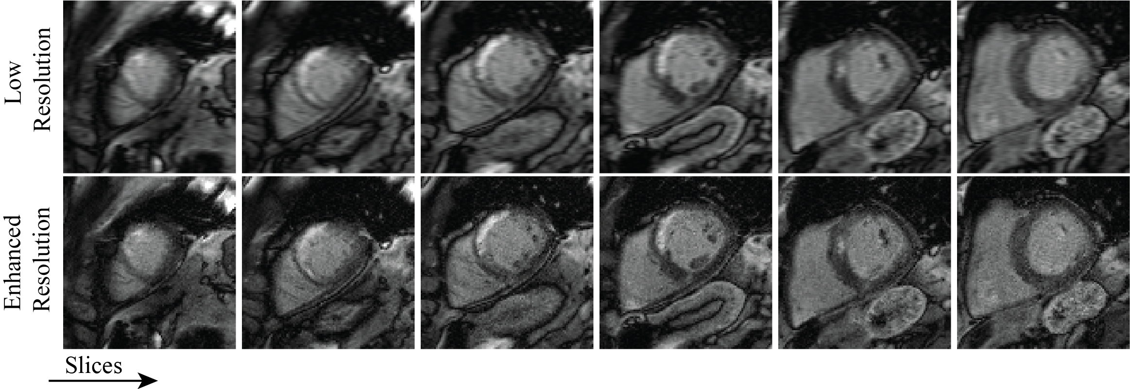
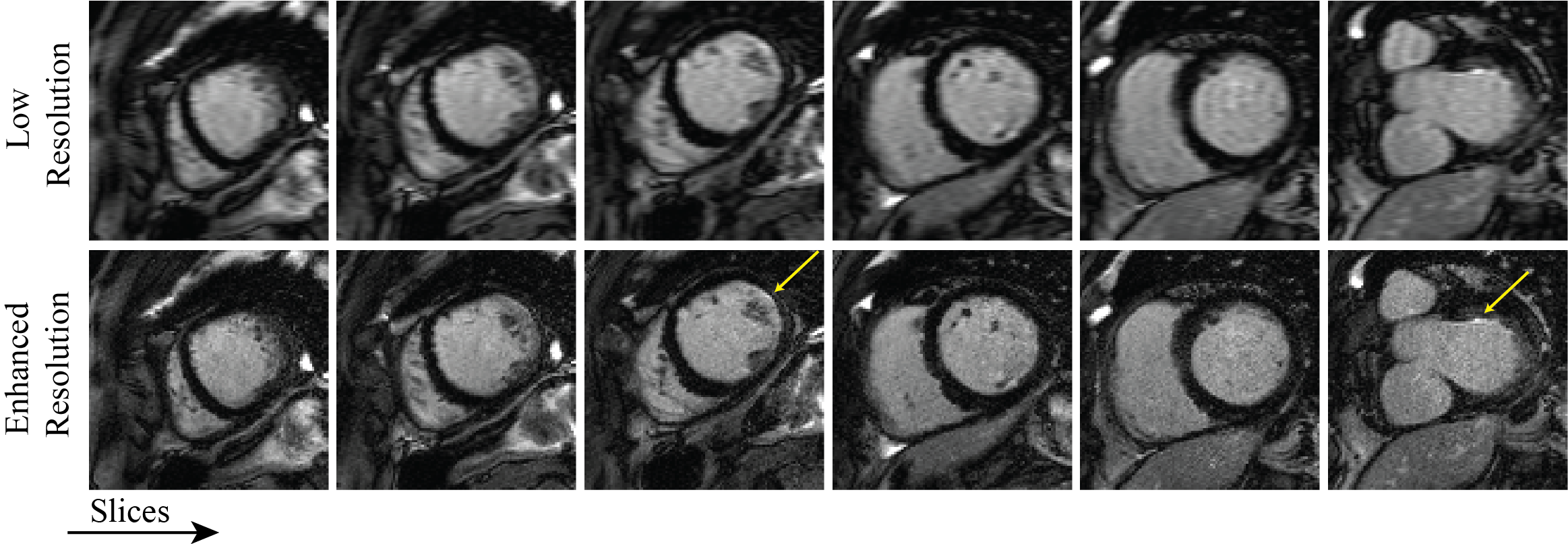
Two subjects with no LGE presence are depicted in Fig. 2. REGAIN reconstruction visually improves image quality and enhances spatial resolution. Fig. 3 shows one subject with LGE in the mid to apical walls. Enhanced images exhibit improved image quality at high resolution, leading to better delineation of scar tissues without additional artifacts. Another subject with subendocardial LGE in the mid-anterolateral wall is depicted in Fig. 4, where REGAIN successfully enhances the scar.CONCLUSION
4.7-fold accelerated, REGAIN-reconstructed single-beat LGE enables full LV coverage in 20 heartbeats without breath-holding. Further evaluation is necessary to investigate the impact of acceleration on the quantification of scar burden.Acknowledgements
Funding: This work is supported by the National Institutes of Health.References
1) Yoon S, Nakamori S, Amyar A, Assana S, Cirillo J, Morales MA, et al. Accelerated Cardiac MRI Cine with Use of Resolution Enhancement Generative Adversarial Inline Neural Network. Radiology 2023;307(5).
2) Chow K, Kellman P, Xue H. Prototyping Image Reconstruction and Analysis with FIRE. In: proc. SCMR. Virtual Scientific Sessions; 2021. p. 838972.
Figures