1504
Rapid 2D Myocardial Late Gadolinium Enhancement Imaging with Resolution Enhancement Generative Adversarial Inline Neural Network1Department of Medicine (Cardiovascular Division), Beth Israel Deaconess Medical Center and Harvard Medical School, Boston, MA, United States, 2Siemens Medical Solutions USA, Boston, MA, United States, 3Cardiovascular MR R&D, Siemens Healthcare Ltd., Calgary, AB, Canada, 4Radiology, Beth Israel Deaconess Medical Center and Harvard Medical School, Boston, MA, United States
Synopsis
Keywords: Myocardium, Cardiovascular
Motivation: 2D high-resolution late gadolinium enhancement (LGE) imaging can benefit from shorter scan times. Current acceleration techniques lead large signal-to-noise (SNR) penalties, reducing diagnostic quality while longer breath-holds increases scan time and makes the imaging susceptible to artifacts.
Goal(s): To assess the feasibility of a rapid 2D LGE imaging using Resolution Enhancement Generative Adversarial Inline Neural Network (REGAIN).
Approach: Images were acquired with 3.3 and 5.7-fold accelerations, reconstructed using REGAIN, and compared with 1.8-fold GRAPPA acceleration.
Results: REGAIN successfully improved visual image sharpness in LGE images acquired with 3.3-fold (~6-second) and 5.7-fold (~10-second) accelerations. Image quality was comparable to 1.8-fold (~16-second) GRAPPA acceleration.
Impact: REGAIN enables accelerated LGE imaging with significantly reduced breath-hold duration.
INTRODUCTION
Late gadolinium enhancement (LGE) imaging is widely used in cardiovascular magnetic resonance (CMR) to visualize and characterize myocardial fibrosis. 2D LGE is typically collected using 10-12 breath-holds (2-4 min of scan time). Increasing in-plane spatial resolution increases breath-hold duration, thereby burdening patients with repeated breath-holds. To reduce breath-hold duration, parallel imaging or compressed sensing can be used1,2; however, these techniques have limited utility in 2D LGE and reduce diagnostic image quality due to SNR penalties. In this study, we sought to develop and evaluate an accelerated 2D LGE imaging technique using resolution enhancement generative adversarial inline neural network (REGAIN)3.METHODS
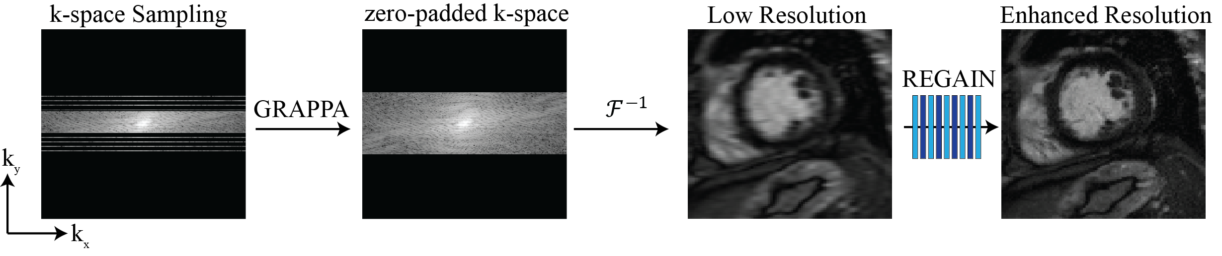
Fig. 1 shows the proposed image acquisition and reconstruction schematic for rapid 2D LGE imaging. Images are acquired by reducing the spatial resolution along the phase-encoding direction to reduce breath-hold duration, producing a reduced k-space sampling pattern. 25-50% of the k-space is collected using standard 2-fold GRAPPA acceleration followed by zero-filling to the intended matrix size. This leads to 3.3-5.7-fold acceleration that reduces the total acquisition time from 16 seconds to only 6 seconds. REGAIN is subsequently used to reconstruct these low-resolution images.REGAIN Reconstruction:
REGAIN is a deep learning reconstruction technique built on the enhanced super-resolution generative adversarial inline neural network (ESRGAN). It utilizes densely connected convolutions with residual blocks for its generator. Furthermore, it employs a discriminator with a relativistic average GAN technique to generate realistic textures. Additionally, REGAIN maps images into a spatial frequency domain using $$$\ell_1$$$ fast-Fourier transform as an additional constraint in its loss function. REGAIN was previously trained using cine images3. For this study, we did not modify the original model and applied the same model for LGE without any modification.
Imaging Experiments:
We prospectively recruited 80 patients (56 ± 14 years, 52 males) referred for a clinical CMR exam for scar evaluation. In each patient, we collected three sets of LGE images: (a) 1.8-fold GRAPPA with spatial resolution = 1.5 × 1.5 mm2, acquisition matrix: 240 × 240, total scan time = ~16 seconds, (b) 3.3-fold REGAIN with spatial resolution = 1.5×3 mm2, acquisition matrix: 240×120, total scan time=~10 seconds, (c) 5.7-fold REGAIN with spatial resolution = 1.5 × 6 mm2, acquisition matrix: 240×60, total scan time = ~6 seconds. All patients were imaged at 3T (MAGNETOM Vida Siemens Healthineers, Erlangen, Germany) and shared the following imaging parameters: TR/TE = 5.6/2.55 ms, α = 20°, FOV = 360 × 360 mm2, acquisition window = 100 ms, slice-thickness = 8 mm. All acquisitions were acquired with 24 ACS lines and 2-fold in-plane acceleration, first reconstructed with GRAPPA and zero-filled to the intended dimensions corresponding to the low-resolution image. Subsequently, these LGE images were reconstructed inline4 using REGAIN to enhance images to 1.5 × 1.5 mm2 resolution from their 1.5 × 3-6 mm2 counterparts.
RESULTS
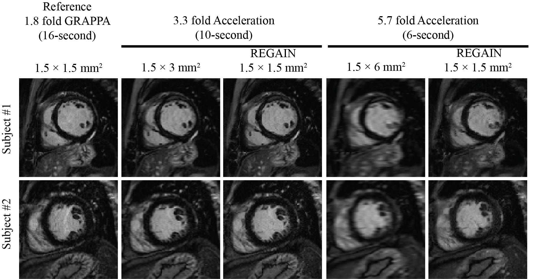
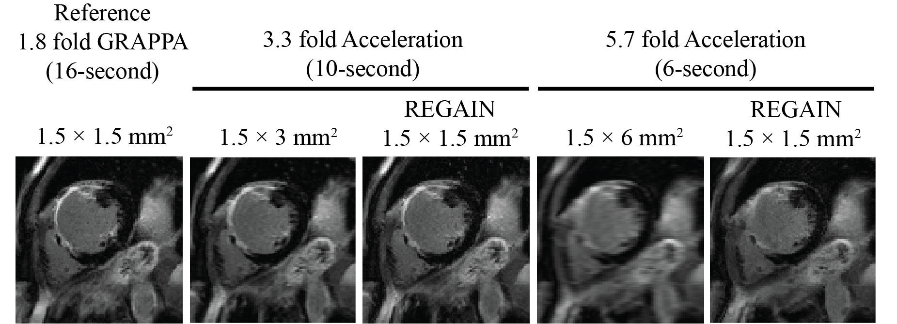
Fig. 2 shows two subjects with no LGE using 1.8-fold acceleration reconstructed with GRAPPA, and 3.3-fold and 5.7-fold accelerations reconstructed with REGAIN. REGAIN visually enhances the image quality at both acceleration rates. 2D LGE images in the mid slice in a patient with myocardial scars are depicted in Fig. 3. At 3.3 and 5.7-fold accelerations, REGAIN enhances the spatial resolution and shows visual improvements in sharpness compared to original low-resolution images. However, we observed that higher acceleration could negatively impact scar detection.CONCLUSION
REGAIN improves image sharpness of accelerated 2D LGE images, resulting in reduced breath-hold duration. Further studies are warranted to investigate how much acceleration can be achieved without affecting diagnostic image quality and its impact on quantification of scar burden.Acknowledgements
Funding: This work is supported by the National Institutes of Health.References
1) Griswold MA, Jakob PM, Heidemann RM, Nittka M, Jellus V, Wang JM, et al. Generalized Autocalibrating Partially Parallel Acquisitions (GRAPPA). Magnetic Resonance in Medicine 2002;47(6):1202-10.
2) Lustig M, Donoho DL, Santos JM, Pauly JM. Compressed sensing MRI. Ieee Signal Processing Magazine 2008;25(2):72-82.
3) Yoon S, Nakamori S, Amyar A, Assana S, Cirillo J, Morales MA, et al. Accelerated Cardiac MRI Cine with Use of Resolution Enhancement Generative Adversarial Inline Neural Network. Radiology 2023;307(5).
4) Chow K, Kellman P, Xue H. Prototyping Image Reconstruction and Analysis with FIRE. In: proc. SCMR. Virtual Scientific Sessions; 2021. p. 838972.
Figures