1500
Predicting Late Gadolinium Enhancement of Acute Myocardial Infarction in Contrast-free Cardiac Cine MRI using Deep Generative Learning1School of Biomedical Engineering, ShanghaiTech University, Shanghai, China, 2Shanghai Clinical Research and Trial Center, Shanghai, China, 3Department of Radiology, Ren Ji Hospital, School of Medicine, Shanghai Jiao Tong University, Shanghai, China
Synopsis
Keywords: Myocardium, Machine Learning/Artificial Intelligence
Motivation: Although late Gadolinium Enhancement (LGE) imaging is widely used for diagnosing myocardial infarction (MI), contrast-free approaches are in need for patients with gadolinium contraindications.
Goal(s): To develop Cine Generated Enhancement (CGE), a novel technique that uses contrast-free cine images to predict images resembling LGE.
Approach: A deep generative model was trained to translate cine images into LGE images of acute MI exploiting the different motion dynamics between heathy and infarcted myocardium.
Results: Realistic enhancement images can be generated for acute MI patients using cine images unseen during training. The scar size and transmurality estimated with CGE agreed well with LGE.
Impact: This study presents an effective, non-invasive, and contrast-free method for predicting LGE in acute MI, potentially reducing the use of gadolinium-based contrast agents and shortening cardiac MR examinations.
Introduction
Acute Myocardial Infarction (AMI) is one of the leading causes of cardiovascular-related deaths1. Late Gadolinium Enhancement (LGE) imaging is a widely used non-invasive method to detect MI, based on which, the estimated infarction parameters are crucial for evaluating AMI prognosis2–4. However, LGE imaging is not applicable to patients with gadolinium contraindications5,6. To fulfill this gap, promising deep learning methods have been proposed to evaluate MI from non-contrast cardiac MR. Some methods use motion-assisted deep recurrent learning based on cardiac cine MRI for myocardial scar segmentation7,8, which however requires manually segmented labels for supervised training, which are laborious and may introduce noise due to annotation variability9. Another type of methods termed as virtual native enhancement (VNE) proposes to generate LGE-like images from native cardiac T1 and cine images10,11. VNE methods do not require manual annotations, but the requirement of native T1 images adds hurdle to the network training as these images may not be acquired in acute patients due to limited examination times. In this work, we explored the feasibility of generating LGE images using only contras-free cine images, where a novel technique, Cine Generated Enhancement (CGE), was developed and validated in AMI patients. The scar size and transmurality were quantified in CGE images and compared with LGE.Methods
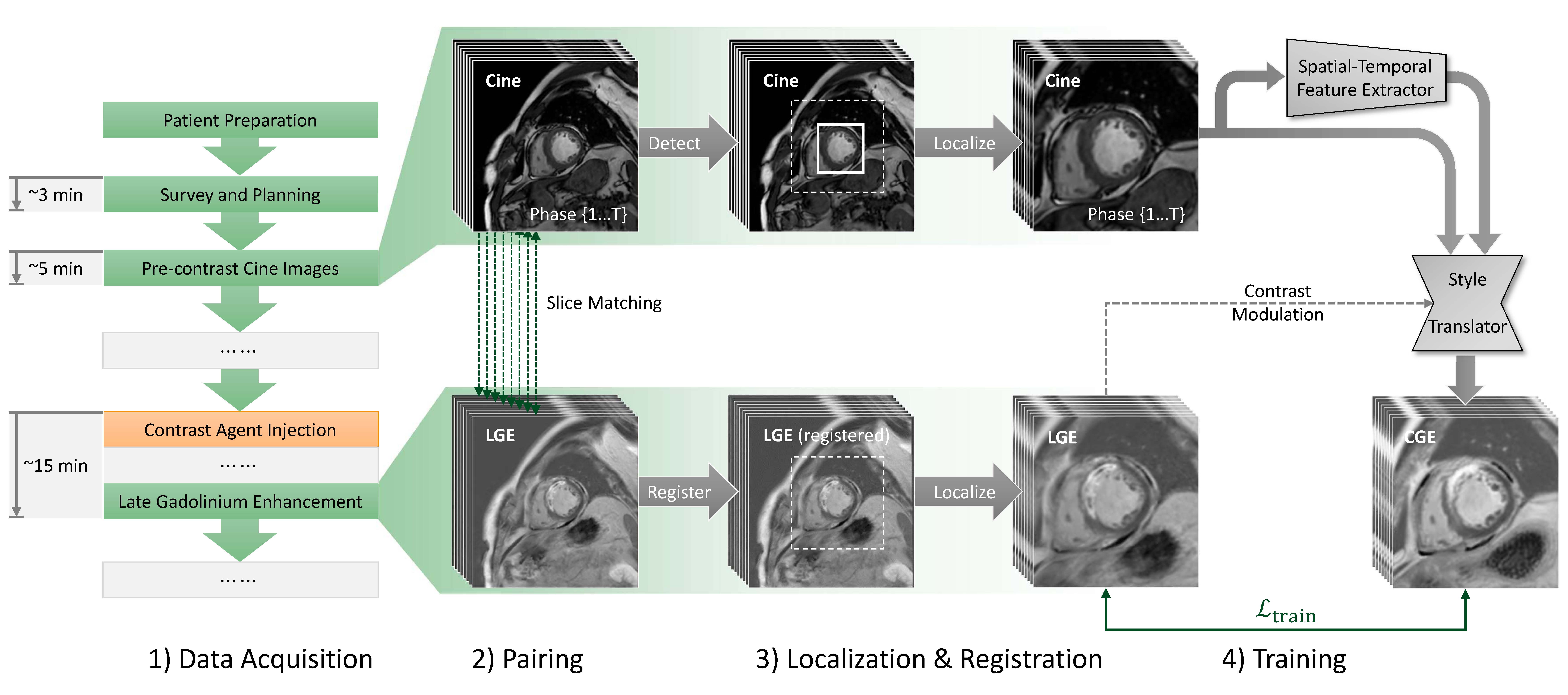
CGE modelFigure 1 illustrates the entire pipeline. We retrospectively enrolled patients diagnosed with AMI. The native cine images and LGE images acquired on a clinical 3.0T scanner were collected. A crucial step for successful training is to align the cine and LGE images. Briefly, the data pre-processing involved automatic slice pairing based on the imaging geometry parameters, localization of the heart region with a pre-trained detection network12 , and registration between the cine and LGE images. After a manual quality control process, excluding unmatched image datasets, 251 AMI patients were included, where 221 patients were used for training and the remining 30 patients were used for testing. A generative adversarial neural network was trained to translate cine images into CGE. In the generator, the network extracted spatial-temporal features from the cine images to translate the source image, which is one of the cine frames closest to the LGE image in cardiac shape, into a CGE. To address the varying contrasts in LGE, induced by the variability of the manually tuned inversion recovery time during LGE imaging, a contrast modulation approach was developed. A clustering algorithm was applied to classify the LGE contrasts, and then the contrast label was utilized to modulate the CGE generation process. For network training, a composite loss consisting of the generative adversarial loss and the loss term measuring the similarity between CGE and the corresponding LGE was adopted. Once trained, the network was employed to generate enhancement images of user-specified LGE contrast with input of native cine images.
CGE analysis
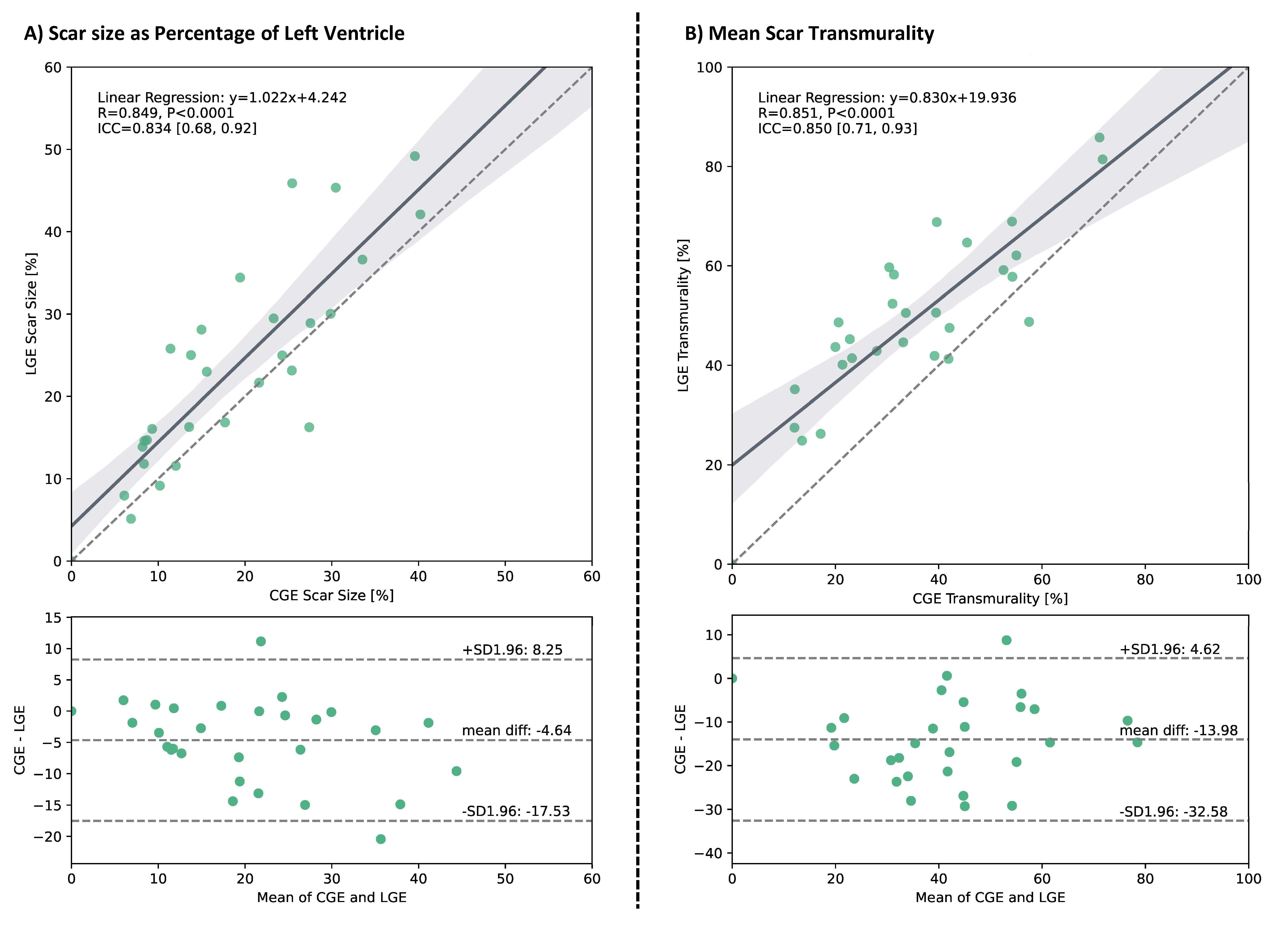
155 CGE-LGE image pairs from the testing AMI patients were used to assess CGE. Epicardial and endocardial contours were delineated manually respectively in CGE and LGE images by an experienced cardiologist who was blinded to the image types. Then the scar region of interests were segmented using the FWHM method2, from which the scar volume fraction and mean scar transmurality11 were calculated. The correlation and consistency of scar measurements with CGE and LGE were evaluated using Linear regression, Bland-Altman analysis, and Intraclass Correlation Coefficient (ICC)13.
Results
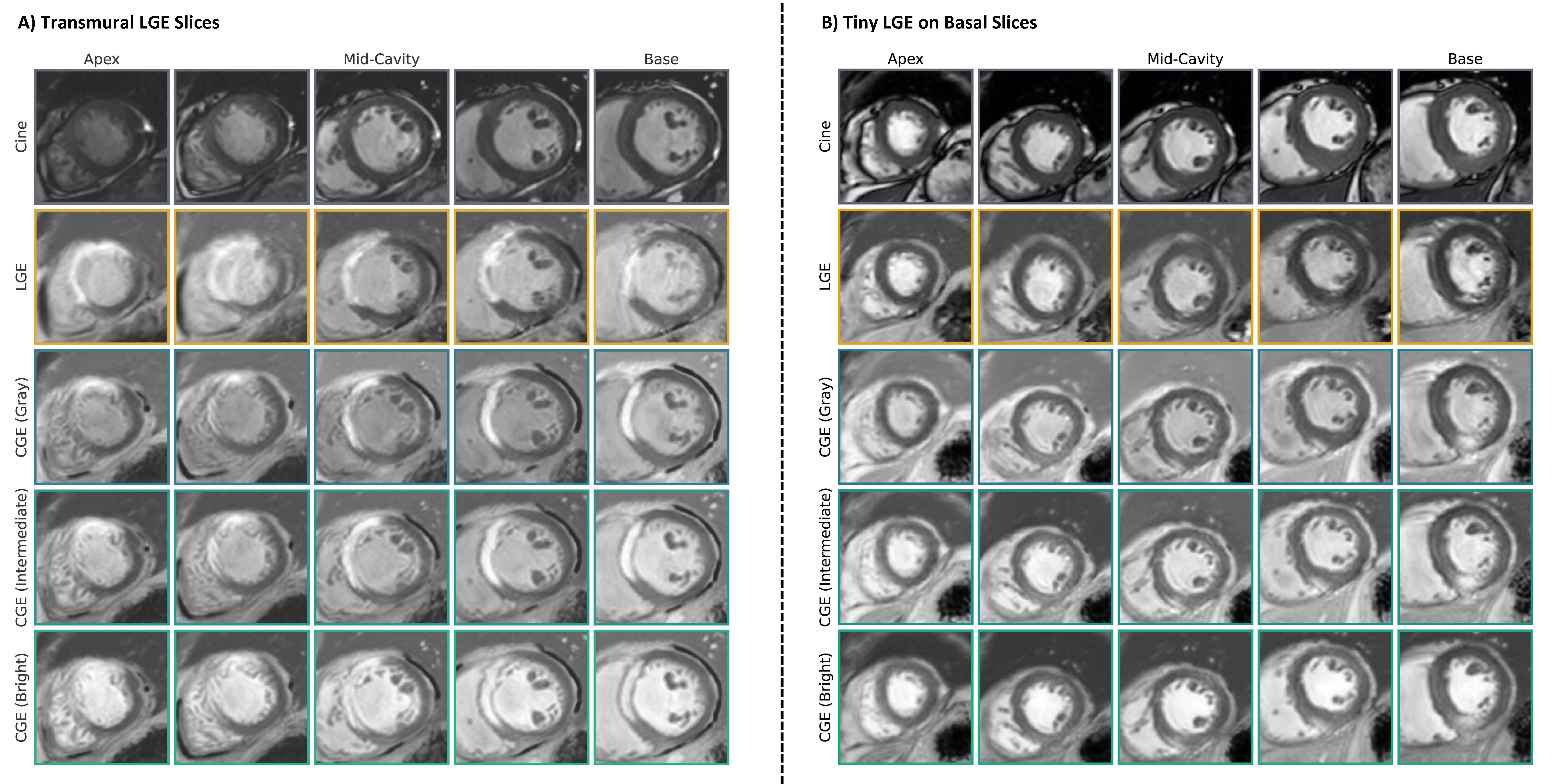
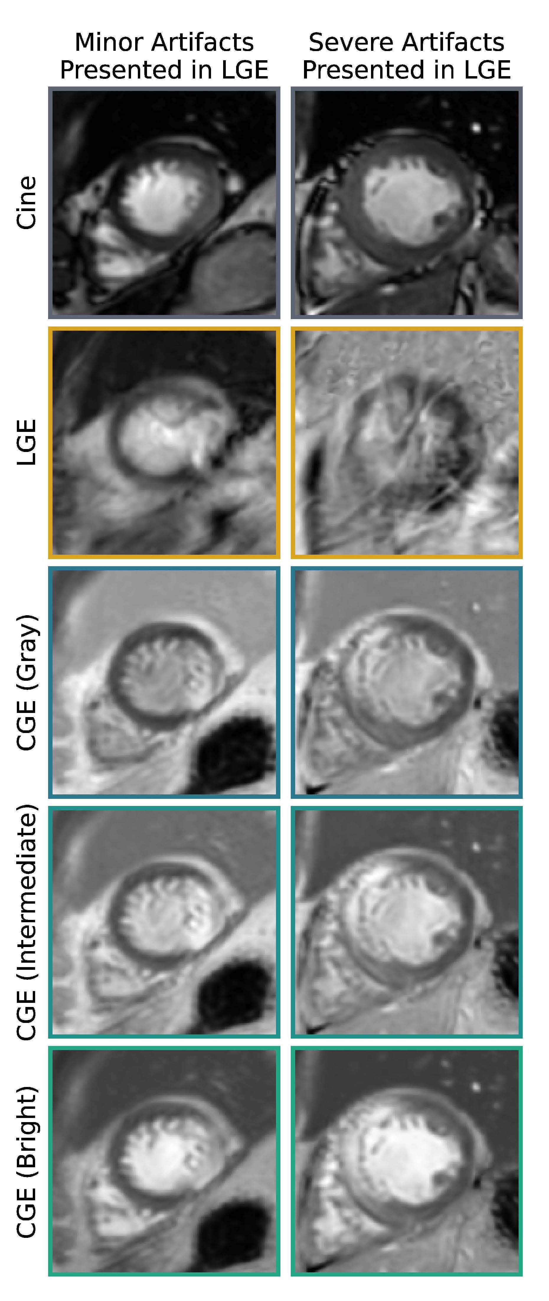
Figure 2 presents two representative patients, highlighting the good agreement of enhanced myocardium between CGE and LGE across the left ventricle for various CGE contrasts. Overall, CGE closely resembles LGE for the two patients, while part of the scar in the apical slices of one patient (Fig. 2A) was only moderately enhanced. Figure 3 illustrates two subjects with minor and severe respiratory motion artifacts in LGE images, while the CGE images are free of motion artifacts and display the enhancement area clearly. The statistical analysis (Fig. 4) indicates a significant (p<0.0001) correlation between CGE and LGE for measuring scar size and transmurality. The Bland-Altman analysis indicates the CGE images slightly underestimate the scar size and transmurality.Discussion
This study presents a novel cine generated enhancement approach, which has shown superior performance in predicting LGE of AMI. The proposed method requiring only cardiac cine images which are widely acquired, holds potential for clinical integration to reduce contrast usage and shorten cardiac MR examinations. Future work will explore post-processing corrections to address the scar underestimation of CGE and expand the patient population for further validation.Acknowledgements
No acknowledgement found.References
1. Roth, G. A. et al. Global Burden of Cardiovascular Diseases and Risk Factors, 1990–2019. Journal of the American College of Cardiology 76, 2982–3021 (2020).
2. Amado, L. C. et al. Accurate and objective infarct sizing by contrast-enhanced magnetic resonance imaging in a canine myocardial infarction model. Journal of the American College of Cardiology 44, 2383–2389 (2004).
3. Roes, S. D. et al. Comparison of myocardial infarct size assessed with contrast-enhanced magnetic resonance imaging and left ventricular function and volumes to predict mortality in patients with healed myocardial infarction. The American journal of cardiology 100, 930–936 (2007).
4. Wu, E. et al. Visualisation of presence, location, and transmural extent of healed Q-wave and non-Q-wave myocardial infarction. The Lancet 357, 21–28 (2001).
5. Ledneva, E., Karie, S., Launay-Vacher, V., Janus, N. & Deray, G. Renal Safety of Gadolinium-based Contrast Media in Patients with Chronic Renal Insufficiency. Radiology 250, 618–628 (2009).
6. Behzadi, A. H., Zhao, Y., Farooq, Z. & Prince, M. R. Immediate Allergic Reactions to Gadolinium-based Contrast Agents: A Systematic Review and Meta-Analysis. Radiology 286, 471–482 (2018).
7. Zhang, N. et al. Deep Learning for Diagnosis of Chronic Myocardial Infarction on Nonenhanced Cardiac Cine MRI. Radiology 291, 606–617 (2019).
8. Xu, C. et al. Segmentation and quantification of infarction without contrast agents via spatiotemporal generative adversarial learning. Medical Image Analysis 59, 101568 (2020).
9. Flett, A. S. et al. Evaluation of techniques for the quantification of myocardial scar of differing etiology using cardiac magnetic resonance. JACC: cardiovascular imaging 4, 150–156 (2011).
10. Zhang, Q. et al. Toward Replacing Late Gadolinium Enhancement With Artificial Intelligence Virtual Native Enhancement for Gadolinium-Free Cardiovascular Magnetic Resonance Tissue Characterization in Hypertrophic Cardiomyopathy. Circulation 144, 589–599 (2021).
11. Zhang, Q. et al. Artificial Intelligence for Contrast-Free MRI: Scar Assessment in Myocardial Infarction Using Deep Learning–Based Virtual Native Enhancement. Circulation 146, 1492–1503 (2022).
12. Liu, W. et al. Ssd: Single shot multibox detector. in European conference on computer vision 21–37 (Springer, 2016).
13. Koo, T. K. & Li, M. Y. A Guideline of Selecting and Reporting Intraclass Correlation Coefficients for Reliability Research. J Chiropr Med 15, 155–163 (2016).
Figures