1498
Patch-based Motion-compensated Image Filter to Improve SNR of Free-Breathing Whole Heart Cardiac MR Late Gadolinium Enhancement Images at 0.55T1The Ohio State University, Columbus, OH, United States, 2Siemens Healthineers, Columbus, OH, United States
Synopsis
Keywords: Myocardium, Myocardium, Late Gadolinium Enhancement , infarction
Motivation: Low-field wide-bore MRI scanners are cost-effective but suffer from lower SNR, impacting myocardial LGE image quality.
Goal(s): To introduce and evaluate a novel imaging strategy that offers full heart coverage and reduces image count by 50%.
Approach: Developed a sequence acquiring 45-55 overlapped short-axis 2-D slices with patch-based motion compensated filtering to enhance SNR.
Results: The new technique improved SNR in whole heart coverage LGE imaging at 0.55T, though further studies with scarred myocardium are needed.
Impact: This study advances cardiac MRI by demonstrating that a novel LGE imaging technique coupled with MC-KW patch filtering substantially enhances SNR in low-field wide-bore scanners, promising improved myocardial scar detection at reduced costs.
Introduction
Low-field wide-bore MRI scanners present a potentially cost-effective complement to their smaller bore, higher field counterparts. Yet, the inherent reduction in signal-to-noise ratio (SNR) can compromise image quality, particularly in myocardial late gadolinium enhancement (LGE). Post-reconstruction motion correction (MOCO) followed by averaging is used conventionally to boost SNR [1]. The conventional free-breathing LGE protocol includes the acquisition of 10 to 12 slices, each with a thickness of 8 mm and an interslice gap of 2 mm, along with 8 to 12 repetitions per slice, requiring a total of 80 to 144 images, and 160 to 288 heartbeats to complete. Therefore, this method prolongs data acquisition time and limits the number of slices covering the whole heart. In this study, we introduce and assess an innovative strategy that can cover the whole heart without gap while concomitantly diminishing the aggregate image count by approximately 50%. Our innovative technique acquires a stack of overlapped, single-shot, short-axis 2-D slices (a total of 45 to 55 images) covering the whole heart, and incorporates a patch-based motion compensated filtering method based on random matrix theory to improve the SNR of 0.55T LGE images.Methods
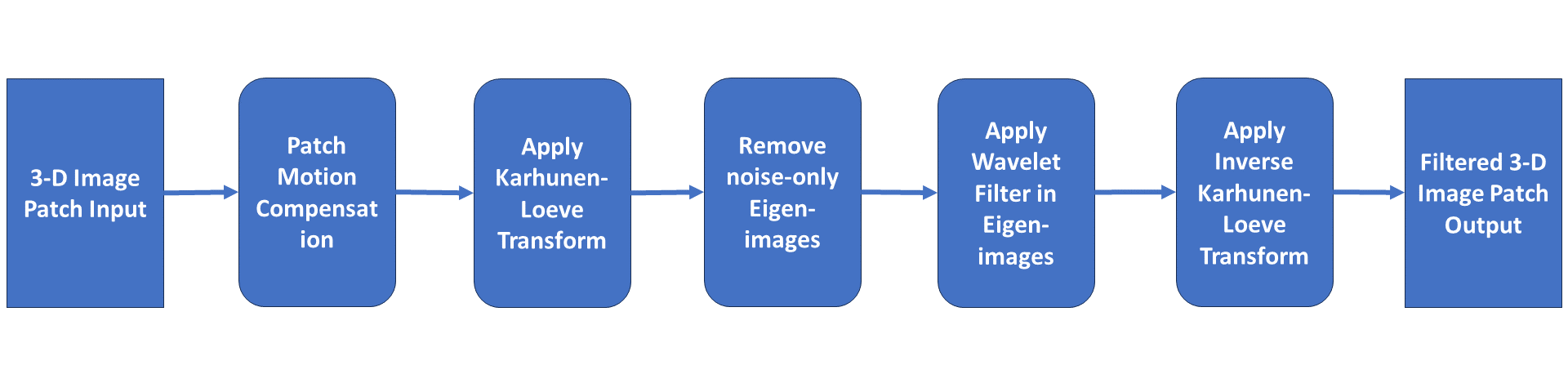
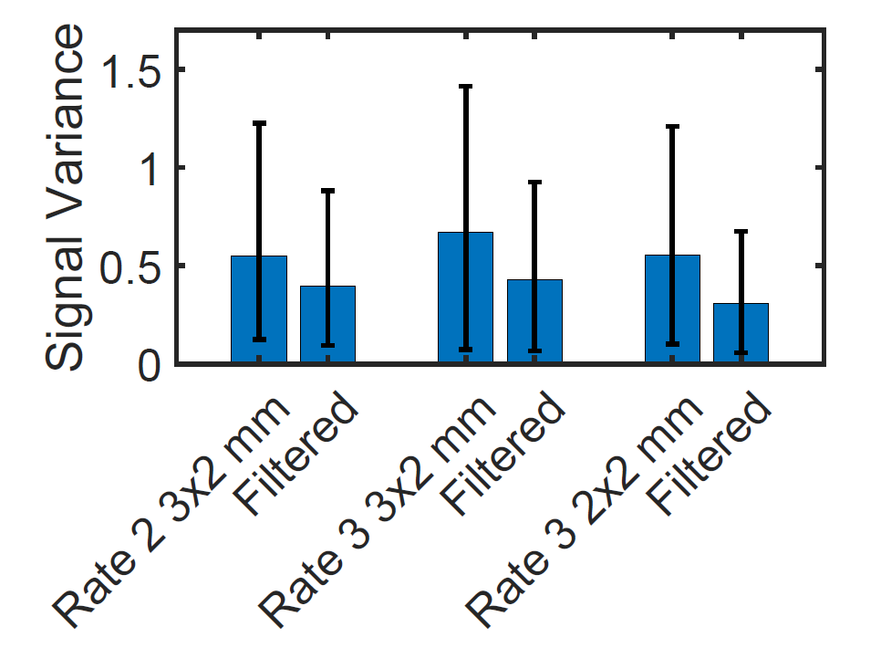
All data were acquired at 0.55T (MAGNETOM Free.Max, Siemens Healthcare, Erlangen, Germany) using an inversion recovery-prepared bSSFP research sequence with GRAPPA reconstruction. Single-shot, single acquisition, short-axis stacks of 2D LGE images encompassing the left ventricle were acquired in four healthy volunteers and one patient with known myocardium scar tissue; 75% slice overlap (8mm slices overlapped by 6mm) was used to augment cross-slice correlation. The reduced gradient performance of this system resulted in TE/TR of 2.0ms/4.6ms and shot time ranging from 290 to 308 ms. For every subject, three sets of 2D LGE image stacks were acquired using the following settings to evaluate SNR at different spatial resolutions and acceleration rates: voxel dimensions of 2.0x2.0x8 mm with GRAPPA rate 3, 2.0x3.0x8 mm with GRAPPA rate 2 and rate 3. The proposed filter (Figure 1) applies to 3D patches of a stack of 2D images, combining rigid motion compensation [2], the Karhunen-Loeve Transform (KLT) [3,4], random matrix Marchenko-Pastur law eigen-mode selection [4], and the spatial wavelet filter [5]. This approach is termed the Motion Compensated KLT Wavelet (MC-KW) patch filter [6]. The proposed filter was retrospectively applied to all 15 LGE image series. The myocardial signal variability was assessed and compared between the filtered and unfiltered LGE images, utilizing the standard deviation of the signal over the mean signal within a myocardial region of interest as a measure of noise. A t-test was employed to evaluate the statistical significance of the observed differences.Results
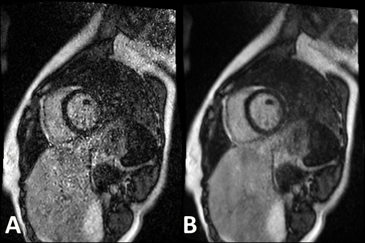
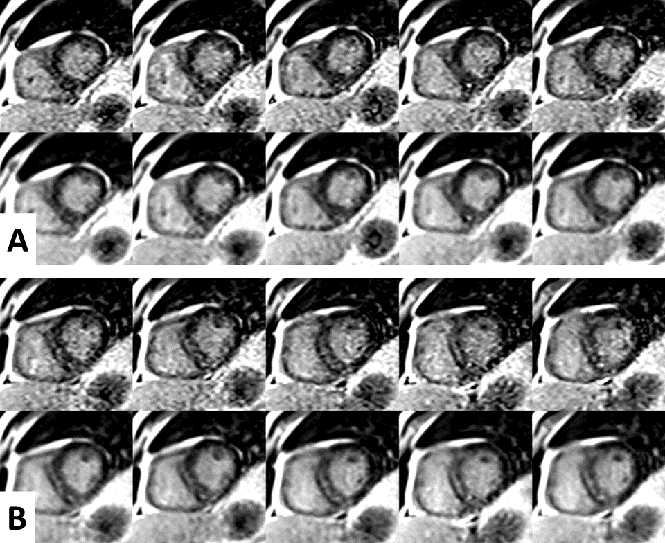
The image SNR exhibited a noticeable enhancement through filtering, as evident in Figure 2. Figure 3 presents a comparative analysis of unfiltered and filtered images of ten consecutive slices, highlighting the enhanced visualization of scar tissue within the interventricular septum. The mid-myocardial basal to mid antero-septal and infero-septal enhancement aligns with clinically documented findings from a prior 3T exam for this patient. Figure 4 shows the myocardial signal variation in healthy volunteers before and after filtering, indicating significantly lower noise in the filtered images.Conclusion
This investigation elucidates that a fast, free-breathing, single-shot, single-acquisition LGE with whole heart coverage technique, when combined with a MC-KW patch filtering approach, accomplishes superior SNR at 0.55T. Our dataset predominantly focused on healthy subjects without myocardial scars, necessitating further evaluation in patients exhibiting infarct scars and fibrosis.Acknowledgements
No acknowledgement found.References
[1] Kellman, Peter, et al. "Motion‐corrected free‐breathing delayed enhancement imaging of myocardial infarction." Magnetic Resonance in Medicine: An Official Journal of the International Society for Magnetic Resonance in Medicine 53.1 (2005): 194-200.
[2] Chen, Xiao, et al. "Motion‐compensated compressed sensing for dynamic contrast‐enhanced MRI using regional spatiotemporal sparsity and region tracking: Block low‐rank sparsity with motion‐guidance (BLOSM)." Magnetic resonance in medicine 72.4 (2014): 1028-1038.
[3] Ding, Yu, et al. "Application of the Karhunen–Loeve transform temporal image filter to reduce noise in real-time cardiac cine MRI." Physics in Medicine & Biology 54.12 (2009): 3909.
[4] Ding, Yu, et al. "A method to assess spatially variant noise in dynamic MR image series." Magnetic Resonance in Medicine 63.3 (2010): 782-789.
[5] Chang, S. Grace, Bin Yu, and Martin Vetterli. "Adaptive wavelet thresholding for image denoising and compression." IEEE transactions on image processing 9.9 (2000): 1532-1546.
[6] Ding, Yu, Palaniappan, P., & Simonetti, O. P. (2016). U.S. Patent No. 9,269,127.
Figures