1487
Free-breathing 3D whole-heart simultaneous bright- and black-blood anatomical imaging and T1/T2 mapping at 0.55T1Department of Diagnostic and Interventional Radiology, Klinikum rechts der Isar, Technical University of Munich, Munich, Germany, 2Institute for Advanced Study, Technical University of Munich, Munich, Germany, 3School of Biomedical Engineering and Imaging Sciences, King's College London, London, United Kingdom, 4Millenium Institute for intelligent Healthcare Engineering, Santiago, Chile, 5Institute for Biological and Medical Engineering, Pontificia Universidad Católica de Chile, Santiago, Chile, 6MR Research Collaborations, Siemens Healthcare Limited, Camberley, United Kingdom, 7School of Engineering, Pontificia Universidad Católica de Chile, Santiago, Chile
Synopsis
Keywords: Myocardium, Heart, Bright- and black-blood imaging, T1/T2 mapping
Motivation: To overcome the limitations of clinical examinations, which require several sequential acquisitions under multiple breath-holds, and to make cardiac MR more accessible and affordable at lower field strength.
Goal(s): To develop a novel 3D free-breathing sequence for simultaneous assessment of cardiovascular anatomy via bright- and black-blood imaging and myocardial tissue quantification in an easy to use one-click-scan at 0.55T.
Approach: Implementation of a novel iNAV-based 5-heartbeat interleaved sequence (proACTION) with distinct IR and T2 preparation modules and non-rigid motion correction at 0.55T.
Results: proACTION provides good delineation of cardiac and vascular structures with accurate joint T1/T2 parametrical mapping in healthy subjects.
Impact: Comprehensive 3D whole-heart evaluation of cardiovascular anatomy and tissue characterization with T1/T2 maps can be obtained in an efficient, free-breathing, and easier to use one-click-scan with proACTION at 0.55T.
Introduction
Clinically established cardiovascular MR (CMR) methods enable the non-invasive analysis of cardiovascular anatomy, function, and myocardial tissue characterization1. Conventional CMR2 involves multiple 2D scans acquired in different orientations under breath-hold conditions, resulting in long examination times and patient fatigue. To overcome these challenges, a free-breathing motion-compensated 3D sequence for simultaneous Assessment of whole-heart Cardiovascular anaTomy via bright- and black-blood Imaging and myocardial tissue quantificatiON (ACTION3) was proposed at a 1.5T. On the other hand, low-field MRI is promising to make CMR more accessible and affordable. However, there is limited experience with 3D whole-heart anatomical imaging and parametric mapping at 0.55T. Recently, a 3D sequence providing good visualization of cardiac vessels was proposed at low-field4. In this study, we propose an imPROved ACTION method (proACTION) on a clinical 0.55T scanner, exploiting the shorter T1 relaxation times, reduced specific absorption rate and fewer B0/B1 inhomogeneities at this field strength.Methods
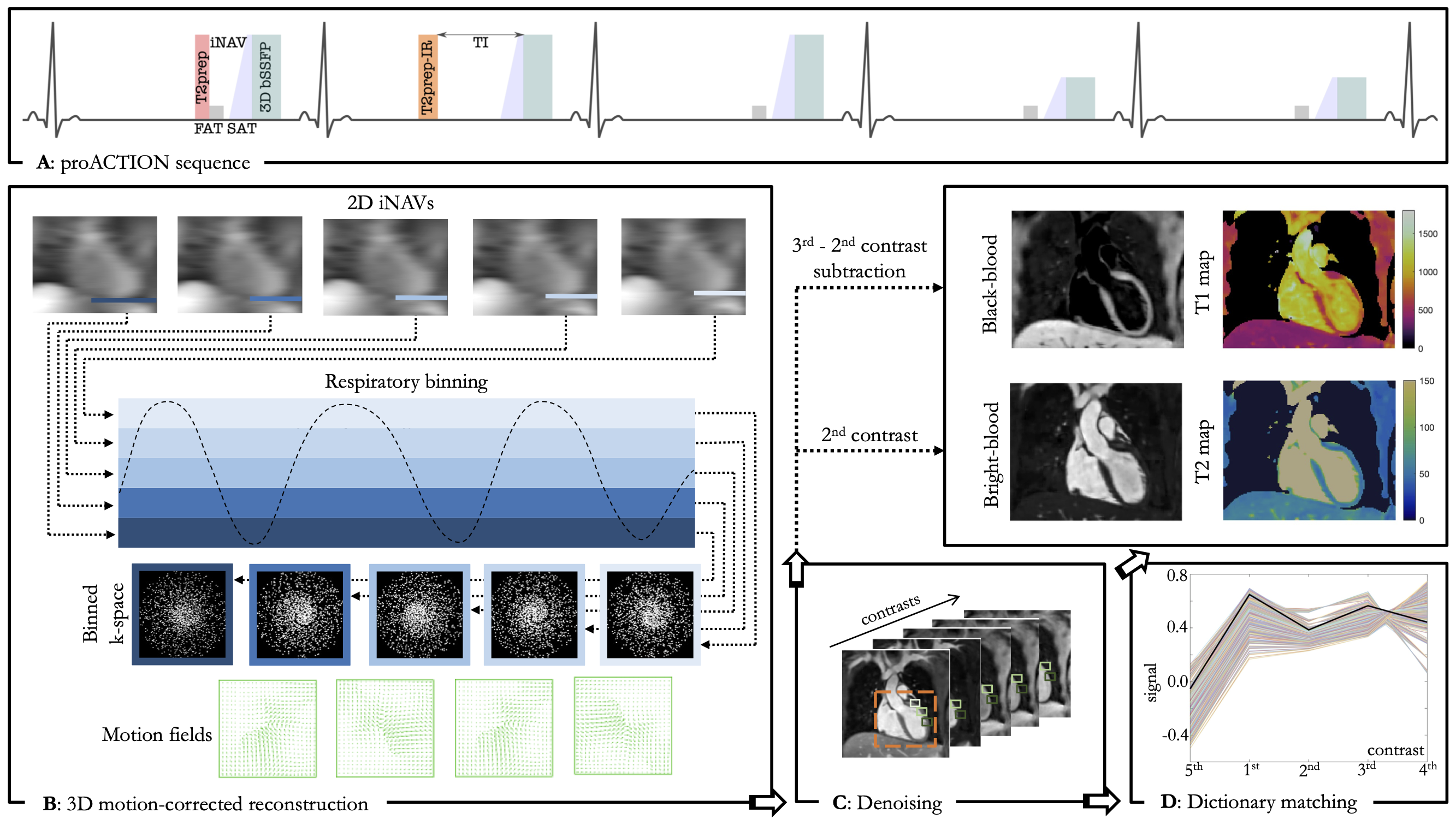
The proposed research sequence proACTION consists of a repeating set of five interleaved heartbeats (HB) with two T2 preparation (T2prep) modules, an inversion recovery pulse and acquisition with varying flip angles (FA) (Fig.1). 2D low-resolution image-based navigators (iNAV5) are acquired prior to each HB by spatially encoding the ramp-up pulses to perform respiratory data binning and beat-to-beat intra-bin translational motion correction. A variable-density 3D cartesian trajectory with spiral profile order6 and golden angle step is employed to obtain a 5-fold undersampled data with 100% respiratory efficiency resulting in a predictable scan time of 12min for a normal heart-rate (HR) of 60bpm. Images are obtained with 3D non-rigid motion corrected reconstruction7 in combination with patch‐based low-rank regularization (HD-PROST8). A short inversion time TI of 90ms was chosen to enable epicardial fat suppression in the 2ndHB. In the remaining HBs, fat suppression was performed with frequency selective inversion recovery (SPIR) fatsat pulses (flip angle = 180°). The 2ndHB provides the bright-blood image, while the black-blood volume is derived by a direct magnitude subtraction of the 2nd from the 3rdHB with a positive contrast window. To obtain high contrast between blood and myocardium, the duration of the second T2prep was optimized by solving Bloch simulations using KomaMRI.jl11 resulting in a T2prep duration of 50ms (Fig. 3B). Together with another T2prep of 40ms in the 1stHB, precise T2 quantification was achieved. To allow for appropriate signal-to-noise, FA was set to 80° for the HBs involved in the anatomical imaging, while lower FA of 40° was employed to enhance T1-sensitivity for remaining HBs. Joint T1/T2 maps were generated voxel-wise by maximizing the inner product of the measured signal with a previously generated dictionary using the extended phase graph (EPG) method. Images were acquired on a clinical 0.55T scanner (MAGNETOM Free.Max, Siemens Healthineers, Erlangen, Germany) with an ECG-triggered bSSFP readout (TR/TE=4.90/2.45ms, FOV=312x312x108-120mm, isotropic spatial resolution=1.5mm) in coronal orientation, utilizing subject dependent trigger delay and acquisition window of 140ms. Imaging was performed during the mid-diastolic rest period to minimize cardiac motion. The proposed proACTION framework was evaluated in the standardized T1MES phantom9 and five healthy subjects (27+-2 years, 1 female, heart-rates of 55-77 bpm). Bull’s-eye plots were generated for in-vivo T1 and T2 maps with 16 segments AHA model across the short-axis.Results
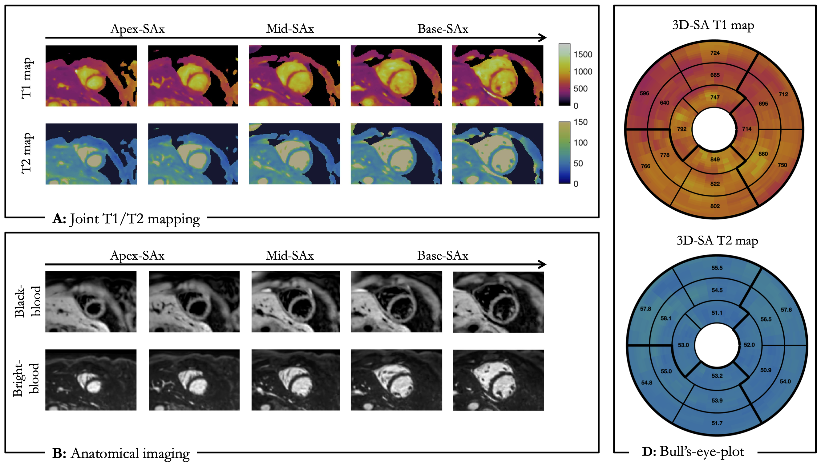
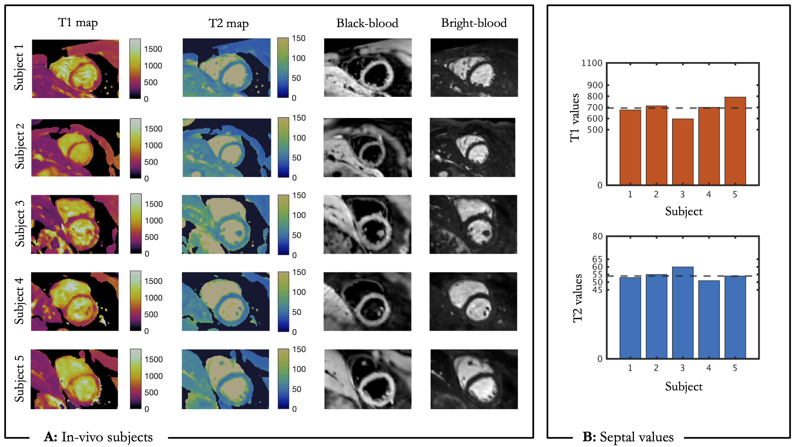
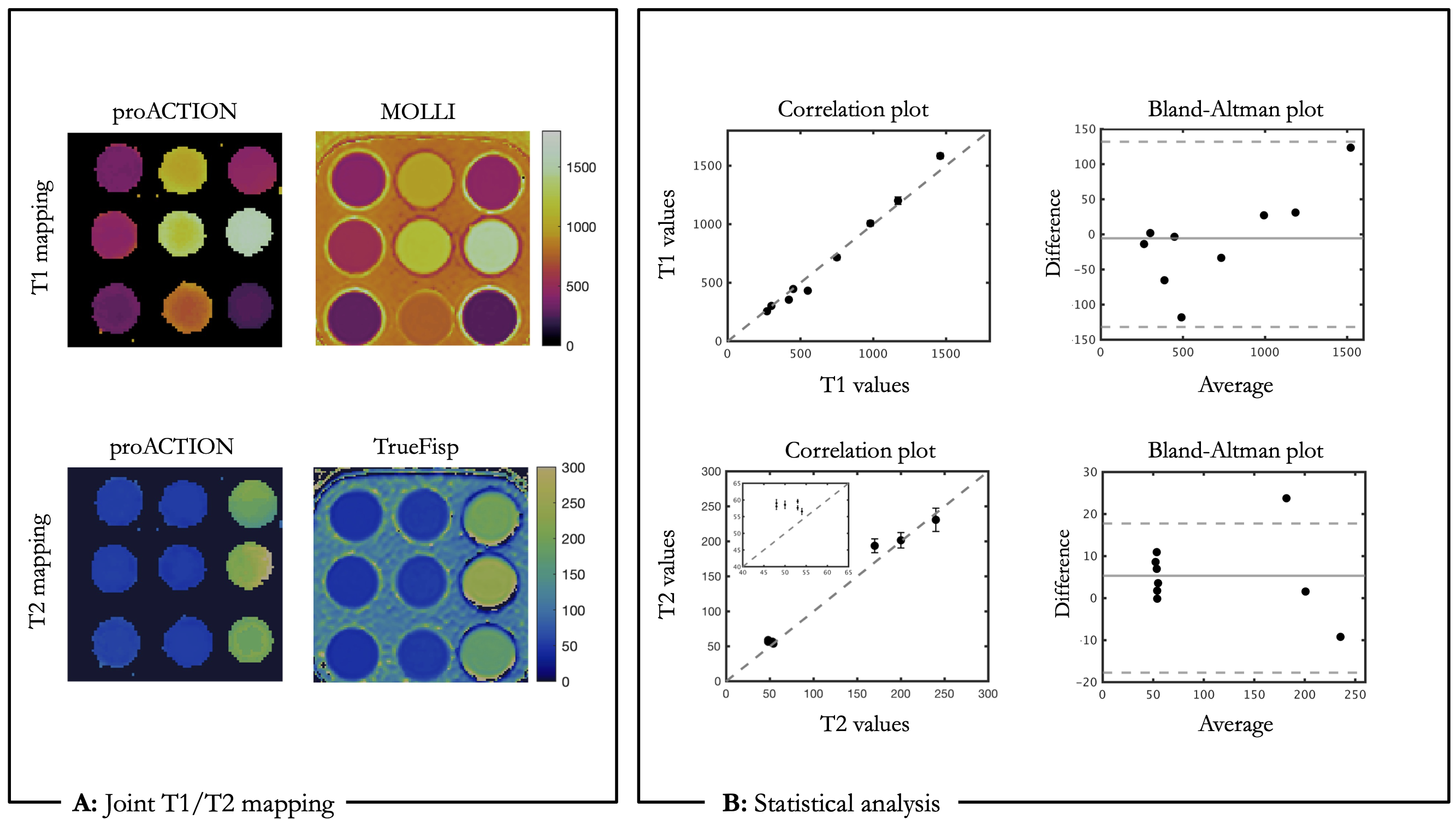
Phantom: proACTION phantom results are shown in Figure 2. Good T1 agreement was observed for T1<1200ms, while T2 values have a bias of 4ms compared to the reference values measured with spin-echo sequences.Healthy subjects: Anatomical images are displayed in Figure 3 for the five healthy subjects in coronal orientation, showing good delineation of cardiac and vascular structures. proACTION bright- and black-blood images alongside corresponding T1 and T2 maps are shown in basal, mid-cavity and apical short-axis (SA) slices for a representative subject in Figure 4. Bull’s-eye plots with mean values of 684ms/55ms and spatial variability of 72ms/3ms were obtained for T1/T2 respectively (Fig.4C). T1/T2 septal values in the mid-cavity SA slices are shown in Figure 5 for five subjects, providing comparable parametric values of 694±71ms for T1 and 54±3ms for T2 to the literature11 at 0.55T (T1=701±25ms, T2=58±6ms).
Discussion and further work
The proposed 3D whole-heart multi-contrast proACTION sequence allows acquisition of bright- and black-blood volumes together with myocardial tissue quantification in a single fast scan at 0.55T. Preliminary results demonstrated good agreement with reference values in the T1/T2 phantom and promising results in healthy subjects. Future work will focus on validation on a larger cohort of healthy subjects and patients with suspected cardiovascular disease.Acknowledgements
The authors acknowledge financial support from: (1) BHF RG/20/1/34802 (2) EPSRC EP/V044087/1 (3) Wellcome EPSRC Centre for Medical Engineering (NS/A000049/1), (4) ANID Millennium Institute iHEALTH, ICN2021_004; Fondecyt 1210637 and 1210638; Fondequip Mayor EQY210003; Basal Funding, IMPACT, FB210024 and (6) the Technical University of Munich – Institute for Advanced Study.References
[1] Nicole Seiberlich, Vikas Gulani, Adrienne Campbell-Washburn, Steven Sourbron, Mariya Ivanova Doneva, Fernando Calamante, Houchun Harry Hu. (2020). Quantitative Magnetic Resonance Imaging. Elsevier.
[2] Grizzard, J. D., Judd, R. M., & Kim, R. J. (2008). Cardiovascular MRI in practice a teaching file approach. Springer.
[3] Ivan Kokhanovskyi, Michael G Crabb, Karl P Kunze, Radhouene Neji, Carl Ganter, Donovan P Tripp, Carlos Castillo-Passi, Dimitrios C Karampinos, Claudia Prieto, and Rene M Botnar. (2023). Multiparametric 3D cardiac MR for simultaneous bright- and black-blood imaging and joint T1/T2 myocardial tissue mapping. Proc. Intl. Soc. Mag. Reson. Med. 31, 1687.
[4] Carlos Castillo-Passi, Michael G. Crabb, Camila Muñoz, Donovan Tripp, Karl P. Kunze, Radhouene Neji, Pablo Irarrazaval, Claudia Prieto, and Rene M. Botnar (2023). Simultaneous 3D Whole-Heart Bright-Blood and Black-Blood Imaging with iNAV-based Non-Rigid Motion-Corrected Reconstruction at 0.55T. Proc. Intl. Soc. Mag. Reson. Med. 31, 1687.
[5] Henningsson M, Koken P, Stehning C, Razavi R, Prieto C, Botnar RM. Whole-heart coronary MR angiography with 2D self-navigated image reconstruction. Magn Reson Med. 2012 Feb;67(2):437-45. doi: 10.1002/mrm.23027. Epub 2011 Jun 7. PMID: 21656563.
[6] Prieto C, Doneva M, Usman M, Henningsson M, Greil G, Schaeffter T, Botnar RM. Highly efficient respiratory motion compensated free-breathing coronary MRA using golden-step Cartesian acquisition. J Magn Reson Imaging. 2015 Mar;41(3):738-46. doi: 10.1002/jmri.24602. Epub 2014 Feb 27. PMID: 24573992.
[7] Cruz G, Atkinson D, Henningsson M, Botnar RM, Prieto C. Highly efficient nonrigid motion-corrected 3D whole-heart coronary vessel wall imaging. Magn Reson Med. 2017 May;77(5):1894-1908. doi: 10.1002/mrm.26274. Epub 2016 May 25. PMID: 27221073; PMCID: PMC5412916.
[8] Bustin A, Lima da Cruz G, Jaubert O, Lopez K, Botnar RM, Prieto C. High-dimensionality undersampled patch-based reconstruction (HD-PROST) for accelerated multi-contrast MRI. Magn Reson Med. 2019 Jun;81(6):3705-3719. doi: 10.1002/mrm.27694. Epub 2019 Mar 4. PMID: 30834594; PMCID: PMC6646908.
[9] Gabriella Captur et al. “A medical device-grade T1 and ECV phantom for global T1 mapping quality assurance—the T1 Mapping and ECV Standardization in cardiovascular magnetic resonance (T1MES) program”. In: Journal of Cardiovascular Magnetic Resonance 18 (Sept. 2016). doi: 10.1186/s12968-016-0280-z.
[10] Castillo-Passi, C, Coronado, R, Varela-Mattatall, G, Alberola-López, C, Botnar, R, Irarrazaval, P. KomaMRI.jl: An open-source framework for general MRI simulations with GPU acceleration. Magn Reson Med. 2023; 1- 14. doi: 10.1002/mrm.29635
[11] Campbell-Washburn AE, Ramasawmy R, Restivo MC, Bhattacharya I, Basar B, Herzka DA, Hansen MS, Rogers T, Bandettini WP, McGuirt DR, Mancini C, Grodzki D, Schneider R, Majeed W, Bhat H, Xue H, Moss J, Malayeri AA, Jones EC, Koretsky AP, Kellman P, Chen MY, Lederman RJ, Balaban RS. Opportunities in Interventional and Diagnostic Imaging by Using High-Performance Low-Field-Strength MRI. Radiology. 2019 Nov;293(2):384-393. doi: 10.1148/radiol.2019190452. Epub 2019 Oct 1. PMID: 31573398; PMCID: PMC6823617.
Figures
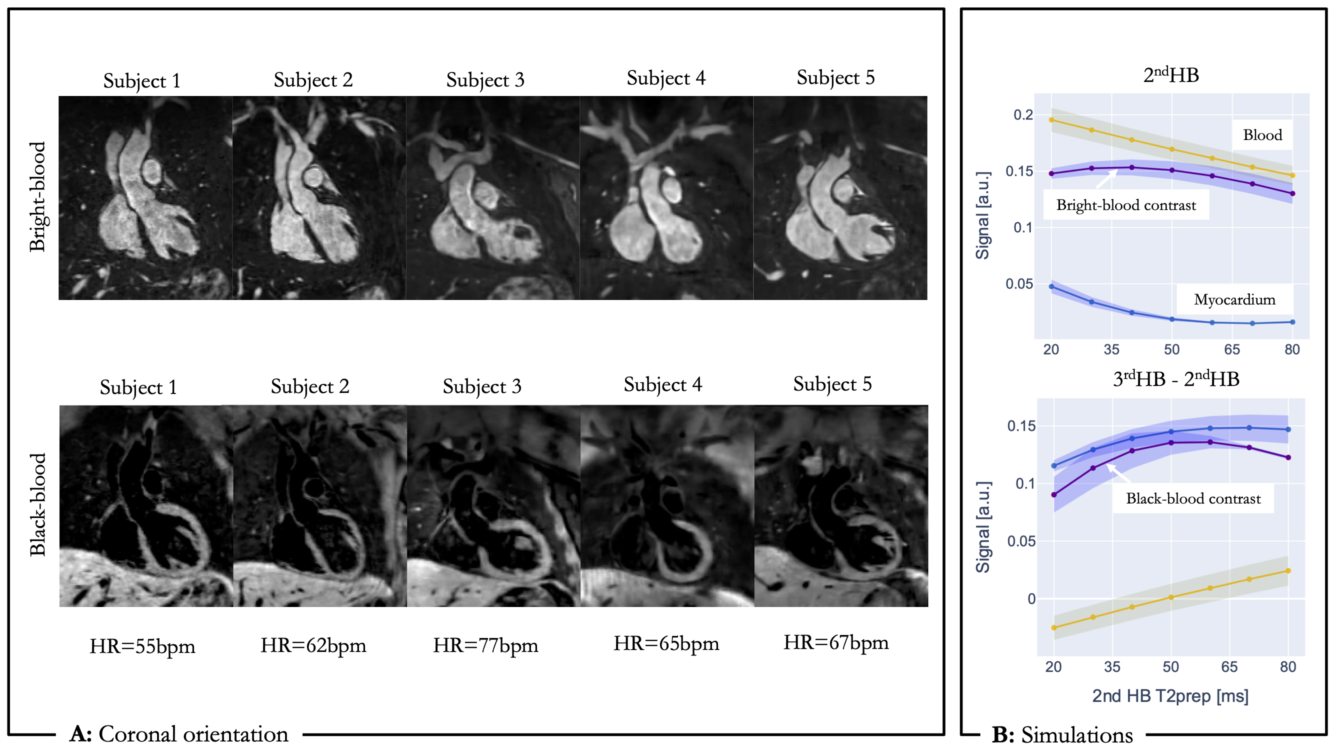
A: Bright- and black-blood images obtained in coronal orientation for five healthy subjects with different HRs. B: Simulation results using for the optimization on T2prep in the 2ndHB for healthy myocardium (T1=701ms, T2=58ms) and arterial blood (T1=1122ms, T2=263ms) with FA of 80°, TI=90ms and HR=50-70bpm. The optimal value was found to be 50ms.