1484
Cardiovascular MR Multitasking T1-T2 Joint Mapping in Healthy Subjects and Cardiomyopathy Patients on both 1.5T and 3T1Applied Science Lab West, GE HealthCare, Menlo Park, CA, United States, 2Biomedical Imaging Research Institute, Cedars-Sinai Medical Center, Los Angeles, CA, United States, 3McGill University Health Centre, Mentreal, QC, Canada, 4Department of Bioengineering, University of California, Los Angeles, Los Angeles, CA, United States, 5Department of Radiological Sciences, David Geffen School of Medicine, University of California, Los Angeles, Los Angeles, CA, United States, 6Area 19, Montreal, QC, Canada, 7Departments of Medicine and Radiology, Stanford University, Palo Alto, CA, United States, 8Applied Science Lab Europe, GE HealthCare, Munich, Germany
Synopsis
Keywords: Myocardium, Quantitative Imaging, Free-breathing, Multi-parametric Mapping
Motivation: CMR Multitasking has shown promise for non-ECG and free-breathing myocardial T1/T2 mapping in the heart, primarily on a single vendor 3T, but 2D T1-T2 CMR Multitasking has not yet been shown for different vendors (e.g., GE) and field strengths (e.g., 1.5T).
Goal(s): To extend 2D T1-T2 CMR Multitasking to 1.5T and 3.0T GE MR systems.
Approach: We implemented a 2D T1-T2 CMR Multitasking sequence on GE MR systems and evaluated its performance in healthy volunteers and patients at two sites.
Results: 2D T1-T2 CMR Multitasking generated T1 and T2 maps with image quality comparable to the reference measurements.
Impact: 2D T1-T2 CMR Multitasking provides an efficient and subject friendly (free-breathing, non-ECG) option for quantitative CMR assessment across sites and vendors.
Introduction
Quantitative biomarkers (e.g., T1, T2) in cardiovascular magnetic resonance (CMR) imaging are promising for assessing focal and diffuse myocardial pathologies. Conventional T1/T2 mapping approaches frequently encounter challenges related to unreliable mechanisms for accommodating physiological motion, such as ECG triggering and breath-holding. CMR Multitasking1-4 has shown promise for non-ECG and free-breathing quantitative imaging in the heart, primarily on a single vendor 3T system. In this study, we extended the applicability of CMR Multitasking by introducing a 2D single-slice T1-T2 joint mapping technique, referred to as 2D T1-T2 CMR Multitasking, designed for GE scanners. The aim of this study was to test the clinical utility of 2D T1-T2 CMR Multitasking in a mixed healthy volunteer and patient population on both 1.5T and 3.0T GE MR systems.Methods
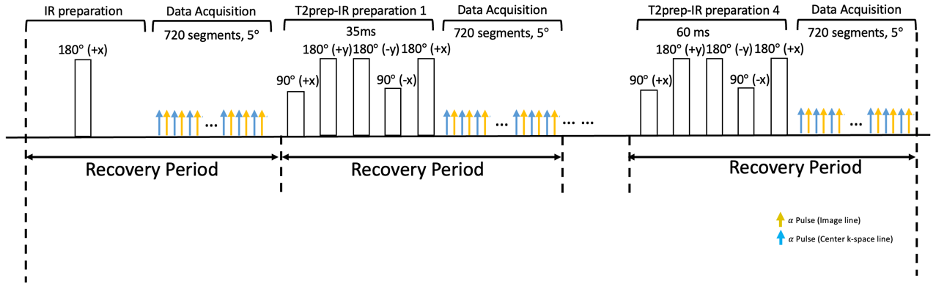
Sequence Design: The 2D T1-T2 CMR Multitasking GE sequence matches the original published Multitasking sequence1. Radial trajectory ordering collected low-rank tensor training data interleaved with imaging data. A golden angle (111.25°) increment was used between consecutive imaging readouts, with the training data acquired at 0° at every other readout. Five different T2prep-IR durations (0, 35ms, 40ms, 50ms, 60ms) were employed to generate both T1 and T2 contrasts, as shown in Fig. 1. After each preparation pulse, FGRE readouts were collected continuously with the excitation flip angle set to 5°3-5.Image Reconstruction and T1-T2 Estimation: The images acquired in the 2D T1-T2 CMR Multitasking framework can be represented as a 5-way tensor 𝒜 with voxel location index r=(x,y,z), a T1 recovery dimension, a T2 weighting dimension, a cardiac motion dimension, and a respiratory motion dimension. The standard Multitasking reconstruction pipeline1-4 was followed. The signal equation at the kth recovery period of the 2D T1-T2 CMR Multitasking sequence is
$$s(A,B,T_1,T_2)=A\frac{1-e^{-TR/T_1}}{1-e^{-TR/T_1}cos(\alpha_k)} \cdot [1+(Be^{-\tau/T_2}-1)(e^{-TR/T_1}cos(\alpha_k))^n] \cdot sin(\alpha_k)$$
with amplitude factor A, IR/T2prep-IR pulse efficiency B, FGRE readout interval TR, flip angle for the kth recovery period $$$\alpha_k$$$, and recovery time point n=1,2,...N, ( N is the number of FGRE pulses in a recovery period). A nonlinear fitting approach4,5 is used to determine the parameter maps.
Data Collection: We compared methods in healthy volunteers, who were scanned on a 1.5T GE MR450w scanner, and patients with cardiomyopathy (CMP), who underwent their scans on a 3.0T GE Signa Premier. All healthy volunteers and CMP patients were consented under respective and approved IRB protocols. 2D T1-T2 CMR Multitasking data were acquired at the base, mid, and apical level on short-axis slices. The sequence parameters were: TR/TE=3.6 ms/1.8 ms, recovery period=2.5 s, spatial resolution=1.7x1.7x8.0 mm3, scan time=1:32min, 20 cardiac bins, 6 respiratory bins. Reference 2D MOLLI T1 maps (2.0x2.0x8.0 mm3) and 2D DIR Multi-echo T2 maps (2.0x2.0x8.0 mm3) were acquired using the same SAX slice positioning. Six echoes were used for T2 mapping (9.6ms, 28.8ms, 48ms, 67.2ms, 86.4ms, 105.6ms). Whole myocardium T1/T2 values from healthy volunteers and septal T1/T2 values from patients were quantified for statistical analysis.
Results
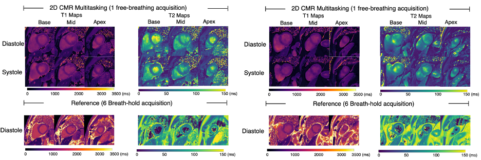
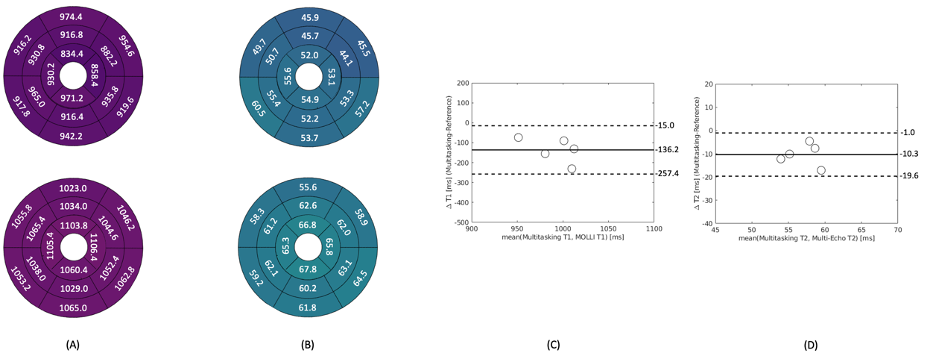
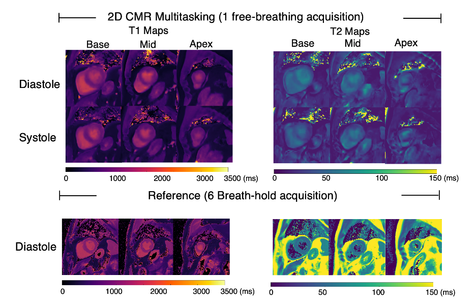
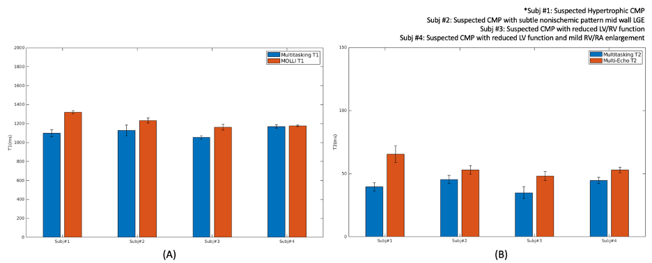
We enrolled five healthy volunteers (all male) and four patients (3 male, 1 female) with suspected non-ischemic CMP. An example of 2D Multitasking T1/T2 and reference images is shown in Fig. 2. Fig. 3 depicts the 16 semgent AHA plots and the Bland-Altman plots for whole myocardium T1/T2 values in five healthy volunteers in Multitasking and references. On average, Multitasking T1 and MOLLI T1 among the five healthy volunteers are 922.9±27.1ms and 1059.1±49.8ms, respectively; Multitasking T2 and Multi-echo T2 values are 51.9±3.3ms and 62.2±3.4ms, respectively. Fig. 4 shows an example of Multitasking and reference T1/T2 maps in a CMP patient, and Fig. 5 further shows the bar plot of the septal T1/T2 values in 4 CMP patients.Discussion
2D T1-T2 CMR Multitasking demonstrated the capability to generate T1 and T2 maps with image quality comparable to that of the existing vendor provided cardiac T1/T2 measurements. We noted the presence of B1 inhomogeneity artifacts in certain Multitasking T2 maps at 3.0T. This suggests the need for further improvements to the existing T2prep module to mitigate B1 inhomogeneity and off-resonance effects on T2 maps. To better estimate T1, a dual flip angle scheme5 and a longer scan time (~2mins) can be explored to mitigate the under-sampling artifacts. In addition, deep learning based radial reconstruction6,7 has the potential to enhance the SNR without extending the scan time.Conclusion
This study shows the potential clinical use of CMR Multitasking to quantitatively assess T1 and T2 in both healthy volunteers and CMP patients at both 1.5T and 3.0T without breath-holding or ECG gating, with comparable image equality. The free-breathing and non-ECG feature has clinical value, and clinical studies with larger sample sizes are warranted.Acknowledgements
No acknowledgement found.References
[1] Christodoulou, A. G., Shaw, J. L., Nguyen, C., Yang, Q., Xie, Y., Wang, N., & Li, D. (2018). Magnetic resonance multitasking for motion-resolved quantitative cardiovascular imaging. Nature biomedical engineering, 2(4), 215-226.
[2] Cao, T., Wang, N., Kwan, A. C., Lee, H. L., Mao, X., Xie, Y., ... & Li, D. (2022). Free‐breathing, non‐ECG, simultaneous myocardial T1, T2, T2*, and fat‐fraction mapping with motion‐resolved cardiovascular MR multitasking. Magnetic Resonance in Medicine, 88(4), 1748-1763.
[3] Serry, F. M., Ma, S., Mao, X., Han, F., Xie, Y., Han, H., ... & Christodoulou, A. G. (2021). Dual flip‐angle IR‐FLASH with spin history mapping for B1+ corrected T1 mapping: Application to T1 cardiovascular magnetic resonance multitasking. Magnetic Resonance in Medicine, 86(6), 3182-3191.
[4] Mao, X., Lee, H. L., Hu, Z., Cao, T., Han, F., Ma, S., ... & Christodoulou, A. G. (2022). Simultaneous Multi-slice Cardiac MR Multitasking for Motion-Resolved, Non-ECG, Free-Breathing T1-T2 Mapping. Frontiers in Cardiovascular Medicine, 267.
[5] Mao X, Lee HL, Eyre K, Delso G, Li D, Christodoulou A, Friedrich M, Salerno M, & Janich M. (2023). Free-breathing, ECG-Free, Myocardial T1 Mapping: Initial Feasibility Experiments of Cardiac MR Multitasking on GE Systems. in Proceedings of International Society of Magnetic Resonance and Medicine (ISMRM) 2023.
[6] Chen, Z., Chen, Y., Xie, Y., Li, D., & Christodoulou, A. G. (2022, March). Data-Consistent non-Cartesian deep subspace learning for efficient dynamic MR image reconstruction. In 2022 IEEE 19th International Symposium on Biomedical Imaging (ISBI) (pp. 1-5). IEEE.
[7] Ryu, K., Li, Z., Sandino, C. M., & Vasanawala, S. S. (2022) Accelerated free-breathing radial cine imaging via GROG-interpolated DL-ESPIRiT. in Proceedings of International Society of Magnetic Resonance and Medicine (ISMRM) 2022.
Figures