1483
Free breathing Dual-excitation flip-angle 3D T1 mapping using randomized stack of spiral acquisition1Stanford University, Stanford, CA, United States, 2Beijing University of Posts and Telecommunications, Beijing, China, 3University of California, San Francisco, San Francisco, CA, United States
Synopsis
Keywords: Myocardium, Quantitative Imaging
Motivation: Cardiac MR (CMR) imaging is a widely used technique that provides important diagnostic and prognostic information in cardiomyopathy. T1 maps and LGE can provide tissue characterization to detect myocardial fibrosis.
Goal(s): Our group has developed a 2D free-breathing and self-gated cine and T1 mapping acquisition CAT-SPARCS. This work is to extend the acquisition to 3D T1 mapping.
Approach: The proposed acquisition is enabled in a single free-running randomized stack of spiral sequence with a 3-minute acquisition and T1 map is reconstructed by dictionary learning method.
Results: The T1 values from the proposed method are comparable to the clinically standard MOLLI sequence.
Impact: Our technique could substantially shorten the clinical scan time providing both cine and T1 mapping images.
Introduction:
Cardiac MR (CMR) imaging is a widely used technique that provides important diagnostic and prognostic information in cardiomyopathy. A particular advantage of CMR is that it can evaluate cardiac function and perform tissue characterization using T1 mapping and LGE. Parametric mapping of myocardial native T1 has shown the potential to assess a variety of myocardial pathologies and guide therapy. Our group has developed a 2D simultaneous free-breathing and self-gated acquisition for T1 map and CINE images [1]. This work aims to extend the acquisition to 3D T1 mapping in a single free-running randomized stack of spiral sequence [2] with a 3-minute Acquisition.Methods:
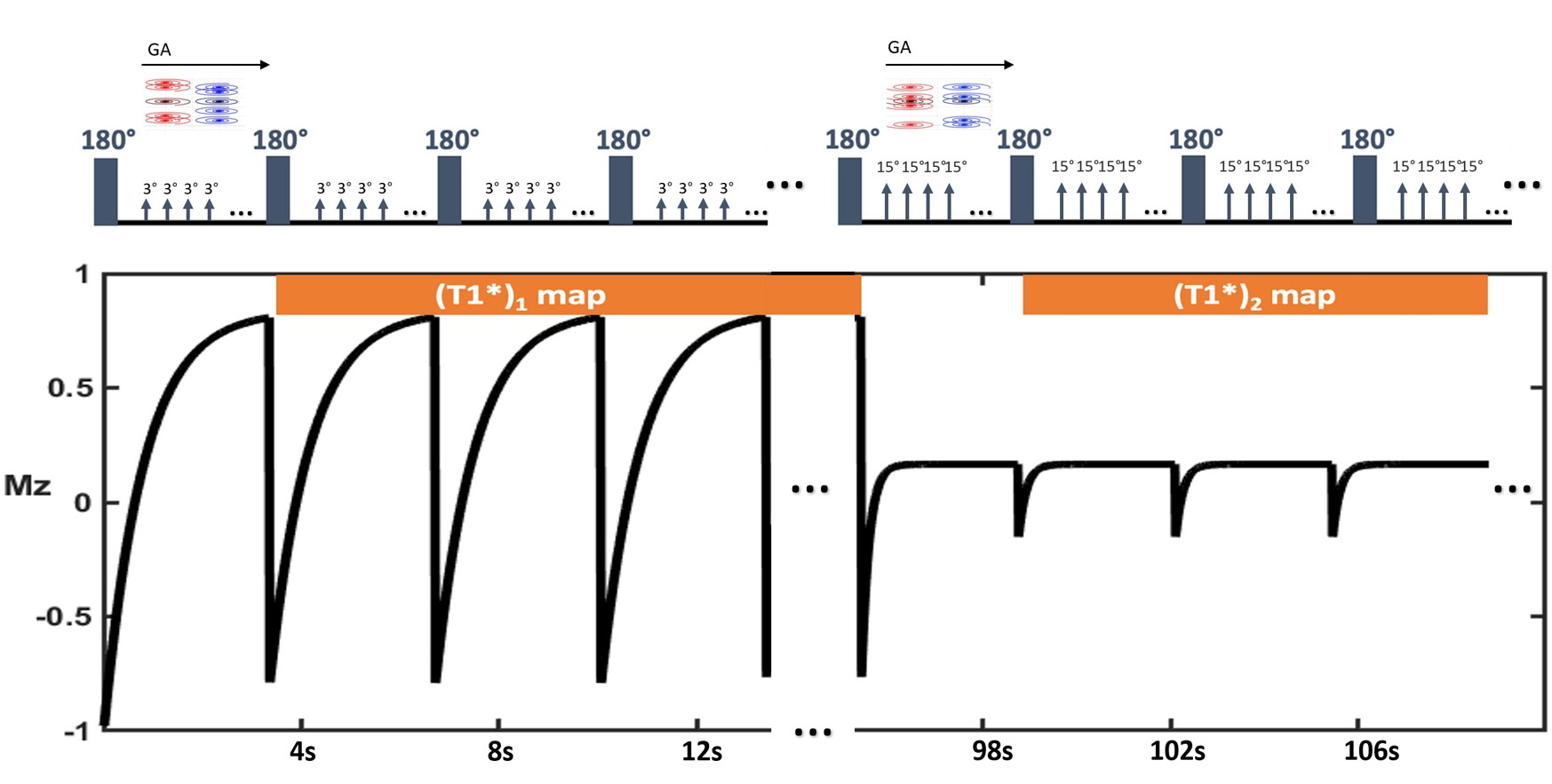
3 volunteers and 5 patients were imaged on a 3T scanner (MAGNETOM Prisma/Skyra, Siemens Healthcare, Erlangen, Germany) without contrast. Data were acquired continuously on a 3T scanner using a golden-angle gradient-echo stack of spiral pulse sequence, with an inversion RF pulse applied every 3.8 seconds. Flip angles of 3° and 15° were used for readouts after the first half of and second half of inversions. We used prospective ECG triggering to bin the diastolic data into different inversion time and used a dictionary learning approach for image reconstruction. The T1 maps were fit using a projection onto convex sets approach from images reconstructed for each flip angle using dictionary learning.We developed a strategy (shown in Figure 1) consisting of a continuous acquisition with an inversion-recovery RF pulse applied every 3.8 seconds, using one excitation flip angle (FA1) for the first half of the inversion pulses and another excitation flip angle (FA2) for the second half of the inversion pulses. For resolving cardiac motion, we exploit ECG signal as reference to bin the data into different cardiac phases. For T1 fitting, we generate the dictionary based on two flip angles by K-singular value decomposition from the Bloch simulation time courses with the acquisition parameters. By fitting the three-parameter model of equation (1) using the dictionary learning–reconstructed images, two apparent T1* maps for two flip angles can be obtained.
$$ M(t)=M_{z_{+}}-\left(M_{z+}+E_{I R} M_{z_{+}}\right) e^{-t / T_1^*} (1)$$
,where $$$M_{z_{+}}$$$ is the signal immediately before the next inversion pulse; $$$E_{I R}$$$ is the IR efficiency; and $$$T_{1}^{*}$$$ is the apparent T1.
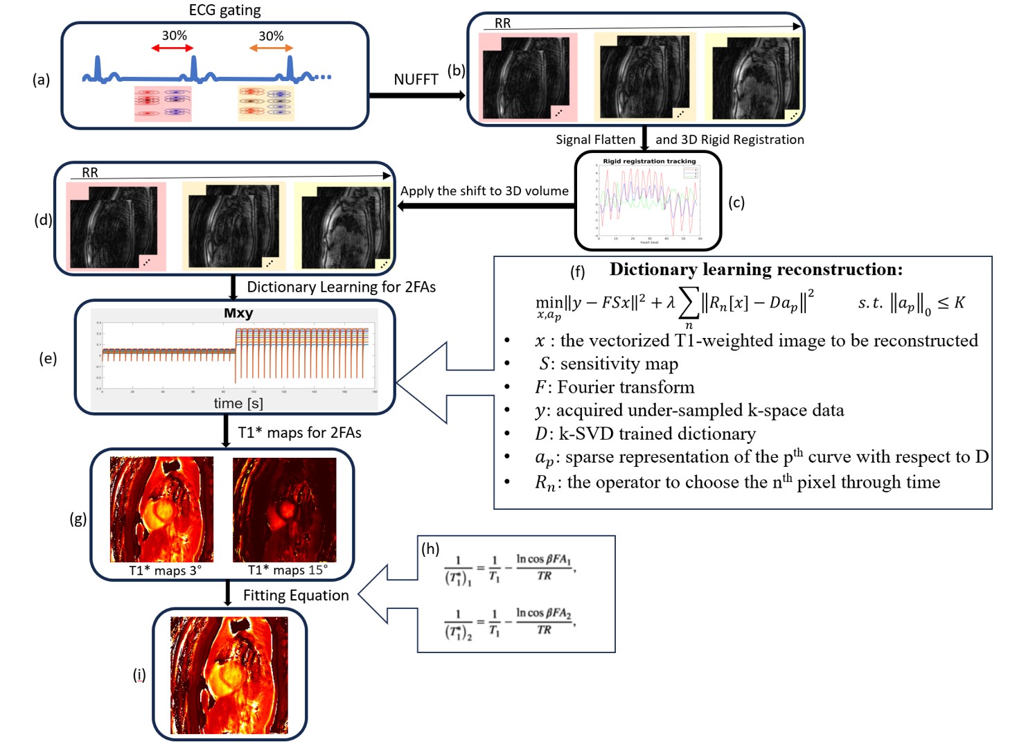
For T1 mapping (Shown in figure 2 ), in each R-R interval every stack of spirals volume was combined into a diastolic acquisition window of 30% of the RR interval. A dictionary was generated using the acquisition parameters and with T1 ranging from 100 to 3000 ms, and IR efficiency from 0.9 to 1. Dictionary learning [3,4] was performed by using OMP and LSQR to solve (Each term defination shows in Figure 3(f)):
$$ \min _{x, a_p}\|y-F S x\|^2+\lambda \sum_n\left\|R_n[x]-D a_p\right\|^2 \quad \text { s.t. }\left\|a_p\right\|_0 \leq K $$
The parameters for dictionary learning reconstruction were initially chosen based on previous studies[3,4], and then tuned empirically for three data sets. The threshold for stopping criterion was ε = 0.00001, the number of dictionary atoms was 1000, the sparsity level (K) was 3, the regularization parameter (λ) was 8, and the maximum iteration number was 15. Then, based on the relationship between T1* and T1, the T1 map can be recovered by simultaneously solving two equations:
$$ \frac{1}{\left(T_1^*\right)_1}=\frac{1}{T_1}-\frac{\ln \cos \beta F A_1}{T R}, \\ \frac{1}{\left(T_1^*\right)_2}=\frac{1}{T_1}-\frac{\ln \cos \beta F A_2}{T R}, $$
where $$$\beta$$$ is the scale factor between the nominal flip angle and the actual flip angle, and the two apparent T1 maps $$$(T_1^*)_1$$$ and $$$(T_1^*)_2$$$ are obtained for two flip angles by fitting Equation. Along with the T1 map, this fitting scheme can also provide a flip-angle scale $$$\beta$$$ map.
Results:
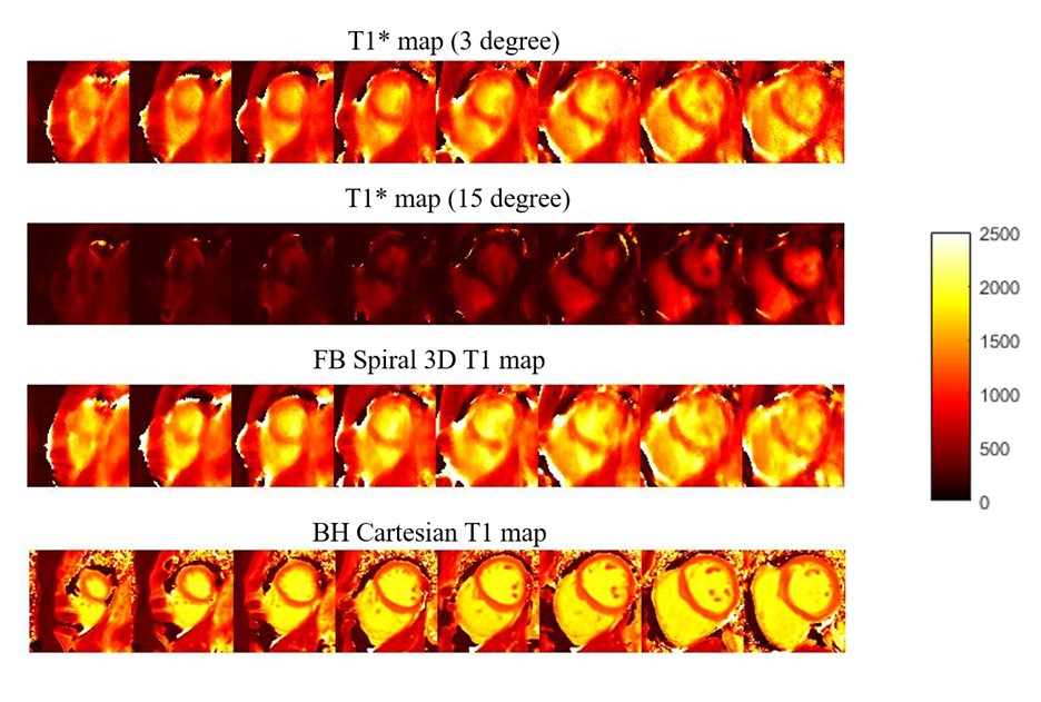
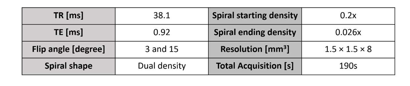
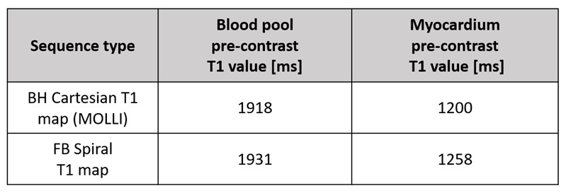
Figure 3 (a) shows the apparent T1 maps $$$(T_1^*)_1$$$ and $$$(T_1^*)_2$$$ corresponding to 3 degrees and 15 degrees. (b) shows the T1 mapping compared to the standard breath-hold Cartesian MOLLI T1 mapping from one subject.Table 2 shows the pre contrast T1 values for myocardium and blood compared with T1 value from standard breath-hold Cartesian MOLLI T1 mapping.
Conclusion and Discussion:
We proposed a strategy to acquire 3D T1 maps with free-running dual-excitation flip-angle sequences. The T1 values from the proposed method are comparable to the clinically standard MOLLI sequence. In the future, this continuous-acquisition dual-excitation flip-angle sequence can also acquire cine images and T1 maps simultaneously by sorting data into different cardiac phases and different contrast. Cine images can be reconstructed from the steady-state portion of 15°. Also, we will extend our technique to self -gating with an additional navigator and will further optimize our motion correction and image reconstruction strategies.Acknowledgements
This work is supported by NIH R01 H155962.References
1. Zhou, R., Weller, D. S., Yang, Y., Wang, J., Jeelani, H., Mugler III, J. P., & Salerno, M. (2021). Dual-excitation flip-angle simultaneous cine and T1 mapping using spiral acquisition with respiratory and cardiac self-gating. Magnetic Resonance in Medicine, 86(1), 82–96.
2. Wang X, Wang J, Zhou R, Salerno M.Rapid Free-breathing 3D SPirAl Respiratory and Cardiac Self-gated (SPARCS) Cine Acquisition Using an Undersampled Stack-of-Spirals. In Proceedings of the ISMRM 30th Annual Scientific Sessions, London, England, UK, 2022
3. Ravishankar S, Bresler Y. MR image reconstruction from highly undersampled k-space data by dictionary learning. IEEE Trans. Med. Imaging. IEEE; 2011;30:1028–41.
4. Zhu Y, Kang J, Duan C, Nezafat M, Neisius U, Jang J, et al. Integrated motion correction and dictionary learning for free-breathing myocardial T1 mapping. Magn. Reson. Med. 2019;81:2644–54.
Figures