1482
Feasibility of T2 mapping for the detection of myocardial injury in hypertrophic cardiomyopathy1Fuwai Hospital, Beijing, China
Synopsis
Keywords: Myocardium, Cardiomyopathy
Motivation: The relationship between T2 value and myocardial injury has not been well demonstrated.
Goal(s): To explore the association between T2 value and myocardial injury in HCM patients.
Approach: 50 patients with HCM were prospectively recruited. Hs-cTnI was obtained as a marker of myocardial injury (>0.016ng/mL). ConSept T2 were measured from the maximal value of 16 segments and the middle septum.
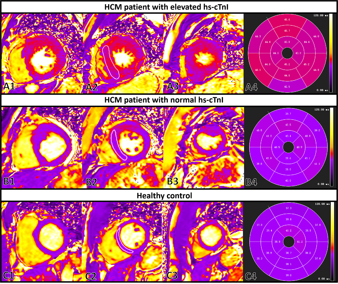
Results: The hs-cTnI elevated group had higher T2 global, ConSept T2, T2 max(P<0.05) than the normal hs-cTnI group. The ConSept T2 value were moderately correlated with hs-cTnI(r=0.52, P<0.001). In logistic regression analysis, ConSept T2 value was significantly associated with elevated hs-cTnI(P<0.01)
Impact: This study provided in-vivo evidence by CMR for the ongoing myocardial injury in HCM.
Purpose
The application of T2 mapping has been introduced in hypertrophic cardiomyopathy (HCM). However, the relationship between T2 value and myocardial injury has not been well demonstrated. This study aims to explore the association between T2 value and myocardial injury in HCM patients.Methods
50 patients with HCM and 20 age- and sex-matched healthy controls were prospectively recruited. All patients underwent laboratory testing and CMR. Hs-cTnI was obtained as a marker of myocardial injury (>0.016ng/mL). Global T2 values were quantified for three left ventricular (LV) short-axis slices. The maximum T2 value and T2 value assessed conservatively within the septum (ConSept T2) were measured from the maximal value of 16 segments and the middle septum, respectively.Results
In the HCM group, elevated hs-cTn I was found in 25 (20 males, 50±13 years) and normal hs-cTn I in 25(17 males, 49±14 years). The hs-cTnI elevated group had higher T2 global, ConSept T2, T2 max, T1 global, ConSept T1 values, and LGE extent(P<0.05) than the normal hs-cTnI group. The ConSept T2 value (r=0.52, P<0.001) and LGE extent (r=0.52, P<0.001) were all moderately correlated with hs-cTnI. Among all parametric parameters, ConSept T2 value showed the best performance in identifying myocardial injury (AUC=0.83). In bivariate logistic regression analyses, after adjusting LV ejection fraction, LV end-diastolic volume index, maximal ventricular wall thickness, LV myocardial mass index, LGE extent, T1 global, and ConSept T1 values, respectively, ConSept T2 value was still significantly associated with elevated hs-cTnI(P<0.01).Conclusions
We confirmed the feasibility of T2 mapping for detecting myocardial injury in HCM. T2 value was associated with an elevated hs-cTnI, which provided in-vivo evidence by CMR for the ongoing myocardial injury in HCM.Acknowledgements
This study was supported by grants from the National Key Research and Development Program of China (No. 2021YFF0501404, No. 2021YFF0501400) and the key projects of the National Natural Science Foundation of China (No. 81930044).References
1. Huang L, Ran L, Zhao P, et al. MRI native T1 and T2 mapping of myocardial segments inhypertrophic cardiomyopathy: tissue remodeling manifested prior to structure changes. Br ] Radiol2019:92(1104):20190634.
2.Arcari L,Hinojar R, Engel l, et al. Native T1 and T2 provide distinctive signatures inhypertrophic cardiac conditions - Comparison of uremic, hypertensive and hypertrophiccardiomyopathy.lnt] Cardiol 2020:306:102-108
3. Rogers T, Dabir D, Mahmoud l, et al. Standardization of T1 measurements with MOLLIindifferentiation between health and disease--the ConSept study.] Cardiovasc Magn Reson2013;15(1):78
Figures