1479
Mapping 1H MR metabolites to transcriptomics and mass spectrometry imaging in PDAC1Division of Cancer Imaging Research, The Russell H. Morgan Department of Radiology and Radiological Science, The Johns Hopkins University School of Medicine, Baltimore, MD, United States, 2Sidney Kimmel Comprehensive Cancer Center, The Johns Hopkins University School of Medicine, Baltimore, MD, United States, 3Departments of Pathology, The Johns Hopkins University School of Medicine, Baltimore, MD, United States, 4Departments of Oncology, The Johns Hopkins University School of Medicine, Baltimore, MD, United States, 5Departments of Medicine, The Johns Hopkins University School of Medicine, Baltimore, MD, United States
Synopsis
Keywords: Cancer, Cancer, PDAC
Motivation: The poor prognosis of pancreatic ductal adenocarcinoma (PDAC) creates an urgent need to identify new targets. PDAC is a metabolically active cancer that is glutamine avid.
Goal(s): We downregulated the glutamine transporter, SLC1A5, in the patient-derived human cancer cell line, Pa04C, and observed significant tumor growth delay.
Approach: High-field, high-resolution 1H MRS was performed of extracts from wild type, empty vector, and SLC1A5 downregulated tumors that was mapped to transcriptomic analysis of the corresponding cells, and to mass spectrometry imaging (MSI) of human normal and PDAC tissue.
Results: Common pathways were identified from the analysis that identify new targets for PDAC.
Impact: This study contributes to our comprehension of how the glutamate transporter SLC1A5 impacts the transcriptomics of pancreatic cancer cells, influences tumor metabolism, and its connection to variations in human PDAC metabolism. These findings could provide new insights into PDAC cancer.
Introduction
PDAC is associated with poor survival. Because PDAC is glutamine avid, targeting the glutamine transporter, SLC1A5, is being explored for treatment. However, the importance of glutamine metabolism in several normal tissue functions causes toxicity. Identifying molecular pathways associated with SLC1A5 and glutamine may provide new targets for PDAC treatment. We therefore downregulated SCL1A5 in Pa04C PDAC cells and characterized cell transcriptomics, tumor growth, and metabolic changes from high-resolution 1H MR tumor spectra. The transcriptomic analysis and the 1H MR spectra were compared to mass spectrometry imaging (MSI) of normal and PDAC human tissue to determine if common pathways were identified from the three analytical methods.Methods
Transcriptomic analysis was performed of wild type (WT), empty vector (EV) and SLC1A5 downregulated Pa04C cells that were inoculated in SCID mice to derive tumors for 1H MRS of extracts performed at 750 MHZ (n=19 per EV/WT group and n=14 per SLC1A5 group). Tumor growth curves were obtained from these cells following subcutaneous inoculation. Human tissue studies were performed with normal pancreas (n=5) and PDAC (n=5) tissues. MALDI imaging was performed on a Bruker RapifleX MALDI TOF/TOF instrument in positive and negative ion modes. Data from all three data sets were analysed using the ConsensusPathDB Reactome data base to determine alterations in molecular pathways.Results and Discussion
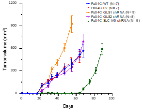
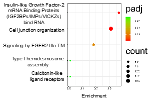
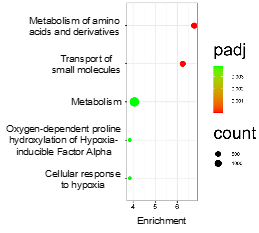
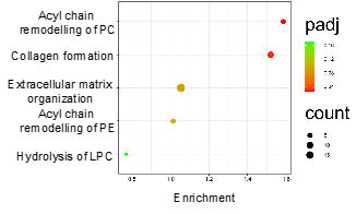
SLC1A5 downregulation resulted in a significant delay of tumor growth (Figure 1). With SLC1A5 downregulation, a significant decrease (P<0.05) of valine, isoleucine, alanine, glutamate, glutathione, aspartate and choline was observed in the 1H MR spectra of tumors. The decrease of glutamate may be due to a compensatory mechanism of glutamine synthetase forming glutamine from glutamate. 19 different molecular pathways were associated with these metabolic changes of which 9, including alanine metabolism and glycerophospholipid catabolism, were common with the transcriptomic analysis. Transcriptomic analysis identified a significant change in 538 genes in the corresponding EV and SLC1A5 downregulated Pa04C cells, with changes in 1155 associated molecular pathways. The most significant 5 changes are presented in Figure 2. The MSI comparison of human normal pancreatic tissue and PDAC (Figure 3) identified significant changes in 32 metabolites, with 85 associated molecular pathways altered. Comparison of transcriptomics and MSI metabolomics data (Figure 4) identified 58 molecular pathways commonly altered in both methods. These results confirm the functionality of the transcriptomic data, and the disruptive effects of SLC1A5 downregulation on PDAC. Included in these 58 pathways were ECM organization, collagen biosynthesis and formation, amino acid transport, glycerophospholipid metabolism, SLC-mediated transmembrane transport, innate immune system, metabolism of lipids, and fatty acid metabolism. Glycerophospholipid catabolism was common to all three data groups highlighting its importance in PDAC, and on the effects of SLC1A5 downregulation in PDAC.Acknowledgements
Support from NIH R35 CA209960 and R50 CA243562 is gratefully acknowledged.References
No reference found.Figures