1472
Fibroglandular tissue segmentation and background parenchymal enhancement quantification in breast MRI using an anatomy-aware loss function1Department of Radiological Sciences, University of California, Los Angeles, Los Angeles, CA, United States, 2Department of Bioengineering, University of California, Los Angeles, Los Angeles, CA, United States, 3Department of Computer Science, University of California, Los Angeles, Los Angeles, CA, United States
Synopsis
Keywords: Segmentation, Machine Learning/Artificial Intelligence, Fibroglandular tissue; Background parenchymal enhancement; Breast cancer
Motivation: Fully automatic segmentation of fibroglandular tissue (FGT) and background parenchymal enhancement (BPE) quantification methods with high generalizability for different FGT levels are still lacking.
Goal(s): We aimed to improve the segmentation accuracy and generalizability across various FGT levels that accurately quantify FGT density and BPE.
Approach: A novel anatomy-aware loss function based on the variations in FGT level was applied in a fully automatic segmentation model training on breast MRIs.
Results: The accuracy of breast tissue segmentation, FGT density estimation, and BPE quantification were improved at various FGT levels.
Impact: The anatomy-aware loss function can help improve the generalization of the breast tissue segmentation model on patients with different breast densities, thereby enabling the model to be more widely used in fibroglandular tissue density estimation and background parenchymal enhancement quantification.
Introduction
Breast density is typically categorized into four levels based on the Breast-Imaging Reporting and Data System (BI-RADS) lexicon, which includes almost entirely fat (about 10% of women), scattered fibroglandular tissue (FGT) (about 40% of women), heterogeneous FGT (about 40% of women), and extreme FGT (about 10% of women). The significant variations in breast density cause the pixel data imbalance between fat and FGT in the deep learning-based breast tissue segmentation problem, leading to low segmentation accuracy. Some segmentation models have been applied to breast tissue segmentation1–5, but few of them have specifically addressed the class of pixel data imbalance problem. Therefore, we proposed a breast image-specific segmentation model with better generalizability for FGT density variation. It can be used to further quantify background parenchymal enhancement (BPE), which is potentially associated with breast cancer risk and treatment response5–8.The purpose of our study is to introduce a fully automated breast tissue segmentation pipeline with a novel loss function that considers the FGT level to address the problem of class imbalance between fat and FGT. We apply the anatomy-aware loss function to improve the accuracy of image segmentation and the downstream task of quantifying BPE at different breast densities. Figure 1 illustrates the segmentation pipeline and downstream tasks.
Methods
Dice loss focuses on the overlap between the prediction and the ground truth9. For images with extremely low breast density, the overlap between the predicted FGT area and the ground truth becomes small, making the Dice loss less effective. Focal loss, on the other hand, aims to address the class imbalance by focusing learning on hard examples and down-weighting easy negatives10,11. We propose the anatomy-aware loss function ($$$L_{aa}$$$) by incorporating the FGT density information into the loss function as a weighted sum of Dice and Focal losses as follows:$$L_{aa}=\alpha_{FGT}L_{Dice}+\left(1-\alpha_{FGT}\right)L_{Focal}$$,
where $$$\alpha_{FGT}$$$ is related to the breast density category and defined as follows:
$$\alpha_{FGT}=\begin{cases}0.25,&\text{if almost entirely fatty}\\0.5,&\text{if scattered FGT}\\0.75,&\text{if heterogeneous FGT}\\1,&\text{if extreme FGT}\end{cases}$$
By up-weighting FGT pixel data in lower FGT level images, this loss function can help reduce the impact of the class imbalance issue.
We collected breast screening MRIs of 180 women between January 2017 and December 2019 at a single institution. Two breast radiologists assessed the FGT level. The fat and FGT were segmented on T1-weighted non-fat-suppressed images by fuzzy-C means clustering method as reference. We developed a fully automatic three-dimensional segmentation pipeline to segment fat and FGT together. We compared the dice score (DSC) and surface distance using different loss functions across the entire cohort and in sub-cohorts at different FGT levels and compared the performance of UNEt TRansformers (UNETR)12 and nnUNet13 as segmentation networks, respectively. To further evaluate the impact of segmentation results on downstream tasks, we calculated FGT density and four quantitative BPE measures using the segmentation masks. These measures included percent enhancement (PE) over FGT and the whole breast, denoted as PEFGT and PEBreast; signal enhancement ratio (SER) over FGT and the whole breast, denoted as SERFGT and SERBreast. We compared the root mean square errors (RMSE) of FGT density and four quantitative BPE measures using different loss functions across the entire cohort and in sub-cohorts at different FGT levels.
Results
When we applied the anatomy-aware loss instead of the Dice loss on both UNETR and nnUNet models, we observed a consistent improvement in the evaluation metrics. As shown in Figure 2, the surface distance of FGT in the whole cohort decreased from 0.93mm to 0.87mm using UNETR and 0.98mm to 0.92mm using nnUNet. Figure 3 presents the surface distance metrics across the entire cohort, as well as subcohorts stratified by FGT levels. We found that the surface distance of FGT notably decreased when using anatomy-aware loss on nnUNet in the “almost entirely fat” subgroup, which also aligns with our original intention of designing this loss function. Figure 4 shows the RMSE of FGT density and four quantitative BPE measures. The RMSE of FGT density and four quantitative BPE measures across the entire cohort also reduced when using anatomy-aware loss, irrespective of the specific neural network employed.Conclusion
We showed that applying the anatomy-aware loss function to a fully automated breast tissue segmentation model can improve the segmentation performance and quantitative BPE measures at various breast density levels. This can provide the improved generalizability of the automatic breast tissue segmentation, creating the conditions for its large-scale use to quantify breast density and BPE in breast cancer risk assessment.Acknowledgements
No acknowledgement found.References
Figures
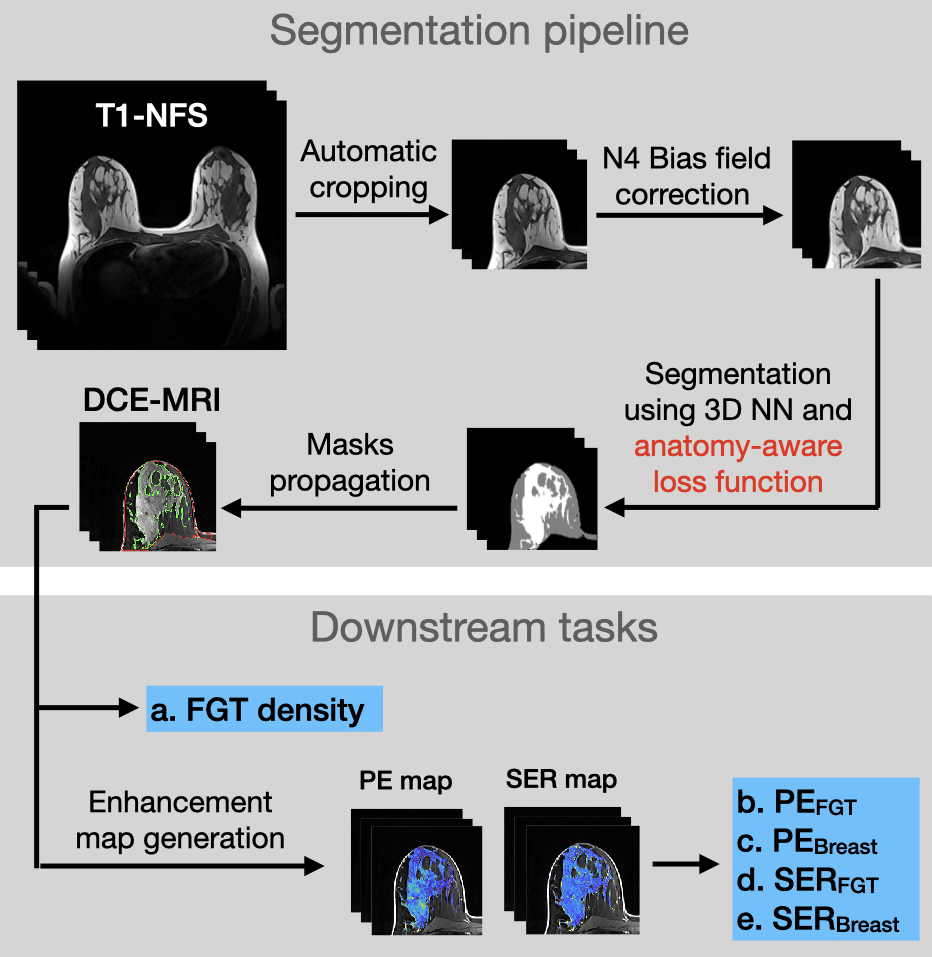
Fig. 1 Illustration of the segmentation pipeline and downstream tasks. T1-NFS, T1-weighted non-fat-suppressed; NN, neural network; PE, percent enhancement; SER, signal enhancement ratio.
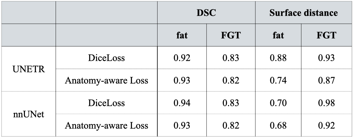
Fig. 2 Comparison of dice score (DSC) and surface distance of fat and FGT using dice loss functions and anatomy-aware loss function across the entire cohort.
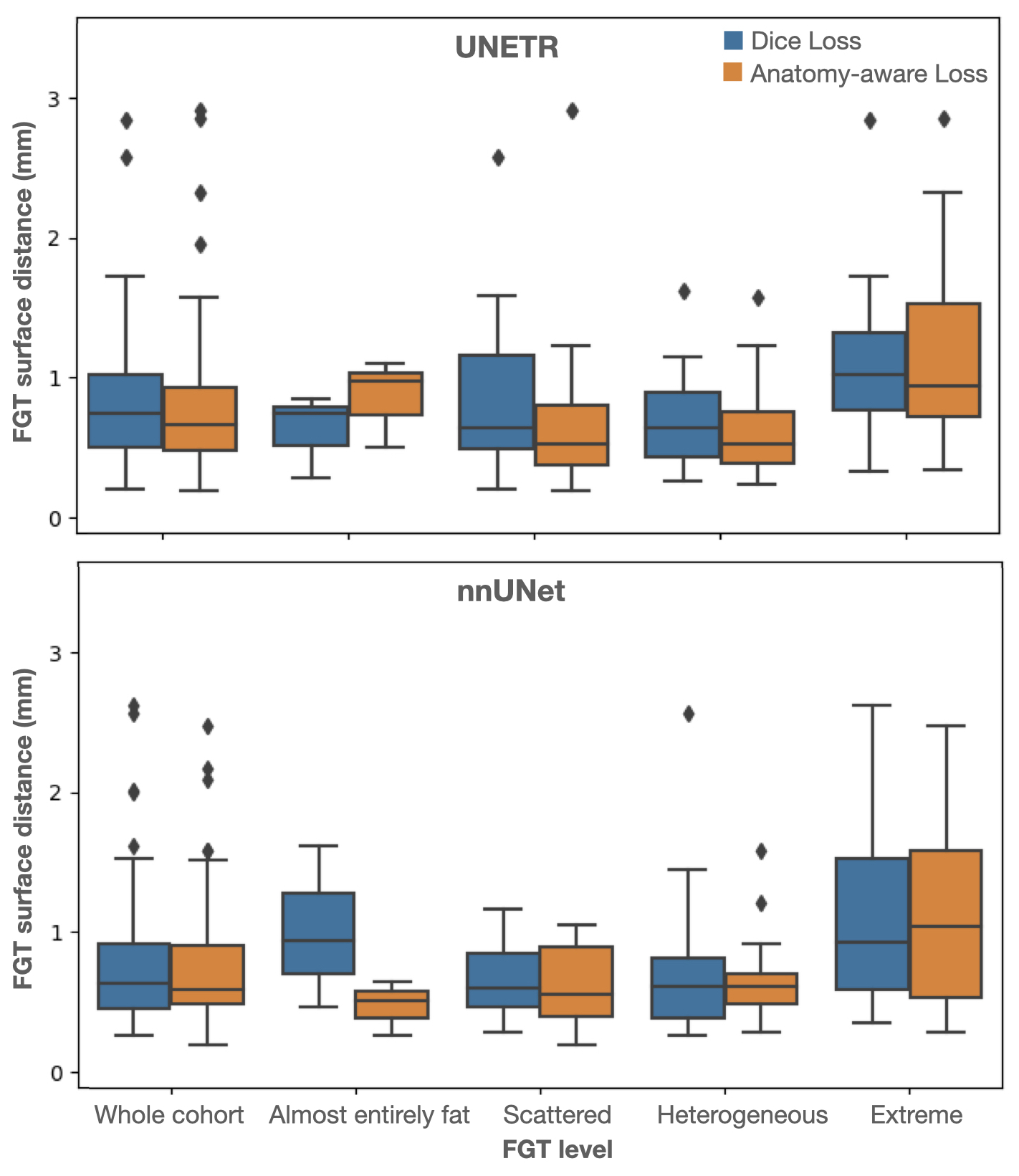
Fig. 3 Comparison of FGT surface distance using different loss functions across the entire cohort and in sub-cohorts at different FGT levels.
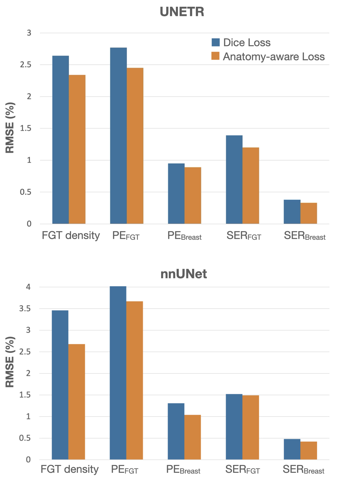
Fig. 4 Comparison of root mean square errors (RMSE) of FGT density and four quantitative BPE measures using different loss functions across the entire cohort. PE, percent enhancement; SER, signal enhancement ratio.