1467
Enhancing Breast Cancer Diagnosis through Deep Learning-Based DWI in Conjunction with Kaiser Score1Department of Magnetic Resonance, The First Affiliated Hospital of Zhengzhou University, Zhengzhou, China, 2MR Research China, GE Healthcare, Beijing, China
Synopsis
Keywords: Breast, Breast, diffusion magnetic resonance imaging; deep learning, Magnetic resonance imaging; kaiser score
Motivation: While the Kaiser score serves a pivotal role in diagnosing breast cancer, it still encounters scenarios where false positives necessitate biopsy confirmation.
Goal(s): This study aims to investigate approaches to enhance the diagnostic efficacy of the Kaiser score through MRI.
Approach: Leveraging deep learning to enhance both the quality of DWI images and diagnosis, we sought more effective indicators in conjunction with the Kaiser score.
Results: ADC values derived from DWI images reconstructed using deep learning, with a b-value of 800 s/mm², in tandem with the Kaiser score, significantly enhance the diagnostic performance nearing 1.
Impact: Integrating DWI under deep learning with the Kaiser score can elevate the accuracy of differentiating between benign and malignant breast cancers to almost 100%, leading to substantial improvements in breast cancer diagnosis and a reduction in unnecessary biopsies.
Introduction:
The Kaiser Score (KS) serves as a highly efficient clinical diagnostic tool employed in the assessment of breast lesions [1]. It adeptly integrates clinical and imaging data for a comprehensive evaluation. Nevertheless, it grapples with the challenge of misdiagnoses. Magnetic Resonance Imaging (MRI), on the other hand, offers a non-invasive means of screening for breast lesions with commendable sensitivity but is hindered by a notable rate of false positives. A revolutionary advancement in the medical realm, deep learning reconstruction[2,3] has demonstrated its prowess in enhancing image quality and signal-to-noise ratios, even when scanning times are abbreviated. While it has received extensive validation across various anatomical regions, its application in breast imaging under Diffusion-Weighted Imaging (DWI) reconstruction awaits our own validation. We postulate that by synergizing the Kaiser Score with a suitable DWI model, we can achieve superior diagnostic outcomes.Methods
Over the course of January 2023 to October 2023, a cohort of 111 cases with breast lesions was enrolled, comprising 72 malignant and 36 benign lesions, all of which were pathologically confirmed. MRI scans were conducted using a 3.0 Tesla MRI scanner (SIGNA Premier, GE Healthcare, WI, USA). Two radiologists conducted assessments using the Kaiser Score without prior knowledge of the pathological findings. Lesions with a score of ≥5 were categorized as malignant. Receiver Operating Characteristic (ROC) curve analysis was employed to establish the optimal threshold values for distinguishing benign from malignant lesions. In the context of a combined diagnostic approach, lesions with a Kaiser Score (KS) of ≥5 and Apparent Diffusion Coefficient (ADC) values falling below a predetermined threshold were classified as malignant, while those failing to meet these criteria were deemed benign. Sensitivity, specificity, and the Area Under the Curve (AUC) were assessed and compared between the Kaiser Score, DWI, and the combined diagnostic method. All MRI scans were performed using a 3T scanner, and both conventional b-values (800 s/mm²) and high b-values (3000 s/mm²) were explored to identify an optimal DWI model.Results and discussion
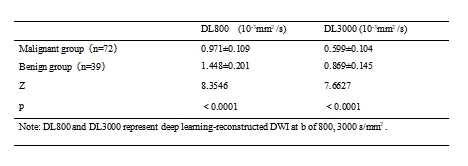
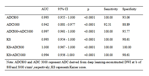
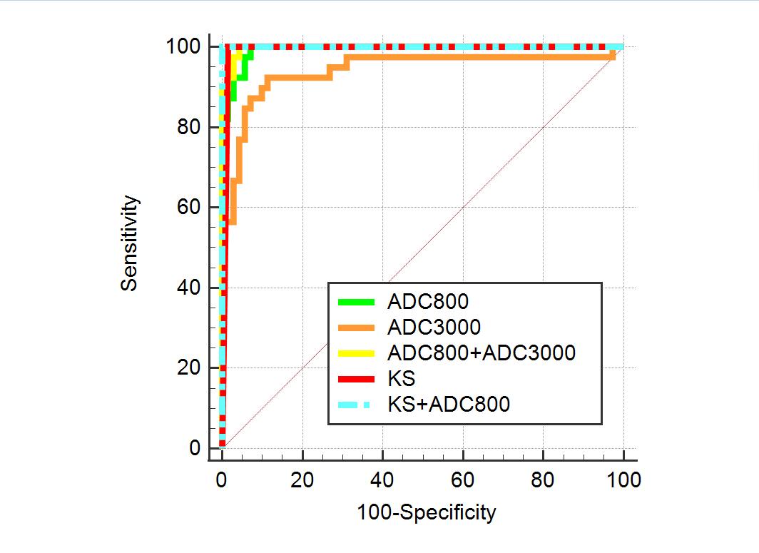
In our research, we thoroughly examined 111 cases of breast lesions, with 72 identified as malignant, spanning various types including invasive carcinoma, intraductal papillary carcinoma, ductal carcinoma in situ, invasive carcinoma combined with ductal carcinoma in situ, and lobular carcinoma in situ. Furthermore, the 39 benign cases encompassed conditions such as fibroadenoma, adenosis with fibroadenoma, adenosis with ductal changes, intraductal papilloma, inflammation, adenosis, and adenomyoepithelioma. Notably, the ADC values exhibited distinct patterns within these breast lesions.Specifically, the malignant lesions demonstrated discernibly lower ADC values. The ADC value derived from deep learning reconstructed DWI at a b-value of 800 s/mm² was measured at 0.971×10^-3 mm²/s, while at a b-value of 3000 s/mm², it was recorded at 0.599×10^-3 mm²/s. These values consistently presented notably lower measurements when compared to the benign group, as outlined in Table 1. Importantly, these differences were statistically significant (P < 0.05), emphasizing a meaningful distinction between the two groups. While the area under the curve (AUC) (Table 2 and Fig. 1) for the KS matched that of DWI800, its specificity was notably higher at 0.986 compared to DWI800. Additionally, the AUC of DWI800 surpassed that of DWI3000. After combining KS with DWI800, the AUC notably improved from KS's original 0.993 to 1, while the AUC achieved a high 0.994 when KS was combined with DWI3000. This suggests that in routine scans, utilizing only a deep learning reconstruction DWI with a b-value of 800 s/mm² alongside KS can approach near 100% accuracy in diagnosing breast lesions as benign or malignant.Conclusions
In conclusion, the diagnostic performance of the KS in distinguishing between benign and malignant breast lesions outperforms standalone deep learning DWI. Augmenting KS with deep learning DWI with b=800 s/mm² significantly enhances its diagnostic efficacy.Acknowledgements
No acknowledgements.References
[1] Aydan Avdan Aslan, et al. "Diagnostic performance of Kaiser score in patients with newly diagnosed breast cancer: Factors associated with false-negative results." European Journal of Radiology, 2023, Volume 164, Page 110864. DOI: 10.1016/j.ejrad.2023.110864.
[2]Minjae Kim, et al. "Thin-Slice Pituitary MRI with Deep Learning–based Reconstruction: Diagnostic Performance in a Postoperative Setting." Radiology, 2021, Volume 298, Number 1.
[3]Kevin M. Koch, et al. "Analysis and Evaluation of a Deep Learning Reconstruction Approach with Denoising for Orthopedic MRI." Radiology, 2021, Volume 3, Number 6.
Figures