1465
Quantitative MRI parameters of amide proton transfer and diffusion kurtosis imaging in the evaluation of breast cancer1Yan 'an Hospital, kunming, China, 2Yunnan Cancer Hospital, kunming, China, 3Philips Healthcare, kunming, China
Synopsis
Keywords: fMRI Analysis, Breast
Motivation: Limited clinical studies have explored the potential of Amide Proton Transfer Weighted Imaging (APTWI) in discriminating between benign and malignant breast lesions and molecular subtypes of breast cancer.
Goal(s): This study aims to assess the clinical utility of APTWI and DKI in the evaluation of benign and malignant breast diseases and the determination of molecular subtypes of breast cancer.
Approach: We quantitatively analyzed lesions in breast MRI scans of patients prior to surgery and evaluated the diagnostic value of each quantitative parameter.
Results: APTWI did not exhibit superior diagnostic efficacy compared to DKI and ADC in characterizing the molecular subtypes of breast cancer.
Impact: Our study underscores that APT imaging, as a novel quantitative magnetic resonance technique, does not confer a diagnostic advantage over DKI and ADC in the context of breast disease assessment.
Objective
This study aims to compare the diagnostic roles of DKI, ADC, and APTWI in distinguishing between benign and malignant breast lesions and in assessing molecular subtypes of breast cancer. Furthermore, it seeks to evaluate the diagnostic significance of the obtained parameters across various prognostic factors related to breast cancer. The objective is to provide novel insights into the diagnosis, treatment, and prognosis assessment of breast cancer.Materials and methods
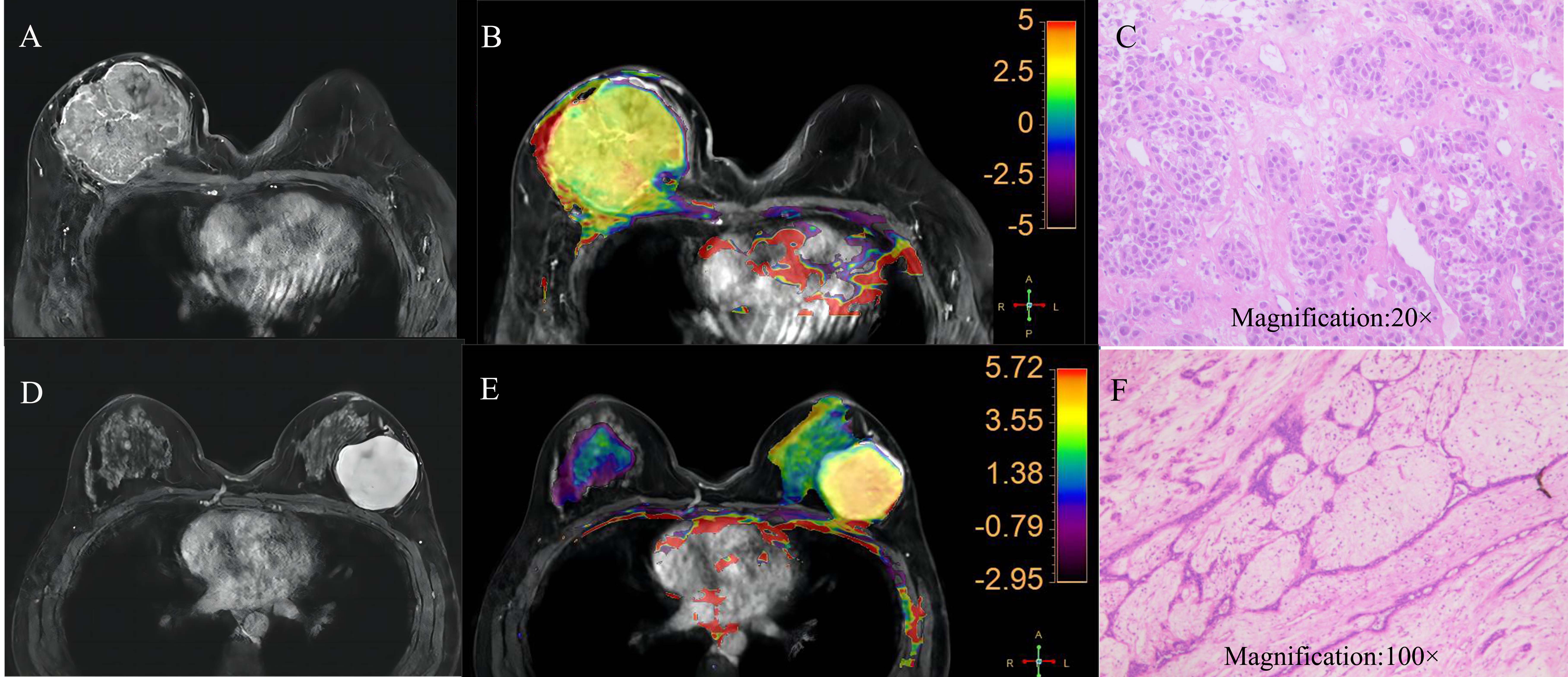
A total of 168 female patients underwent both breast APTWI and DKI scans (Figure 1). Quantitative measurement of APT signal intensity (SI), apparent kurtosis coefficient (Kapp), non-Gaussian diffusion coefficient (Dapp), and ADC values were measured before surgical procedures (Figure 2). Variance analysis was employed to examine variations in these quantitative MRI parameters across different molecular subtypes of breast cancer. The diagnostic efficacy of each quantitative parameter for distinguishing between benign and malignant breast diseases was evaluated using receiver operating characteristic curve (ROC) analysis.Results
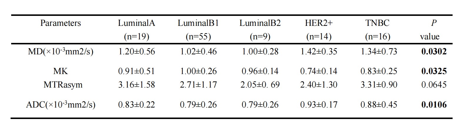
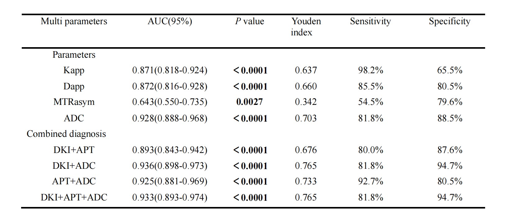
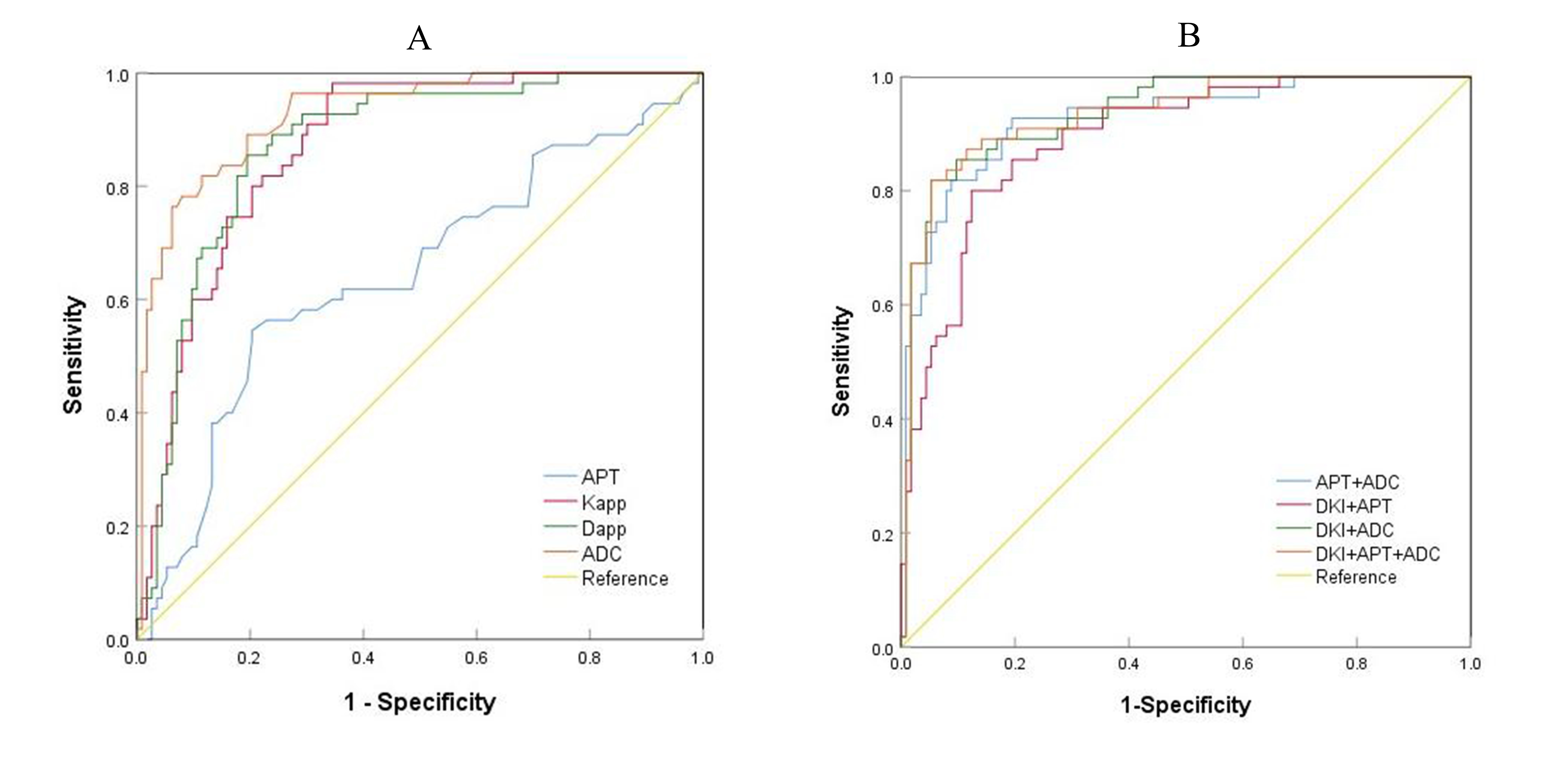
The study found significant differences between benign and malignant breast lesions in various quantitative parameters (summarized in Table 1). Specifically, the Kapp and APT values were notably higher in the malignant group (P < 0.0001 and P < 0.05, respectively) compared to the benign group. Conversely, ADC values (P < 0.0001) and Dapp values (P < 0.0001) were substantially lower in the malignant group than in the benign group. The diagnostic performance was assessed using the area under the curve (AUC) for various parameter combinations. AUC values were as follows: AUC (Kapp) = 0.871, AUC (Dapp) = 0.872, AUC (APT SI)) = 0.643), AUC (DKI+APT) = 0.893, AUC (DKI + ADC) = 0.936, AUC (APT + ADC) = 0.925, AUC (DKI + APT + ADC) = 0.933 (as shown in Table 2 and Figure 3).Discussion
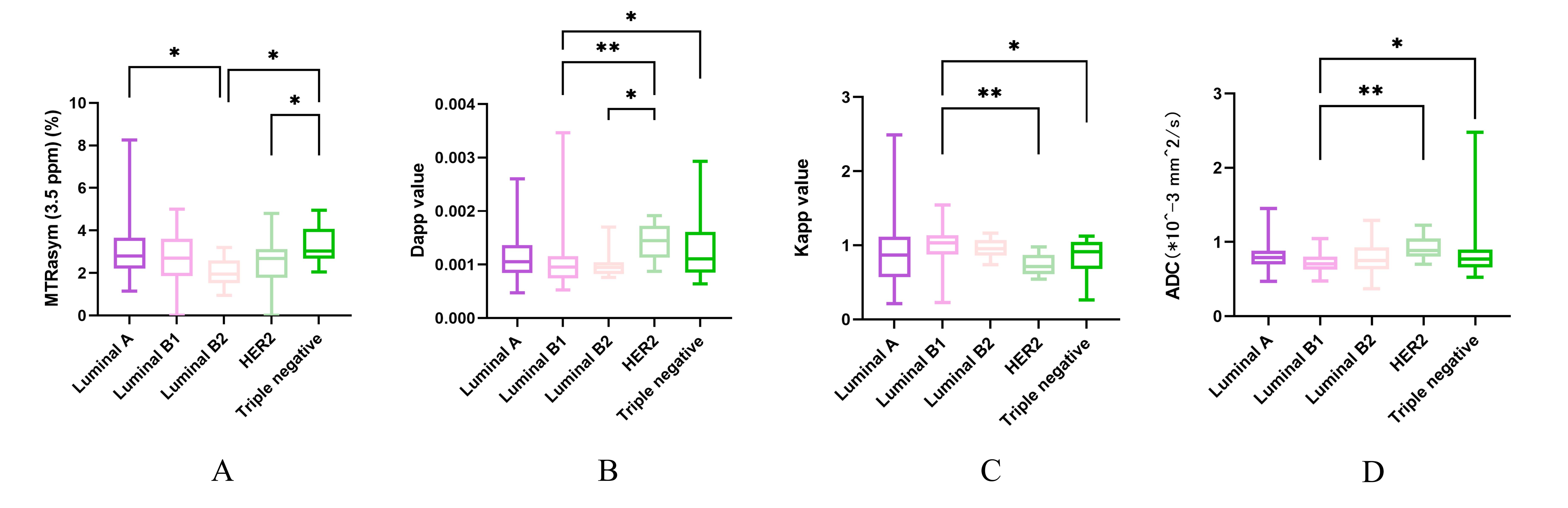
In this study, we observed that the Kapp value and APT SI values were higher in the malignant group compared to the benign group, while the Dapp value and ADC values were lower in the malignant group, consistent with findings from previous studies1-5. Benign tumors typically exhibit low diffusion due to their regular cell arrangement, relatively normal tissue structure, and ample intercellular space, causing minimal hindrance to water molecule diffusion. As a result, benign tumors often yield low ADC values on DWI. Conversely, malignant tumors display disrupted cell arrangements, an absence of normal hierarchy, and reduced intercellular, leading to restricted water molecule diffusion, deviating from the Gaussian distribution. This is a primary reason for the low ADC values in malignant tumors6. In benign lesions, looser cell arrangements, atypical nuclei, and reduced tissue necrosis allow for more accessible water molecule diffusion, approximating Gaussian distribution7. Moreover, the elevated APT SI values in the malignant group suggest that malignant tumors tend to contain higher concentrations of mobile proteins and peptides compared to benign tumors. This phenomenon is attributed to rapid cell proliferation, increased protein expression, and heightened cell density in malignant tumors, leading to a higher concentration of mobile proteins and peptides within the tumor8.Accurate preoperative evaluation of breast cancer’s molecular subtypes is crucial, as different subtypes warrant distinct clinical treatments and prognoses. This study evaluated the diagnostic potential of APTWI, DKI, and ADC in identifying different molecular subtypes of breast cancer. The results show that DKI and ADC values outperformed APTWI in diagnosing molecular subtypes of breast cancer, as well as in distinguishing and evaluating the prognosis of benign and malignant breast lesions. Several factors may contribute to this discrepancy: (1) APTWI image post-processing relies on superior image quality, and the absence of standard parameter APT imaging sequences may impacter performance. (2) Patients with breast diseases often detect breast nodules at an early stage, leading to smaller lesions with lower protein and peptide secretion. (3) Variability in water diffusion ratio among different populations may be more pronounced than difference in protein and peptide content. These factors may explain the observed contrasts between DKI and ADC values and APTSI values in this study.
However, our study has limitations. First, the sample size was relatively small and confined to a single center. Second, the region of interest was delineated only at the level of the largest lesion and adjacent sections, potentially introducing bias. Third, the extended imaging time of APTWI and DKI might have caused discomfort to participants and affected image quality.
Conclusion
APTWI proved effective in distinguishing between benign and malignant breast disease and enhancing the utility of diffusion-weighted MRI. However, it did not exhibit superior diagnostic efficacy compared to DKI and ADC in characterizing the molecular subtypes of breast cancer.Acknowledgements
No acknowledgment found.References
[1]. Li T, Hong Y, Kong D, Li K. Histogram Analysis of Diffusion Kurtosis Imaging Based on Whole-Volume Images of Breast Lesions. J Magn Reson Imaging (2020) 51(2):627-34. Epub 2019/08/07. doi: 10.1002/jmri.26884.
[2]. Meng N, Wang X, Sun J, Han D, Bai Y, Wei W, et al. A Comparative Study of the Value of Amide Proton Transfer-Weighted Imaging and Diffusion Kurtosis Imaging in the Diagnosis and Evaluation of Breast Cancer. Eur Radiol (2021) 31(3):1707-17. Epub 2020/09/06. doi: 10.1007/s00330-020-07169-x.
[3]. Tahmassebi A, Wengert GJ, Helbich TH, Bago-Horvath Z, Alaei S, Bartsch R, et al. Impact of Machine Learning with Multiparametric Magnetic Resonance Imaging of the Breast for Early Prediction of Response to Neoadjuvant Chemotherapy and Survival Outcomes in Breast Cancer Patients. Investigative radiology (2019) 54(2):110-7. Epub 2018/10/26. doi: 10.1097/rli.0000000000000518.
[4]. Ohno Y, Yui M, Koyama H, Yoshikawa T, Seki S, Ueno Y, et al. Chemical Exchange Saturation Transfer Mr Imaging: Preliminary Results for Differentiation of Malignant and Benign Thoracic Lesions. Radiology (2016) 279(2):578-89. Epub 2015/11/21. doi: 10.1148/radiol.2015151161.23. Takayama Y, Nishie A, Togao O, Asayama Y, Ishigami K, Ushijima Y, et al. Amide Proton Transfer Mr Imaging of Endometrioid Endometrial Adenocarcinoma: Association with Histologic Grade. Radiology (2018) 286(3):909-17. Epub 2017/10/31. doi: 10.1148/radiol.2017170349.
[5]. Wang HJ, Cai Q, Huang YP, Li MQ, Wen ZH, Lin YY, et al. Amide Proton Transfer-Weighted Mri in Predicting Histologic Grade of Bladder Cancer. Radiology (2022) 305(1):127-34. Epub 2022/06/29. doi: 10.1148/radiol.211804.
[6]. Gao W, Zhang S, Guo J, Wei X, Li X, Diao Y, et al. Investigation of Synthetic Relaxometry and Diffusion Measures in the Differentiation of Benign and Malignant Breast Lesions as Compared to Bi-Rads. J Magn Reson Imaging (2021) 53(4):1118-27. Epub 2020/11/13. doi: 10.1002/jmri.27435.
[7]. Yuan C, Jin F, Guo X, Zhao S, Li W, Guo H. Correlation Analysis of Breast Cancer Dwi Combined with Dce-Mri Imaging Features with Molecular Subtypes and Prognostic Factors. Journal of medical systems (2019) 43(4):83. Epub 2019/02/28. doi: 10.1007/s10916-019-1197-5.
[8]. Chen W, Li L, Yan Z, Hu S, Feng J, Liu G, et al. Three-Dimension Amide Proton Transfer Mri of Rectal Adenocarcinoma: Correlation with Pathologic Prognostic Factors and Comparison with Diffusion Kurtosis Imaging. Eur Radiol (2021) 31(5):3286-96. Epub 2020/10/31. doi: 10.1007/s00330-020-07397-1.
Figures