1463
The value of amide proton transfer imaging in the diagnosis of malignant and benign urinary bladder lesions: comparison with DWI1Department of Radiology, Tongji Hospital, Shanghai, China, 2Philips Healthcare, Shanghai, China
Synopsis
Keywords: Cancer, Bladder, APT, DWI
Motivation: Conventional MRI has certain limitations in distinguishing between malignant and benign urinary bladder (UB) lesions.
Goal(s): Compare the diagnostic value of Amide proton transfer (APT) imaging with diffusion-weighted imaging (DWI).
Approach: Participants with confirmed UB lesions underwent preoperative multiparametric MRI. The APT signal intensity (represented by asymmetric magnetization transfer ratio, MTRasym), and apparent diffusion coefficient (ADC) values were compared between malignant and benign UB lesions, and their diagnostic performance was evaluated with receiver operating characteristic (ROC) curve analysis.
Results: The MTRasym value was significantly better in differentiating urothelial carcinoma from benign UB lesions than the ADC value.
Impact: Conventional MRI exhibits limitations in accurately distinguishing between malignant and benign urinary bladder (UB) lesions. Amide Proton Transfer (APT) imaging shows promise in effectively discriminating between malignant and benign UB lesions, surpassing the performance of Diffusion-Weighted Imaging (DWI).
Introduction
Conventional MRI has been highly regarded for its ability to detect tumours in the follow-up process and to differentiate malignant from benign bladder lesions.1 However, each of these methods has its own limitations, and generally accepted thresholds for identifying tumour types have not been established.2, 3Amide proton transfer (APT) imaging, a novel MRI technique based on chemical exchange saturation transfer (CEST), provides more information on subcellular protein levels than current standard MR protocols.4
We aimed to investigate the potential of APT imaging in the diagnosis of malignant and benign UB lesions and compare its diagnostic efficacy with that of DWI.
Methods
Eighty patients with UB lesions were enrolled prospectively from a single centre. The MRI scans were conducted on a 3.0T Ingenia system (Philips Healthcare, Best, the Netherlands).APT signal intensity in UB lesions was quantified by the asymmetric magnetization transfer ratio (MTRasym). MTRasym and apparent diffusion coefficient (ADC) values were measured and compared between malignant and benign UB lesions, and their diagnostic performance was evaluated with receiver operating characteristic (ROC) curve analysis.
Results
In this study, 32 patients had pathologically confirmed benign UB lesions, including 2 bladder leiomyomas, 1 submucosal amyloidosis, 1 inflammatory myofibroblastic tumour and 28 inflammatory lesions, and 48 patients had pathologically confirmed urothelial carcinoma.Urothelial carcinomas showed significantly higher MTRasym values (1.53% [0.74%] vs. 0.85% [0.23%]) and lower ADC values (1.24 ± 0.34×10⁻³ mm2/s vs. 1.43 ± 0.22×10⁻³ mm2/s) than benign UB lesions. The MTRasym value (AUC = 0.928) was significantly better in differentiating urothelial carcinoma from benign UB lesions than the ADC value (AUC = 0.722) according to the area under the ROC curve.
Discussion
The present results revealed that urothelial carcinoma showed higher MTRasym values than benign UB lesions. This result may contribute to the higher concentrations of endogenous cellular proteins in malignant UB lesions than in benign UB lesions because malignant UB lesions are characterized by a high proliferation rate and increased cellular density.In addition, we also found that urothelial carcinoma showed lower ADC values than benign UB lesions. The higher density of tissue cells in malignant UB lesions greatly restricts water molecule diffusion, which accounts for the lower ADC value.
We also compared the diagnostic ability of APT imaging and DWI in differentiating urothelial carcinoma from benign UB lesions. We noticed that the AUC of the MTRasym value was significantly higher than that of the ADC value. This result showed that the diagnostic performance of APT imaging in differentiating urothelial carcinoma from benign UB lesions is better than that of DWI.
Malignant UB lesions exhibit aggressive biological behaviours and are more likely present with a large tumour size, high nuclear grade, increased cellular density, and high proliferation rate. Perfusion disorders, hypoxia and acidification are intrinsic properties of altered tumour cell metabolism. Cancer cells usually have higher intracellular pH than normal cells when investigated under similar environmental conditions. The sources of the APT signal and contrast, to a large extent, could be attributed to the concentration of endogenous cellular proteins in tissue and the peptides responsible for exchanging amide protons with water protons (pH dependent). Nuclear atypia of malignant tumours can induce the interaction of macromolecules with hydrophobic cell membranes and promote the release of proteins and peptides, and urothelial carcinoma might show a higher pH value, both of which might be responsible for the increase in the MTRasym value.
The ADC value, a diffusion coefficient reflecting the degree of diffusion of water molecules in tissues, is highly sensitive to cellularity changes. However, the main disadvantage of conventional DWI is that it is nonspecific in terms of microstructure components, and the observed values of the ADC can be affected by, for example, effects of flow apart from the effects of diffusion, the timing of the gradient pulse and the b value. Furthermore, the measured ADC value is acquired by averaging the images collected in a gradient order oriented along three perpendicular directions. The resulting value is only an approximation, which may lead to a large overestimation of the true ADC value in tissues experiencing anisotropic diffusion.
Conclusion
APT imaging has the potential to discriminate malignant from benign UB lesions with a performance superior to that of DWI.Acknowledgements
No acknowledgement found.References
1. Lenis AT, Lec PM, Chamie K, et al. Bladder Cancer: A Review. JAMA 2020; 324: 1980.
2. Avcu S, Koseoglu MN, Ceylan K, et al. The value of diffusion-weighted MRI in the diagnosis of malignant and benign urinary bladder lesions. Br J Radiol 2011; 84: 875–882.
3. Wang H, Pui MH, Guo Y, et al. Diffusion-weighted MRI in bladder carcinoma: the differentiation between tumor recurrence and benign changes after resection. Abdom Imaging 2014; 39: 135–141.
4. Kamimura K, Nakajo M, Yoneyama T, et al. Amide proton transfer imaging of tumors: theory, clinical applications, pitfalls, and future directions. Jpn J Radiol 2019; 37: 109–116.Figures
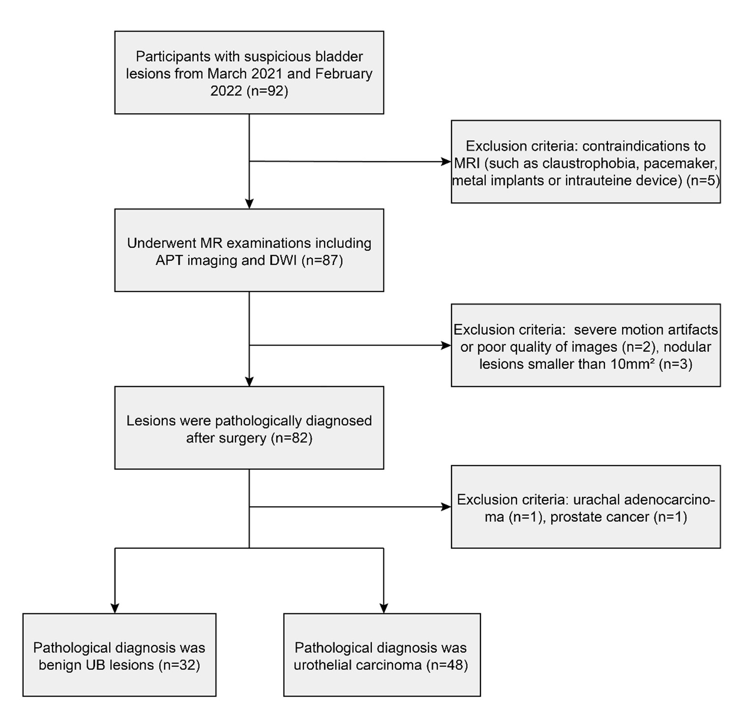
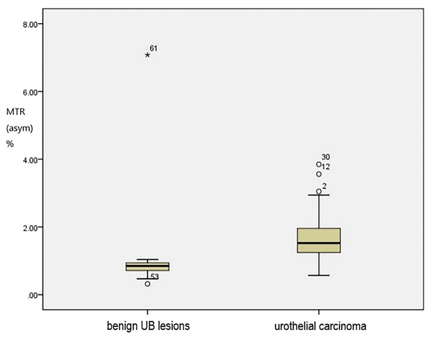
Fig. 2. Comparisons of MTRasym values between benign UB lesions and urothelial carcinoma. Urothelial carcinoma showed significantly higher MTRasym values than benign UB lesions (P < 0.001).
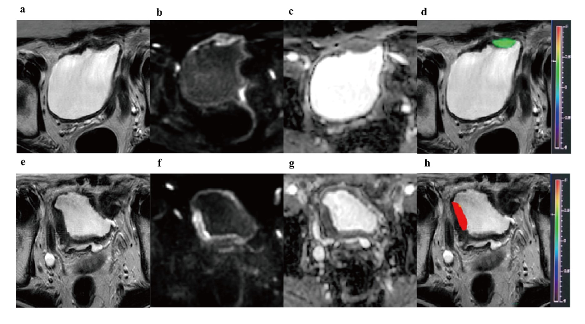
Fig. 3. (a-d) A 58-year-old female with cystitis after TURBT. (e-h) An 82-year-old male with a high-grade invasive bladder tumour invading the adipose tissue around the bladder. (a, e) T2WI. (b, f) DWI (b = 1000 s/mm2). (c, g) ADC image. (d, h) APT image fused with T2WI. The colour bar indicates the MTRasym value. The mean MTRasym and ADC values of cystitis measured by two radiologists were 0.82% and 1.50×10⁻³ mm2/s, respectively. The mean MTRasym and ADC values of high-grade invasive bladder tumours measured by the two radiologists were 2.44% and 0.93×10⁻³ mm2/s, respectively.
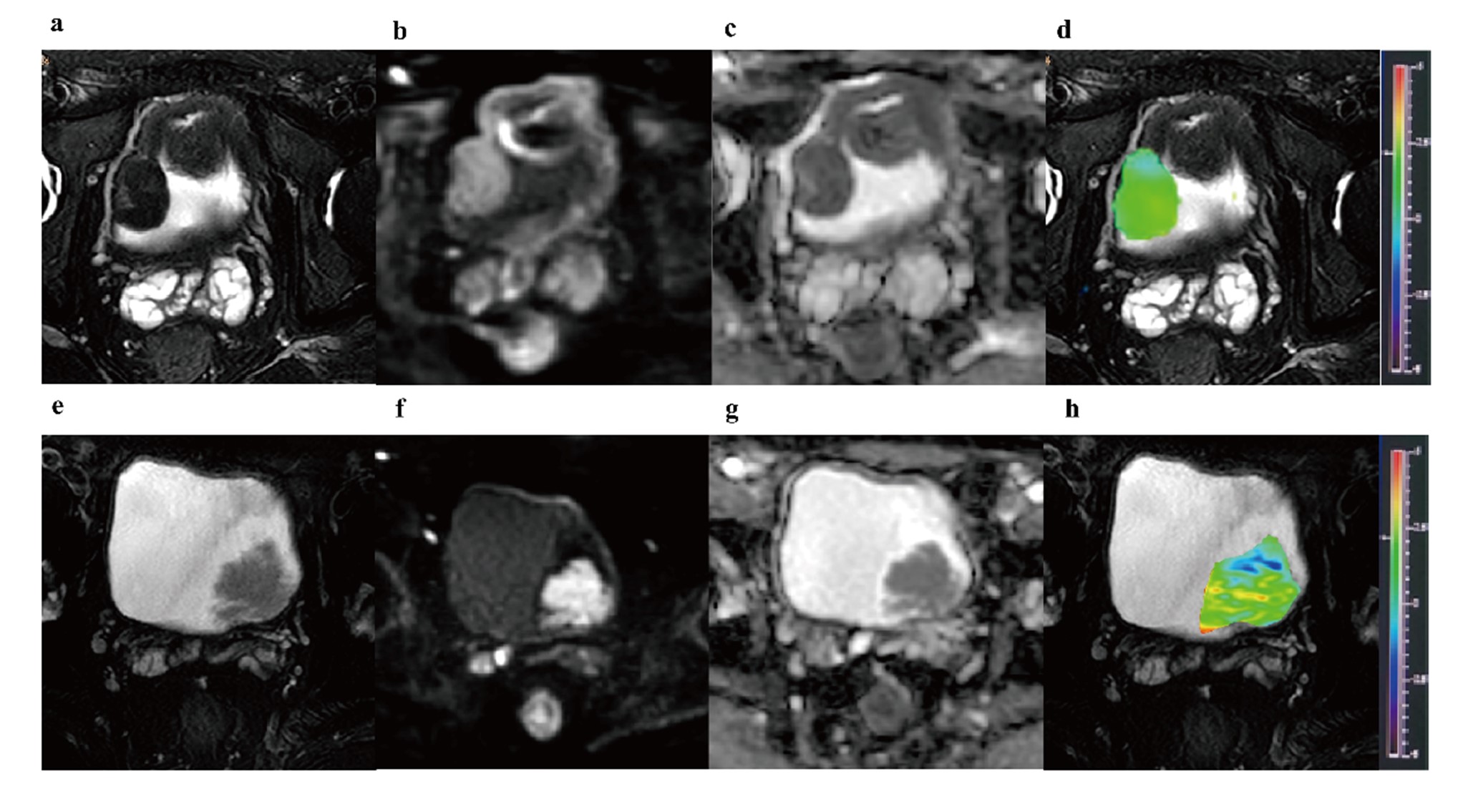
Fig. 4. (a-d) A 76-year-old male with bladder leiomyoma. (e-h) A 48-year-old male with urothelial carcinoma. (a, e) T2WI SPAIR. (b, f) DWI (b = 1000 s/mm2). (c, g) ADC image. (d, h) APT imaging fused with T2WI SPAIR. The colour bar indicates the MTRasym value. The mean MTRasym and ADC values of leiomyoma measured by two radiologists were 0.98% and 1.01×10⁻³ mm2/s, respectively. The mean MTRasym and ADC values of urothelial carcinoma measured by two radiologists were 1.58% and 1.38×10⁻³ mm2/s, respectively.
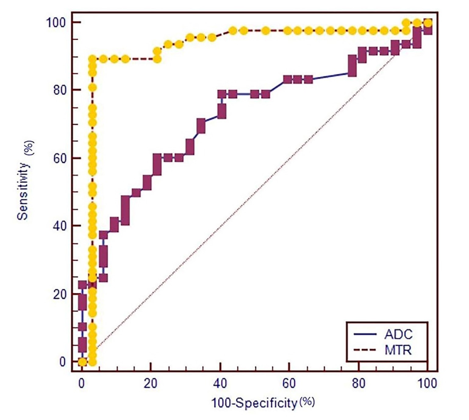
Fig. 5. ROC curves of MTRasym and ADC values in discriminating benign UB lesions from urothelial carcinoma. The AUC of MTRasym (0.928) was significantly larger than that of ADC (0.771) in discriminating benign UB lesions from urothelial carcinoma (P = 0.0015).