1457
Investigation of Texture Features in Head and Neck Cancer: Preliminary Results for Early Radiation Therapy induced Changes1Department of Diagnostic and Interventional Radiology, University of Tuebingen, Section on Experimental Radiology, Tübingen, Germany, 2Institute for Diabetes Research and Metabolic Diseases of the Helmholtz Centre Munich at the University of Tuebingen, Tübingen, Germany, 3German Center for Diabetes Research (DZD), Neuherberg, Germany, 4Institute of Signal Processing and System Theory, University of Stuttgart, Stuttgart, Germany, 5Department of Diagnostic and Interventional Radiology, University of Tuebingen, University Hospital Tuebingen, Tübingen, Germany, 6Department of Radiation Oncology, University Hospital Tübingen, Section for Biomedical Physics, Tübingen, Germany, 7Department of Radiation Oncology, University Hospital Tübingen, University Hospital and Medical Faculty, Tübingen, Germany
Synopsis
Keywords: Cancer, Head & Neck/ENT
Motivation: Radiomic texture features are considered promising biomarkers for tumor’s response to therapy.
Goal(s): To identify texture features that can enhance predictive accuracy regarding tumor treatment outcomes.
Approach: The study included 13 patients with head and neck cancer undergoing radiation therapy, with MRI (T2w, DWI) conducted before treatment and during early-treatment phase. Image processing, tumor segmentation, and feature extraction are performed.
Results: Wilcoxon signed-rank tests with a Holm-Bonferroni correction reveals that Skewness in T2w-images exhibits significant changes during early treatment. This finding suggests that this feature may hold promise for predicting therapeutic responses, although larger studies are needed to confirm these results.
Impact: The study's preliminary findings suggest that Skewness in T2w images may have the potential to provide useful information for early response assessment in head and neck cancer patients undergoing radiation therapy, warranting further investigation to confirm its clinical significance.
Introduction
Great efforts are being made to identify potential magnetic resonance imaging (MRI) biomarkers that enable rapid and accurate prediction of tumor response to therapy. In addition to inherent tissue properties such as apparent diffusion coefficient (ADC) and relaxation times, radiomic texture features are considered promising biomarkers for response monitoring and outcome prediction1-8. Texture features changing significantly between baseline (pre-treatment) and the early treatment phase can potentially provide additional information for an improved prediction of tumor response1,2.The overall aim of this work was to identify texture features that show significant changes in the early phase of radiotherapy (RT) of head and neck cancer (HNC). For this, texture features were extracted on segmented T2-weighted images and calculated ADC maps and compared at baseline and in the early treatment phase.
Methods
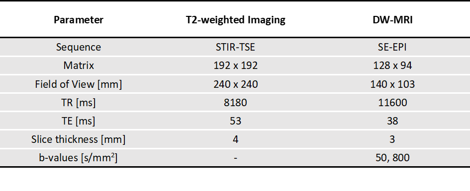
Patient cohort and image acquisitionThe study included 13 patients with locally advanced HNC who underwent radiation therapy (RT). MRI was performed before treatment (baseline) and during the second week of RT using a whole-body, 3.0T clinical system (MAGNETOM Vida, Siemens Healthcare, Erlangen, Germany). T2w-images were acquired in the axial plane with an inversion recovery prepared turbo spin echo sequence (STIR-TSE). Diffusion-weighted MRI (DW-MRI) was performed using a spin-echo echo planar imaging (SE-EPI) sequence with two b-values (DWIb50, DWIb800). Acquisition parameters are listed in Table 1.
MR image preprocessing, segmentation and feature extraction
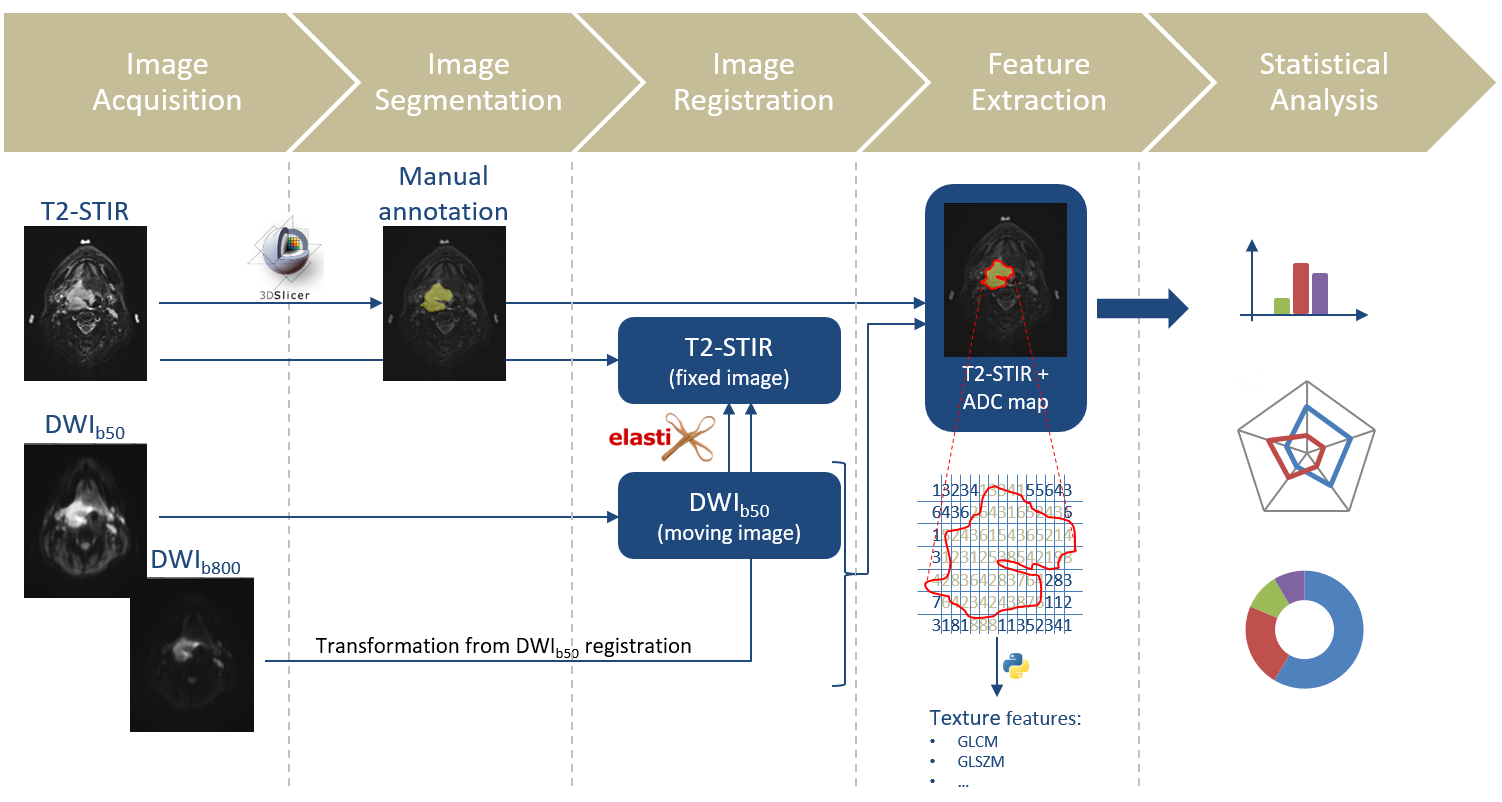
The image processing pipeline is schematically depicted in Figure 1. Tumor segmentation was carried out by an experienced radiologist (JK) using an open-source medical image computing software (3D slicer9,https://www.slicer.org/). For each patient and each examination, the entire tumor volume was manually segmented on T2w images. In addition, T1-weighted dynamic contrast enhanced images were used as a reference for visual co-registration. Due to image distortions in EPI-based diffusion-weighted acquisitions, DW-MRI were registered on the distortion-free T2w image. Therefore, a rigid registration in the region of the tumor segmentation according to Habrich et al.10 was performed using elastix11,12. Due to the higher correspondence of the visible structures between DWIb50 and T2w, DWIb50 was registered and the transformation applied on DWIb800 accordingly. After image registration, ADC maps were calculated using MATLAB® (The MathWorks, Natick, MA, USA). Feature extraction was performed according to IBSI (Image Biomarker Standardisation Initiative) guidelines using the open-source PyRadiomics Python package13. A total number of 93 texture features (First Order (18), Gray Level Cooccurrence Matrix-(GLCM) (24), Gray Level Run Length Matrix-(GLRLM) (16), Gray Level Size Zone Matrix-(GLSZM) (16), Neighbouring Gray Tone Difference Matrix-(NGTDM) (5), Gray Level Dependence Matrix-(GLDM) (14)) were extracted from each segmented tumor in both normalized T2w images and the computed ADC maps. The definitions and mathematical calculations of the texture features can be found on the PyRadiomics documentation site (https://pyradiomics.readthedocs.io/en/latest/features.html).
Statistical analysis
To identify early treatment-induced changes in texture features, a two-sided Wilcoxon signed-rank test with a significance level of α = 0.05 was used for three test cases: I) only T2w, II) only ADC and III) T2w + ADC features. A Holm-Bonferroni correction was applied to compensate for multiple testing. Since some features are volume-dependent14,15, these radiomic features were normalized to the volume in order to determine volume-independent changes. All statistical analyses were performed using MATLAB®.
Results
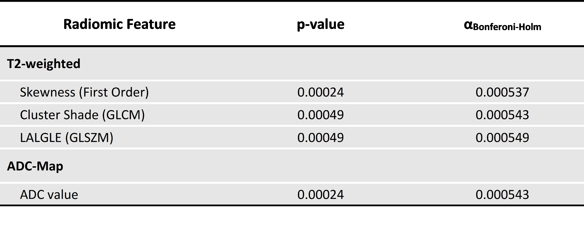
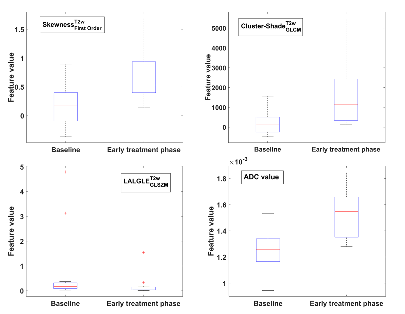
The Holm-Bonferroni-corrected Wilcoxon signed-rank test revealed three texture features on T2w that changed significantly in the early treatment phase: Skewness(First Order), Cluster shade(GLCM), and LALGLE(GLSZM) (Table 2, Figure 2). However, LALGLE(GLSZM) proved to be volume-dependent and showed no significant changes over the course of treatment after applying a volume correction. For test case II (only ADC), there were only significant changes regarding first order features mainly related to the known changes in ADC values after RT16 (Figure 2). In a pooled analysis (test case III), only the Skewness(First Order) in T2w and first order features in ADC are still significant.Conclusion
Skewness(First Order) extracted on T2w was identified as a texture feature that showed statistically significant changes in the course of RT in head and neck cancer with and without additional ADC features. Future studies with larger patient cohorts, which is clearly a limitation of this preliminary study, will investigate whether the skewness will still indicate significant changes and if it can create synergies with established biomarkers (e.g. ADC values, relaxation times) and thus provide useful information about the tumor's response to therapy. Furthermore, an investigation of not yet included higher order features might be also beneficial.Acknowledgements
No acknowledgement found.References
1. Vignati A, Mazzetti S, Giannini V, Russo F, Bollito E, Porpiglia F, Stasi M, Regge D. Texture features on T2-weighted magnetic resonance imaging: new potential biomarkers for prostate cancer aggressiveness. Phys Med Biol. 2015 Apr 7;60(7):2685-701.
2. Rodrigues A, Loman K, Nawrocki J, Hoang JK, Chang Z, Mowery YM, Oyekunle T, Niedzwiecki D, Brizel DM, Craciunescu O. Establishing ADC-Based Histogram and Texture Features for Early Treatment-Induced Changes in Head and Neck Squamous Cell Carcinoma. Front Oncol. 2021 Sep 2;11:708398.
3. Chitalia RD, Kontos D. Role of texture analysis in breast MRI as a cancer biomarker: A review. J Magn Reson Imaging. 2019 Apr;49(4):927-938.
4. Meng J, Liu S, Zhu L, Zhu L, Wang H, Xie L, Guan Y, He J, Yang X, Zhou Z. Texture Analysis as Imaging Biomarker for recurrence in advanced cervical cancer treated with CCRT. Sci Rep. 2018 Jul 30;8(1):11399.
5. Caruso D, Zerunian M, Ciolina M, de Santis D, Rengo M, Soomro MH, Giunta G, Conforto S, Schmid M, Neri E, Laghi A. Haralick's texture features for the prediction of response to therapy in colorectal cancer: a preliminary study. Radiol Med. 2018 Mar;123(3):161-167.
6. Dregely I, Prezzi D, Kelly-Morland C, Roccia E, Neji R, Goh V. Imaging biomarkers in oncology: Basics and application to MRI. J Magn Reson Imaging. 2018 Jul;48(1):13-26.
7. Litvin AA, Burkin DA, Kropinov AA, Paramzin FN. Radiomics and Digital Image Texture Analysis in Oncology (Review). Sovrem Tekhnologii Med. 2021;13(2):97-104.
8. Jethanandani A, Lin TA, Volpe S, Elhalawani H, Mohamed ASR, Yang P, Fuller CD. Exploring Applications of Radiomics in Magnetic Resonance Imaging of Head and Neck Cancer: A Systematic Review. Front Oncol. 2018 May 14;8:131.
9. Fedorov A., Beichel R., Kalpathy-Cramer J., Finet J., Fillion-Robin J-C., Pujol S., Bauer C., Jennings D., Fennessy F.M., Sonka M., Buatti J., Aylward S.R., Miller J.V., Pieper S., Kikinis R. 3D Slicer as an Image Computing Platform for the Quantitative Imaging Network. Magnetic Resonance Imaging. 2012 Nov;30(9):1323-41.
10. Habrich J, Boeke S, Nachbar M, Nikolaou K, Schick F, Gani C, Zips D, Thorwarth D. Repeatability of diffusion-weighted magnetic resonance imaging in head and neck cancer at a 1.5 T MR-Linac. Radiother Oncol. 2022 Sep;174:141-148.
11. S. Klein, M. Staring, K. Murphy, M.A. Viergever, J.P.W. Pluim, "elastix: a toolbox for intensity based medical image registration," IEEE Transactions on Medical Imaging, vol. 29, no. 1, pp. 196 - 205, January 2010.
12. D.P. Shamonin, E.E. Bron, B.P.F. Lelieveldt, M. Smits, S. Klein and
M. Staring, "Fast Parallel Image Registration on CPU and GPU for
Diagnostic Classification of Alzheimer's Disease", Frontiers in
Neuroinformatics, vol. 7, no. 50, pp. 1-15, January 2014.
13. van Griethuysen, J. J. M., Fedorov, A., Parmar, C., Hosny, A., Aucoin, N., Narayan, V., Beets-Tan, R. G. H., Fillon-Robin, J. C., Pieper, S., Aerts, H. J. W. L. (2017). Computational Radiomics System to Decode the Radiographic Phenotype. Cancer Research, 77(21), e104–e107.
14. Roy S, Whitehead TD, Quirk JD, Salter A, Ademuyiwa FO, Li S, An H, Shoghi KI. Optimal co-clinical radiomics: Sensitivity of radiomic features to tumour volume, image noise and resolution in co-clinical T1-weighted and T2-weighted magnetic resonance imaging. EBioMedicine. 2020 Sep;59:102963.
15. Jensen LJ, Kim D, Elgeti T, Steffen IG, Hamm B, Nagel SN. Stability of Radiomic Features across Different Region of Interest Sizes-A CT and MR Phantom Study. Tomography. 2021 Jun 8;7(2):238-252.
16: van Houdt PJ, Yang Y, van der Heide UA. Quantitative Magnetic Resonance Imaging for Biological Image-Guided Adaptive Radiotherapy. Front Oncol. 2021 Jan 29;10:615643.
Figures