1455
Perfusion parameters of DCE-MRI using GRASP correlates with tumour cellularity of lung cancer.1Department of Radiology, Chongqing University Cancer Hospital, Chongqing, China
Synopsis
Keywords: Cancer, Perfusion
Motivation: Tumour cellularity indicates tumour proliferation. Although perfusion MRI parameters have been proposed for non-invasive assessment of tumour cellularity, few studies have validated this theory, particularly in lung cancer. Previous studies provided a free-breathing perfusion MRI method using GRASP.
Goal(s): Therefore, our aim is to investigate the relationship between tumour cellularity and perfusion parameters of MRI using GRASP.
Approach: Pearson correlation coefficients were used to illustrate the relationship between perfusion parameters and cell density.
Results: The findings indicated an inverse correlation between tumour cellularity and Ve. However, the study found no significant correlation between Ktrans and cell density.
Impact: It suggests that GRASP perfusion MRI has potential as a non-invasive technique to assess tumour cellularity in lung cancer.
The primary hallmark of cancer cells is their capacity for prolonged, continual proliferation(1). The evaluation of tumour cellularity, which is indicative of tumour proliferation, presently depends on measurements of histological cell density(2). Perfusion parameters obtained via dynamic contrast-enhanced (DCE)-MRI have demonstrated a correlation with cell density in certain malignant lesions, such as cervical cancer or head and neck squamous cell carcinoma, along with proliferation potential and hypoxia levels(3,4). However, it is yet to be established whether perfusion parameters are linked to tumour cellularity in lung cancer. Possible reasons for the lack of balance between spatiotemporal resolution and volumetric coverage, as well as different types of physiological motion in routine DCE-MRI of the lung(5). Numerous approaches have been introduced for quick free-breathing MR acquisitions to address this, including Golden-angle RAdial Sparse Parallel (GRASP) MRI. Through our prior research, a free-breathing perfusion MRI technique for quantitative assessment of lung cancer has been provided(6-8). Therefore, our hypothesis was that free-breathing perfusion MRI employing GRASP could serve as a useful non-invasive method for assessing tumour cellularity. The aim of this research was to investigate the correlation between perfusion parameters of MRI and cell density in lung cancer patients.
Methods
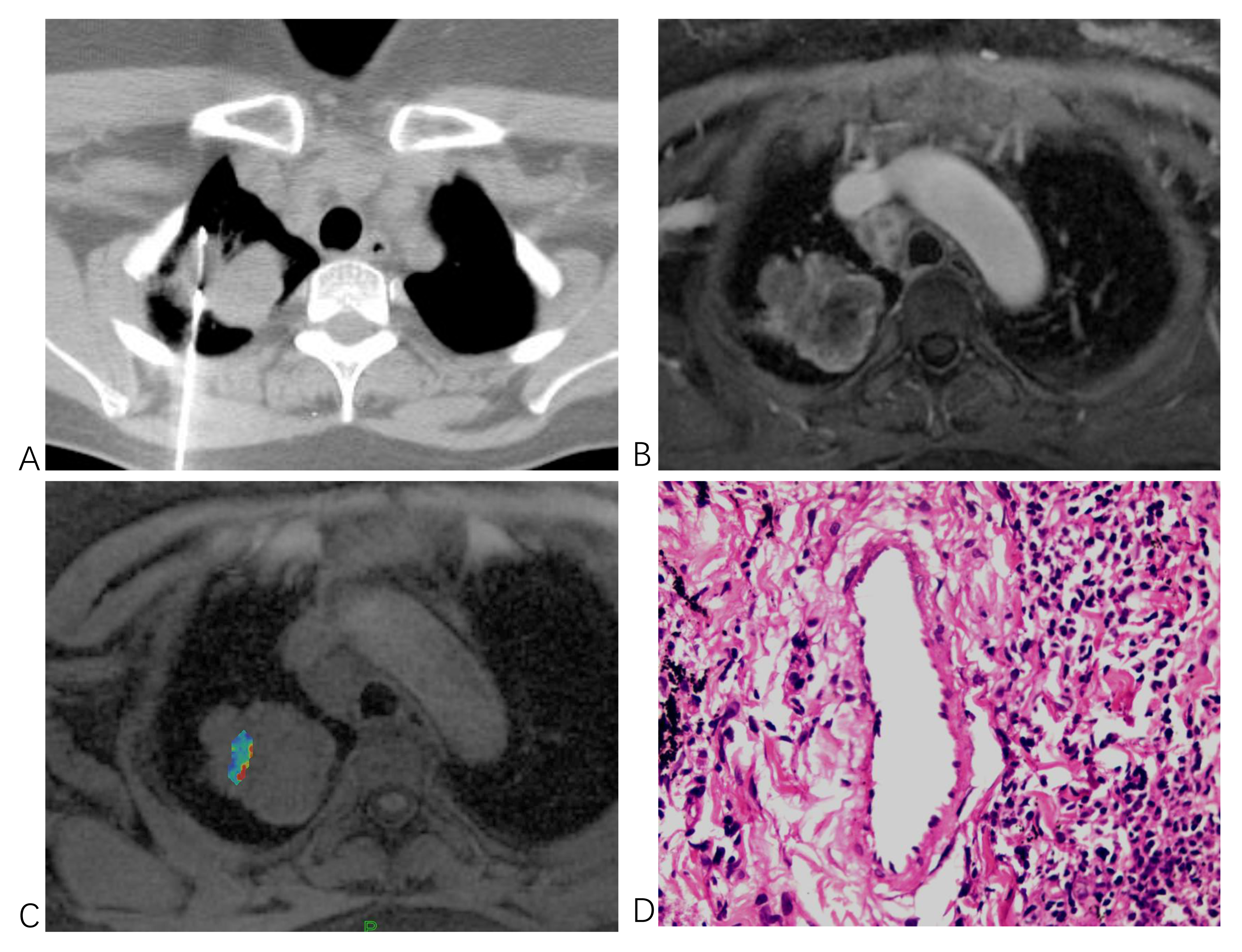
Seventeen consecutive patients (8 males, median age=56 (29,76)) who were diagnosed with non-small cell lung cancer (NSCLC) were retrospectively recruited for this study. Each participant underwent a free-breathing DCE-MRI scan employing a fat-statured T1-weighted stack-of-stars radial sequence. The obtained data underwent GRASP reconstruction with a temporal resolution of 3 second/ phase. Perfusion analysis was conducted using the Tofts model, which yielded Ktrans and Ve parameters. HE-stained tumour specimens were examined for cell density. Repeatability of measurements was determined by intraclass correlation coefficient (ICC), with Pearson correlation coefficients illustrating the relationship between the perfusion parameters and cell density.
Results
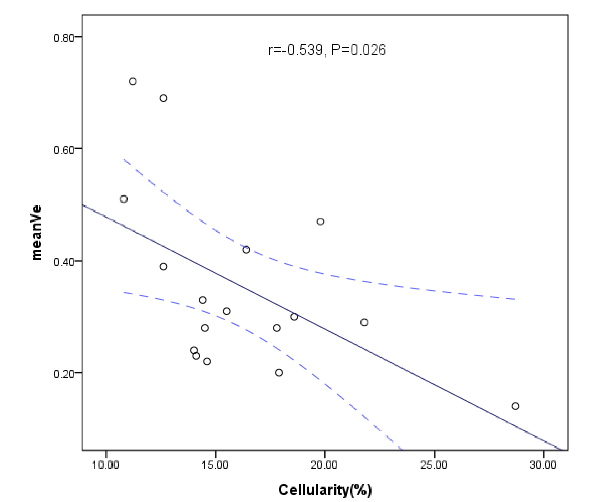
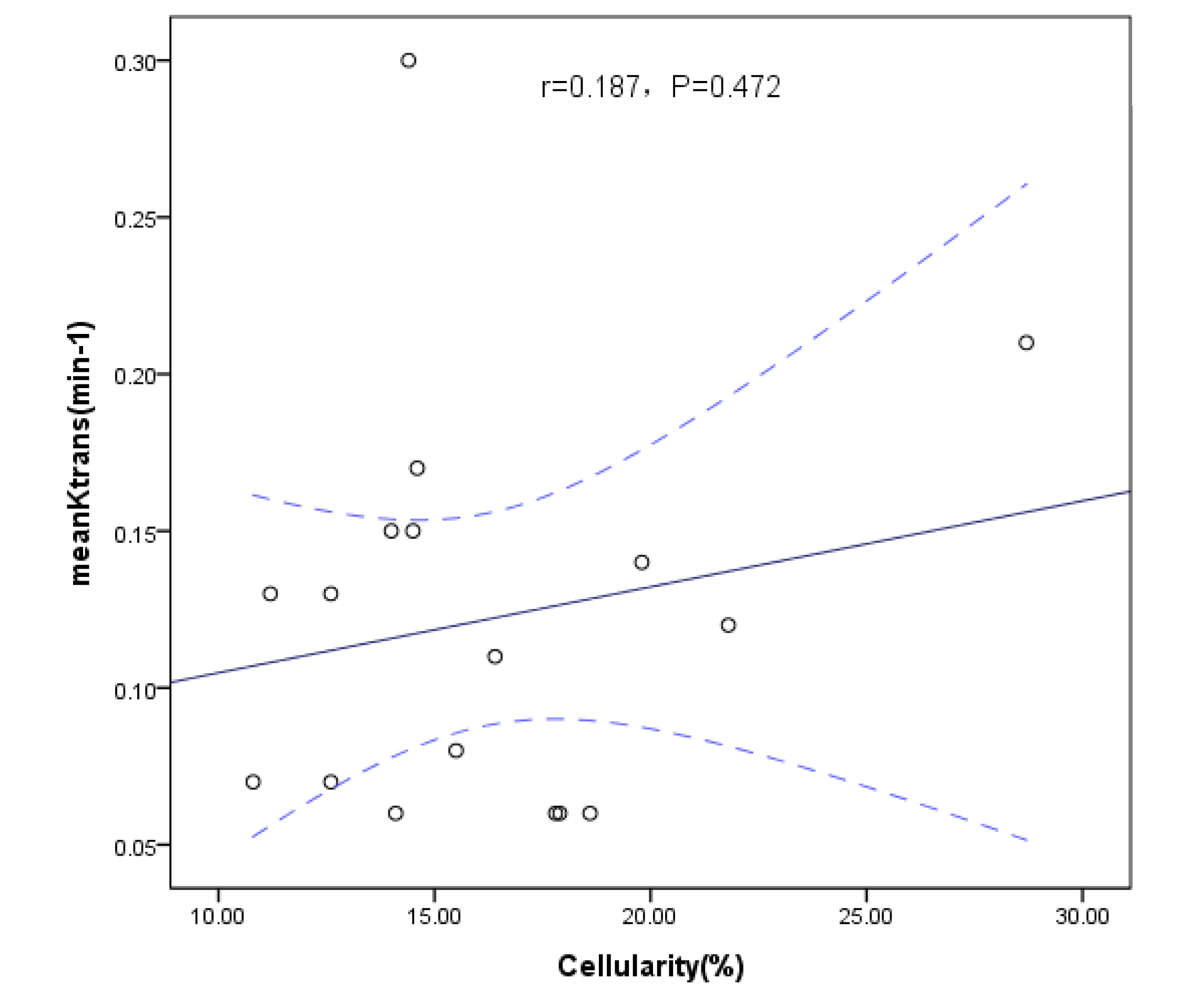
The replicated measurements of Ktrans and Ve demonstrated ICCs of 0.916 and 0.904 respectively. NSCLC had mean values of 0.12±0.06 min-1, 0.35±0.16 and 16.19±4.04% for Ktrans, Ve, and cell density. For adenocarcinoma, these values were 0.12±0.06 min-1, 0.35±0.17 and 14.79±2.66%. The squamous cell carcinoma results showed values of 0.13±0.07 min-1, 0.36±0.13, and 22.77±5.51% for Ktrans, Ve, and cell density, respectively. There was an inverse correlation observed between Ve and cell density, and the data achieved a relatively good fit (r=-0.539, P=0.026). However, the study found no significant correlation between Ktrans and cell density (r=0.187,P=0.472).
Discussion
We conducted correlation analyses on meanVe and medianVe with tumour cellularity and found that both meanVe and medianVe exhibit a negative relationship with tumour cellularity in lung cancer. This outcome is consistent with existing research in cervical cancer or head and neck squamous cell carcinoma(3,4) and suggests that Ve may be a highly valuable biomarker for estimating tumour cellularity in lung cancer and may also be extendable to other cancer types. Although the study did not find a significant correlation between Ktrans and cell density, our past findings indicated a positive association between the Ktrans and microvascular density(8). Thus, these results imply that perfusion MRI could assist in estimating tumor pathophysiology noninvasively, along with tumour cellularity and microvascular density.
In conclusion, the preliminary results of this study suggest that DCE-MRI using the GRASP technique is a promising non-invasive alternative for the quantitative assessment of tumour cellularity in patients with lung cancer.
Acknowledgements
We thank Pro. Feng Li from the New York University School of Medicine for his support in implementing the image reconstruction.References
1. Hanahan D. Hallmarks of Cancer: New Dimensions. Cancer discovery 2022;12(1):31-46.
2. Chen L, Min L, Bao J, et al. The Correlation between Apparent Diffusion Coefficient and Tumor Cellularity in Patients: A Meta-Analysis. Plos One 2013;8(11):e79008.
3. Surov A, Meyer HJ, Gawlitza M, et al. Correlations Between DCE MRI and Histopathological Parameters in Head and Neck Squamous Cell Carcinoma. Translational oncology 2017;10(1):17-21.
4. Hillestad T, Hompland T, Fjeldbo CS, Skingen VE. MRI Distinguishes Tumor Hypoxia Levels of Different Prognostic and Biological Significance in Cervical Cancer. Cancer research 2020;80(18):3993-4003.
5. Ohno Y, Koyama H, Yoshikawa T, Matsumoto S, Sugimura K. Lung Cancer Assessment Using MR Imaging: An Update. Magnetic resonance imaging clinics of North America 2015;23(2):231-244.
6. Chen L, Liu D, Zhang J, et al. Free-breathing dynamic contrast-enhanced MRI for assessment of pulmonary lesions using golden-angle radial sparse parallel imaging. Journal of Magnetic Resonance Imaging 2018;48(2):459-468.
7. Chen L, Zeng X, Ji B, et al. Improving dynamic contrast-enhanced MRI of the lung using motion-weighted sparse reconstruction: Initial experiences in patients. Magnetic resonance imaging 2020;68:36-44.
8. Chen L, Zeng X, Wu Y, et al. A Study of the Correlation of Perfusion Parameters in High-Resolution GRASP MRI With Microvascular Density in Lung Cancer. Journal of magnetic resonance imaging 2019;49(4):1186-1194.
Figures