1454
Dynamic contrast enhanced MRI and susceptibility-weighted imaging for diffierentiation of parotid gland tumors: a pilot study1Radiology, the First Affiliated Hospital of Dalian Medical University, Dalian, China, 2the First Affiliated Hospital of Dalian Medical University, Dalian, China
Synopsis
Keywords: Cancer, Cancer
Motivation: MRI is one of the major methods to diagnose parotid gland tumors. Substantial overlap in the appearance of tumors may be seen on anatomic MR images.
Goal(s): To assess the usefulness of combined DCE with SWI in the differentiation of benign and malignant tumors.
Approach: We assess the value of DCE and ITSS in the differential diagnosis between malignant and benign parotid tumors.
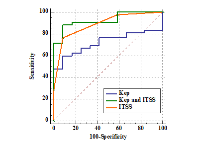
Results: Results showed that the differences between malignant and benign tumors in Kep (p=0.024) and ITSS (p<0.01) were statistically significant. Combined with ITSS, the diagnostic performance of Kep was improved for differentiating malignant from benign tumor (AUC 0.718 vs 0.927).
Impact: Our current study showed that DCE can elucidate perfusion characteristics of parotid tumors. SWI is a new complementary technique that can detect signal intensity changes from both T2WI and susceptibility differences between tissues. These fndings suggest that SWI and DCE quantitative parameters may facilitate the understanding of the pathophysiological characteristics of parotid tumors.
Introduction
MRI is one of the major methods to diagnose parotid gland tumors. However, substantial overlap in the appearance of tumors may be seen on anatomic MR images, thereby limiting the role in characterization. A quantitative evaluation can be made with perfusion parameters including transfer constant (Ktrans), rate constant (Kep), fractional volume of the extravascular extracellular space (Ve) to identify benign and malignant lesions and monitor tumor responses to treatment in the tumors.Susceptibility-weighted imaging (SWI) can detect tissue susceptibility and provide intratumoral information based on the magnetic susceptibility.Methods
The images of 54 patients (including 42 benign lesions and 12 malignant lesions) with complete surgical records and pathological findings were analyzed retrospectively. The intratumoral susceptibility signal (ITSS) and DCE quantitative parameters were evaluated(Krans, Kep, Ve). The Mann-Whitney U-test and ROC curve were conducted for statistical analysis. DCE-MRI was performed using the sequences described below. First, a baseline T1-weighted MRI (TR/TE = 5.08/1.74 ms, FOV = 260 mm × 260 mm, slice-thickness = 5 mm, and flip-angles of 2° and 15°) was used to create two precontrast datasets. At the beginning of the baseline acquisition, a bolus of 0.1 mmol/kg gadolinium (Gd)-DTPA contrast agent was injected intravenously at a rate of 2 ml/s.The detailed parameters of SWI were as follows: TR/TE = 31.0/7.2ms, FOV =230 mm × 189 mm, slice thickness = 2mm, voxel size = 0.6 × 0.6 × 2 mm.Results
The degree of ITSS in the malignant group was significantly higher than those in the benign group (p<0.01). The Kep of malignant group (434.52 [339.08,539.58]) were higher than benign ones (132.64[317.24,130.76]) (p=0.022). The area under the corresponding curve (AUC) of Kep was 0.718. After the combination of ITSS, Kep can improve the AUC (0.927) in the differentiation of the lesions of the two groups (p<0.01).Discussion
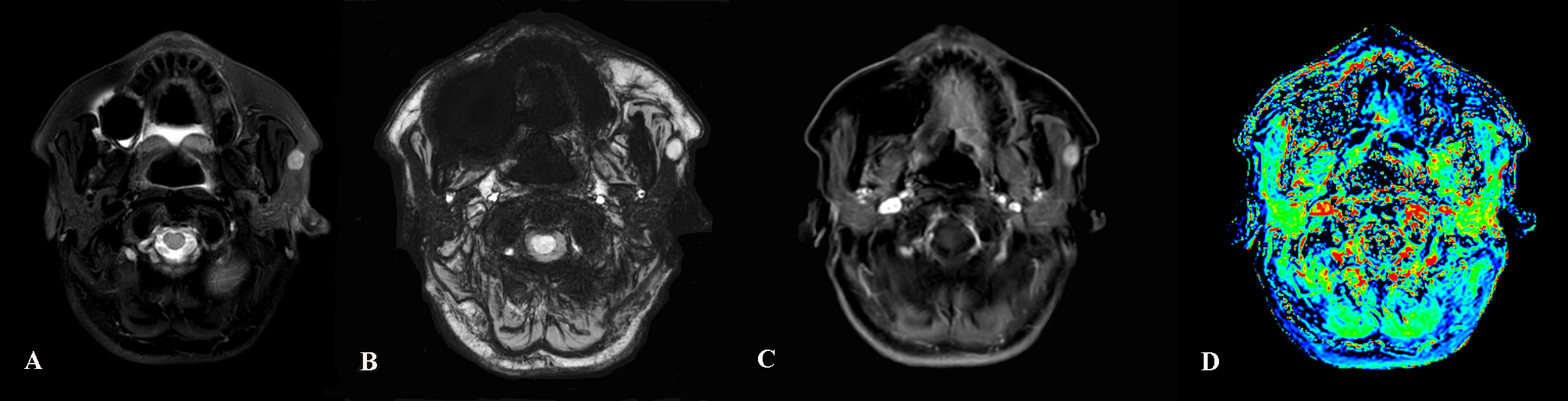
ITSS was defined as hypo-intense signal fine dot-like or linear area and enables semi-quantitative differentiation of benign from malignant lesions. Our study indicated that benign lesions have a significantly lower ITSS grade than malignant ones. The pathophysiology of this result is possible as follows: benign lesions have less hemorrhage because of their rich microvasculature and slow growth and vascular architecture abnormalities of malignant lesions all result in an increase in deoxyhemoglobin4. Quantitative DCE-MRI is a functional MRI modality that analysis the various parameters related to perfusion and permeability within the tumor. Combined with ITSS, the diagnostic performance of DCE was improved for differentiating malignant tumors from benign tumors. Our results suggested DCE combined with SWI could be beneficial to improve the differential diagnosis between malignant and benign parotid gland tumors.Conclusions
The combination of DCE to SWI can improve the efficiency significantly in differentiating benign from malignant parotid gland tumors.Acknowledgements
No acknowledgement found.References
1. Zhang W, Zuo Z, Huang X, et al. Value of Diffusion-Weighted Imaging Combined with Susceptibility-Weighted Imaging in Differentiating Benign from Malignant Parotid Gland Lesions. Med Sci Monit, 2018. 24: p. 4610-4616.2. Kim HS, Jahng GH, Ryu CW, et al. Added value and diagnostic performance of intratumoral susceptibility signals in the differential diagnosis of solitary enhancing brain lesions: preliminary study. Am J Neuroradiol, 2009; 30: 1574–793. Nan Huang, Yu Chen, Dejun She, et al. Difusion kurtosis imaging and dynamic contrast‑enhanced MRI for the diferentiation of parotid gland tumors. Eur Radiol, (2022) 32:2748–2759.4. Bhattacharjee R, Gupta RK, Patir R, et al. Quantitative vs. semiquantitative assessment of intratumoral susceptibility signals in patients with different grades of glioma. J Magn Reson Imaging, 2020. 51(1): p. 225-233.5. Zhai JN,Zuo ZC,Wang P, et al. The Diagnostic Value of Conventional MRI Combined with SWI in the Differential Diagnosis of Benign Parotid Gland Lesions. J Clin Radiol, 2018. 37: p. 1810-1814.Figures