1453
The quantitative parameters based on IVIM andDCE-MRI in predicting the efficacy in patients with nasopharyngeal carcinoma1Radiology, the First Affiliated Hospital of Dalian Medical University, Dalian, China, 2GE Healthcare, MR Research China, Beijing, China
Synopsis
Keywords: Cancer, fMRI
Motivation: This study aims to establish a predictive model based on IVIM and DCE-MRI that could ultimately improve the prognosis of nasopharyngeal carcinoma (NPC) patients.
Goal(s): This research paves the way for more effective and tailored treatment approaches for NPC patients.
Approach: Patients in the CR group and the NCR group underwent IVIM and DCE-MRI experiments and their quantitative parameters were compared.
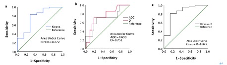
Results: The CR group exhibited higher Ktrans value and lower D and ADC values compared to the NCR group. ROC analysis showed that the AUC values of Ktrans, D and ADC before treatment were approximately 0.772, 0.751, and 0.699, respectively.
Impact: Early prediction of the efficacy of NPC patients can optimize the treatment plans, functional MRI techniques can offer insights into the pathological and physiological state of living tissues before morphological changes, prolong their survival, and hold immense clinical significance.
Purpose
To investigate the potential of quantitative parameters derived from intravoxel incoherent motion (IVIM) and dynamic contrast-enhanced magnetic resonance imaging (DCE-MRI) in predicting the efficacy of radiotherapy and chemotherapy in patients with nasopharyngeal carcinoma (NPC).Introduction
NPC is a common malignant tumor that occurs in the epithelial tissue of the nasopharynx, which is very prevalent in East and Southeast Asia. The primary treatment modality for NPC is radiotherapy, and chemotherapy has been integrated into standard radiotherapy to improve the prognosis of high-risk patients, whether as an adjuvant, neoadjuvant, or concurrent treatment with radiotherapy. Despite these advancements, there remain patients with poor prognosis due to treatment failures [1]. Early prediction of the efficacy of radiotherapy and chemotherapy can significantly optimize the treatment plans for NPC patients, prolong their survival, and hold immense clinical significance.Recent reports have highlighted the critical role of the tumor microenvironment in influencing the effectiveness of tumor treatments [2]. Unfortunately, conventional MRI falls short in assessing the tumor microenvironment. In contrast, functional MRI techniques can offer insights into the pathological and physiological state of living tissues before morphological changes. IVIM-MRI utilizes multiple b-values with significant differences to simultaneously provide information on tumor diffusion and perfusion. This technology has demonstrated its utility in measuring blood microcirculation perfusion, distinguishing cervical lymph node metastasis, early diagnosis of T1-stage nasopharyngeal carcinoma, and predicting and evaluating treatment outcomes [3]. However, previous research has yielded inconsistent results regarding the predictive value of IVIM-MRI perfusion parameters[4].DCE-MRI is another advanced imaging modality that offers insights into tissue microcirculation perfusion at the molecular level and has been reported in the staging and grading of NPC, evaluation of tumor hypoxia, and prediction of therapeutic efficacy [5]. This suggests its potential for clinical applications. Consequently, the combination of IVIM and DCE-MRI is hypothesized to provide a more comprehensive and accurate prediction of NPC treatment efficacy before initiating therapy. This study aims to establish a predictive model that could serve as a basis for personalized treatment strategies, ultimately improving the prognosis of NPC patients.Methods
54 patients with an initial diagnosis of NPC confirmed by pathology were recruited in the study. All patients underwent MRI examinations utilizing a GE Signa HDxt 3.0T MR scanner with an 8-channel head & neck phased array coil, including IVIM and DCE-MRI sequences. Two radiologists independently measured various quantitative parameters, including the rate constant (Kep), volume transfer constant (Ktrans), volume fraction of the extravascular extracellular matrix (Ve), true molecular diffusion coefficient (D), pseudo-diffusion coefficient (D*), perfusion fraction (f) and apparent diffusion coefficient (ADC). Regions of interest (ROIs) were delineated to encompass as much parenchyma of the mass as possible. Patients were followed up with MRI examinations for 6 months after receiving radiotherapy and chemotherapy. Based on the internationally recognized solid tumor efficacy evaluation standard (RECIST) 1.1, patients were categorized into two groups: the complete response group (CR group) and the non-complete response group (NCR group). Independent samples T-tests were employed to assess the differences in quantitative parameters between the CR and NCR groups. The receiver operating characteristic curve (ROC) analysis was utilized to evaluate the diagnostic performance of IVIM and DCE-MRI parameters in predicting treatment outcomes, with statistical significance set at p <0.05.Results
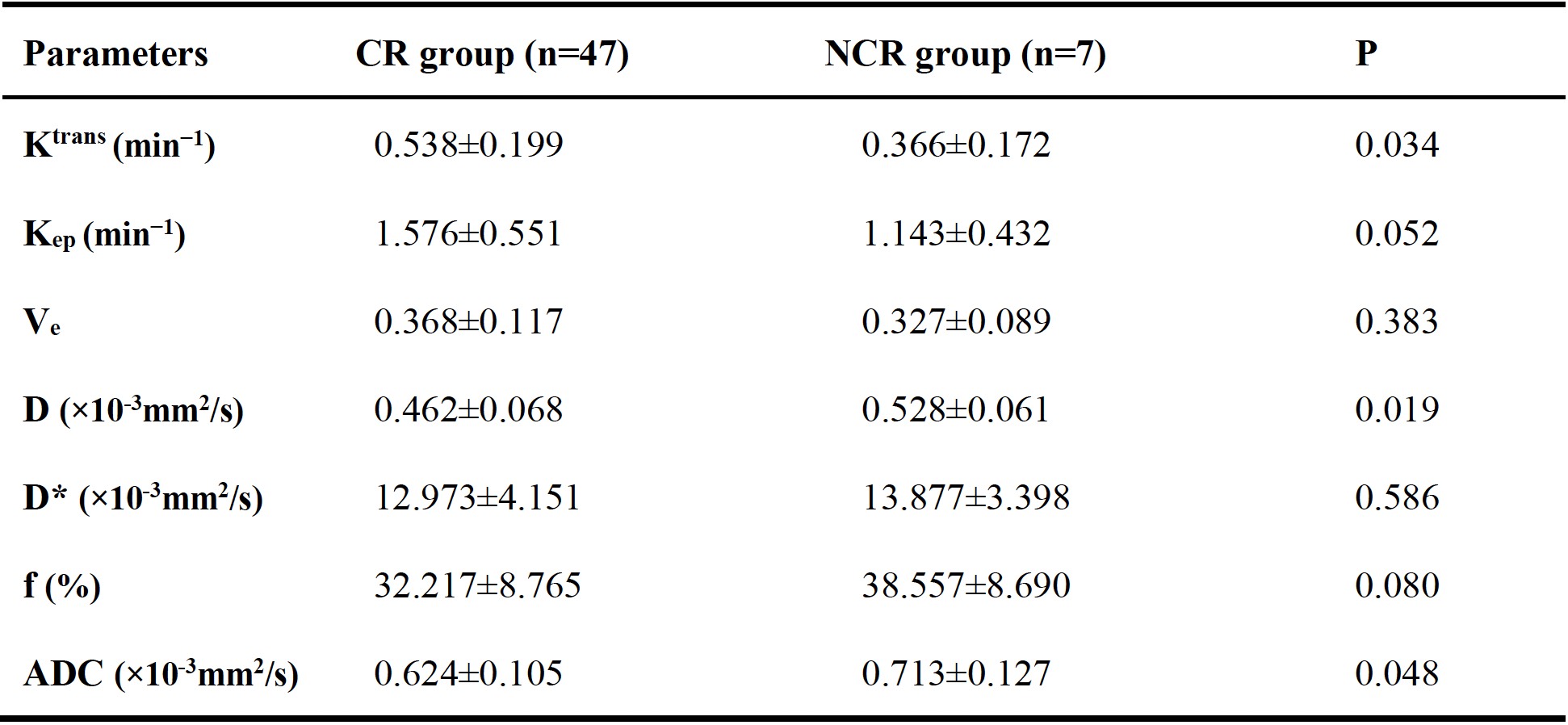
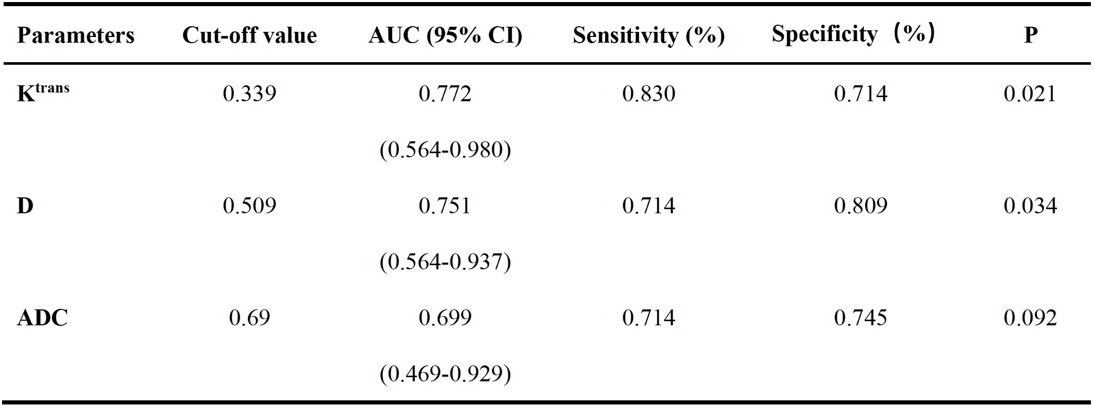
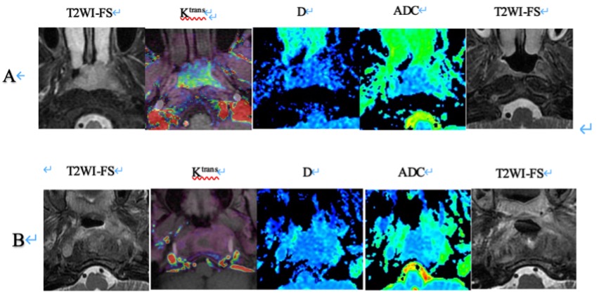
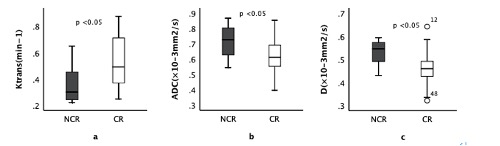
Following six months of radiotherapy and chemotherapy, 47 patients were classified into the CR group, while 7 patients were placed in the NCR group. Pre-treatment IVIM and DCE-MRI parameters revealed notable distinctions between groups. Specifically, the CR group exhibited higher Ktrans value (P=0.034) and lower D and ADC values (P=0.019, 0.048) compared to the NCR group (Figure 1, 2). Conversely, the Kep, Ve, D* and f demonstrated no significant differences between the two groups (Table 1). Furthermore, ROC analysis showed that the area under the curve (AUC) values of Ktrans, D and ADC before treatment were approximately 0.772, 0.751, and 0.699, respectively. The sensitivity and specificity of the parameters are summarized in Table 2. These values indicated a promising predictive potential. Further enhancement was observed when combining Ktrans, and D, with an AUC of 0.845. (Figure 3) (Table 3).Conclusion
In conclusion, the combination of IVIM-MRI and DCE-MRI demonstrates the ability to predict the early treatment efficacy of nasopharyngeal carcinoma. Notably, the D and ADC values obtained through IVIM-MRI, along with the perfusion coefficients Ktrans from DCE-MRI, have the potential to serve as valuable imaging indicators. These indicators guide the treatment strategies for nasopharyngeal carcinoma, offering a path towards personalized care and improved patient prognosis.Acknowledgements
No acknowledgement found.References
[1] Juarez-Vignon Whaley J J, Afkhami M, Onyshchenko M, et al. Recurrent/Metastatic Nasopharyngeal Carcinoma Treatment from Present to Future: Where Are We and Where Are We Heading?[J]. Curr Treat Options Oncol, 2023, 24(9): 1138-1166. DOI: 10.1007/s11864-023-01101-3.[2] Liao C, Liu X, Zhang C, et al. Tumor hypoxia: From basic knowledge to therapeutic implications[J]. Semin Cancer Biol, 2023, 88: 172-186. DOI: 10.1016/j.semcancer.2022.12.011.[3] Paudyal R, Chen L, Oh J H, et al. Nongaussian Intravoxel Incoherent Motion Diffusion Weighted and Fast Exchange Regime Dynamic Contrast-Enhanced-MRI of Nasopharyngeal Carcinoma: Preliminary Study for Predicting Locoregional Failure[J]. Cancers (Basel), 2021, 13(5). DOI: 10.3390/cancers13051128.[4] Liao L, Liu T, Wei B. Prediction of short-term treatment outcome of nasopharyngeal carcinoma based on voxel incoherent motion imaging and arterial spin labeling quantitative parameters[J]. Eur J Radiol Open, 2023, 10: 100466. DOI: 10.1016/j.ejro.2022.100466.[5] Liu L, Hu L, Zeng Q, et al. Dynamic contrast-enhanced MRI of nasopharyngeal carcinoma: correlation of quantitative dynamic contrast-enhanced magnetic resonance imaging (DCE-MRI) parameters with hypoxia-inducible factor 1alpha expression and tumor grade/stage[J]. Ann Palliat Med, 2021, 10(2): 2238-2253. DOI: 10.21037/apm-21-303.Figures