1452
Different MRI-based radiomics models for distinguishing misdiagnosed or ambiguous pleomorphic adenoma and Warthin tumor: A multicenter study1The First People’s Hospital of Yunnan Provence, kunming, China, 2MR Research Collaboration Team, Siemens Healthineers Ltd, shanghai, China
Synopsis
Keywords: Cancer, Cancer
Motivation: Enhance the accuracy of distinguishing misdiagnosed or ambiguous cases of pleomorphic adenoma (PA) and Warthin tumor (WT).
Goal(s): This study aims to construct various MRI-based radiomics models employing different machine learning classifiers to determine the optimal models for identifying misdiagnosed or ambiguous PA and WT cases.
Approach: we evaluate the effectiveness of various MRI-based radiomics models.
Results: A nomogram demonstrates exceptional and consistent diagnostic performance. In routine practice, combining clinical parameters is essential for distinguishing between PA and WT.
Impact: MRI-based radiomics models can effectively differentiate misdiagnosed or ambiguous cases of PA and WT. The nomogram is a valuable tool for preoperatively and non-invasively distinguishing between PA and WT, a task often challenging for clinicians and radiologists before surgery.
Introduction
Pleomorphic adenoma (PA) and Warthin tumor (WT) stand as the most prevalent benign tumors of the parotid gland[1-2], each requiring distinct surgical approaches and having different prognoses[3-4]. Therefore, accurate differentiation between PA and WT is critical for delivering precise and individualized treatment to patients with benign parotid tumors. Conventional MRI-based differential diagnosis has been limited due to substantial overlap in morphological features between PA and WT, particularly in misdiagnosed or ambiguous cases. Furthermore, conventional MRI diagnosis is subjective, relying on the expertise and experience of radiologists[5]. Radiomics provides a comprehensive analysis of medical images, surpassing human visual assessment[6]. While previous studies have analyzed the differentiation of PA from WT[7-8], none have focused on differentiating misdiagnosed or ambiguous cases using radiomics. This study aims to construct various MRI-based radiomics models employing different machine learning classifiers to determine the optimal models for identifying misdiagnosed or ambiguous PA and WT cases. Subsequently, we assess the diagnostic performance of integrated models combining clinical parameters and radiomics to enhance accuracy.Methods
This study included 126 patients from center A and 23 patients from center B who underwent magnetic resonance imaging (MRI) before surgery, using a 3T/1.5T MRI system (3.0T MAGNETOM Prisma, 1.5T MAGNETOM Aera, Siemens Healthcare, Erlangen, Germany). The MRI sequences included transverse T1-weighted imaging (T1WI), T2-weighted imaging (T2WI), and contrast-enhanced T1-weighted imaging (CE-T1WI). The T2WI parameters were TR/TE = 4800/83 ms, 20, FOV = 220×220mm2; scan matrix = 240×320; Slice thickness=4 mm; Slice gap=1 mm. The T1WI parameters were TR/TE =6.18/2.46 ms, FOV = 220×220mm2; scan matrix =259×288; Slice thickness=3 mm; Slice gap=1 mm. The CE-T1WI parameters were TR/TE =6.18/2.46 ms, FOV = 220×220mm2; scan matrix =256×320; Slice thickness=3 mm; Slice gap=0.6 mm. Tumor segmentation was performed using 3D Slicer 4.11.0 software (https://www.slicer.org/). Image preprocessing and feature extraction were accomplished using the open-source Python package Pyradiomics (https://pypi. org/project/pyradiomics/). Significant features from univariate analysis were included in multivariate LR analyses to create a clinical model. Nine mainstream machine learning algorithms were employed to build radiomics models. Model performance was assessed through sensitivity, specificity, accuracy, and the area under the curve (AUC) of the receiver operating characteristic curve (ROC) in internal and external validation groups. The model with the highest average AUC was considered the optimal radiomics model. A nomogram was developed, integrating independent clinical parameters and the radscore was constructed using multivariate LR analysis. Two integrated models were constructed using ensemble and stacking algorithms based on the clinical and optimal radiomics models. Model performance was assessed using AUC.Results
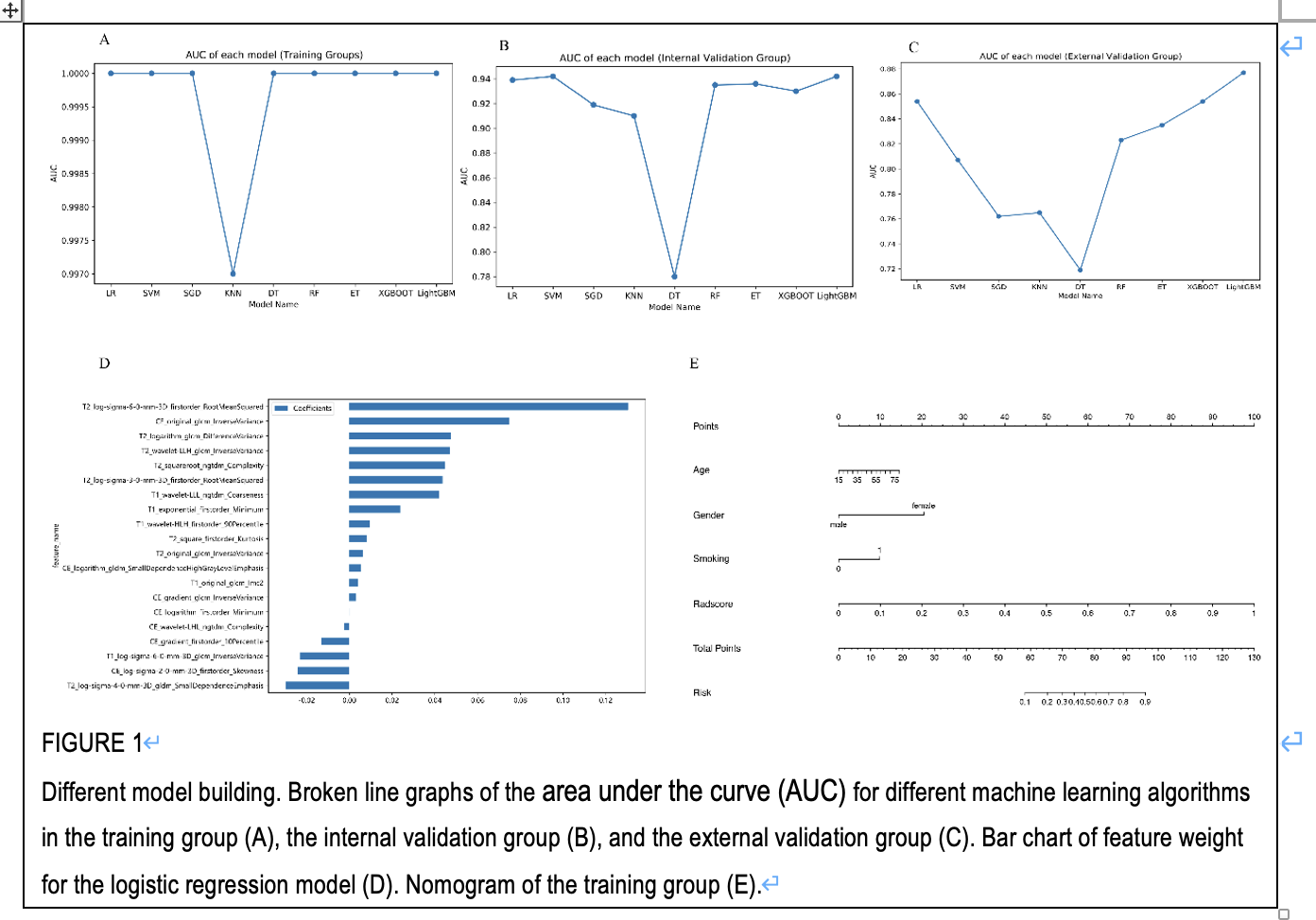
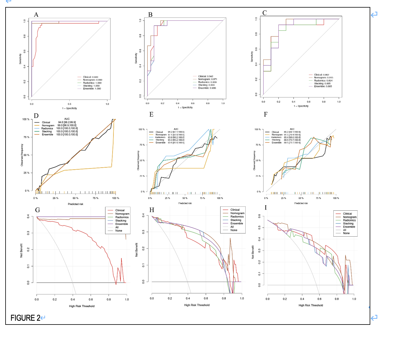
A total of 149 patients were included in the study. Independent clinical predictors were patient gender, age, and smoking history. The LR model emerged as the best radiomics model, achieving the highest average AUC of 0.896 in both internal and external validation groups (0.854). The AUC of the optimal radiomics model and clinical model were 0.896 and 0.902, respectively. The nomogram demonstrated superior discrimination performance, with an AUC of 0.943 in the validation groups. The AUC of the stacking model (0.910) and ensemble model (0.910) were lower than that of the nomogram in the validation groups.Discussion & Conclusions
This study represents a preliminary feasibility assessment of distinguishing misdiagnosed or ambiguous cases of PA and WT using various MRI-based radiomics models with different machine learning classifiers. MRI-based radiomics models can non-invasively differentiate these cases. The nomogram exhibits excellent and consistent diagnostic efficiency. In daily practice, combining clinical parameters is essential for distinguishing between PA and WT.Acknowledgements
No acknowledgement found.References
1.Barnes LEJ, R. P., Sidransky D Pathology and genet-ics of head and neck tumors. IARC Press, Lyon. 2017.2.Luers, J. C.; Guntinas-Lichius, O.; Klussmann, J. P.; Küsgen, C.; Beutner, D.; Grosheva, M. The incidence of Warthin tumours and pleomorphic adenomas in the parotid gland over a 25-year period. Clin otolaryngol all 2016, 41 (6), 793-797.3.Dulguerov, P.; Todic, J.; Pusztaszeri, M.; Alotaibi, N. H. Why Do Parotid Pleomorphic Adenomas Recur? A Systematic Review of Pathological and Surgical Variables. Front Surg 2017, 4, 26.4.Rooker, S. A.; Van Abel, K. M.; Yin, L. X.; Nagelschneider, A. A.; Price, D. L.; Olsen, K. D.; Janus, J. R.; Kasperbauer, J. L.; Moore, E. J. Risk factors for subsequent recurrence after surgical treatment of recurrent pleomorphic adenoma of the parotid gland. Head neck-j sci spec 2021, 43 (4), 1088-1096.5.Maraghelli, D.; Pietragalla, M.; Cordopatri, C.; Nardi, C.; Peired, A. J.; Maggiore, G.; Colagrande, S. Magnetic resonance imaging of salivary gland tumours: Key findings for imaging characterisation. Eur j radiol 2021, 139 , 109716.6.Scapicchio, C.; Gabelloni, M.; Barucci, A.; Cioni, D.; Saba, L.; Neri, E. A deep look into radiomics. Radiol med 2021, 126 (10), 1296-1311.7.Song, L. L.; Chen, S. J.; Chen, W.; Shi, Z.; Wang, X. D.; Song, L. N.; Chen, D. S. Radiomic model for differentiating parotid pleomorphic adenoma from parotid adenolymphoma based on MRI images. BMC Med Imaging 2021, 21 (1), 54.8.Faggioni, L.; Gabelloni, M.; De Vietro, F.; Frey, J.; Mendola, V.; Cavallero, D.; Borgheresi, R.; Tumminello, L.; Shortrede, J.; Morganti, R.; et al. Usefulness of MRI-based radiomic features for distinguishing Warthin tumor from pleomorphic adenoma: performance assessment using T2-weighted and post-contrast T1-weighted MR images. Eur J Radiol Open 2022, 9 , 100429.FiguresFigures