1449
Initial experience in pCASL using a novel neurovascular coil at 7T1Imaging Centre of Excellence, University of Glasgow, Glasgow, Scotland, 2Siemens Healthcare Ltd, Glasgow, Scotland, 3University of Southern California, Los Angeles, CA, United States, 4Siemens Healthineers USA, Los Angeles, CA, United States, 5University of Glasgow, Glasgow, Scotland, 6MR Coil Tech, Glasgow, Scotland
Synopsis
Keywords: High-Field MRI, RF Arrays & Systems
Motivation: Labeling efficiency in pCASL suffers from an inferior drop-off in B1+ leading to low SNR perfusion-weighted images which can affect CBF quantification.
Goal(s): To assess the feasibility of pCASL in healthy volunteers using a custom-built head and neck coil at 7T.
Approach: Volunteers were scanned with pCASL on two coils: a normal head coil and a head and neck coil with circularly polarized shim weights, and the results were compared in terms of CBF and tSNR.
Results: pCASL in healthy volunteers using a novel neurovascular coil at 7T gave an average GM/WM CBF ratio of 1.51, comparable to a conventional head coil.
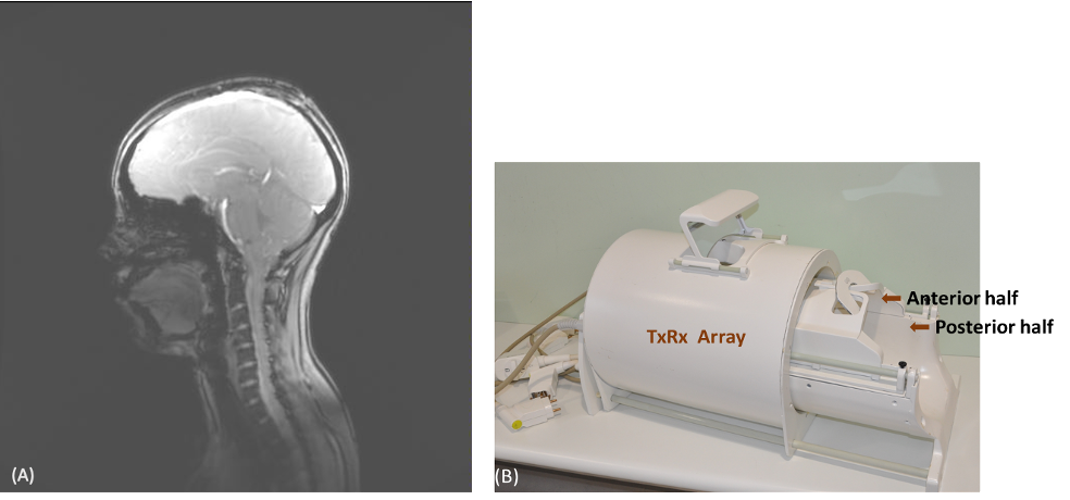
Impact: This proof-of-concept study shows the feasibility of pCASL using a novel 8TxRx56Rx neurovascular (head and neck) coil at 7T and compares the performance against a conventional 8Tx64Rx head coil.
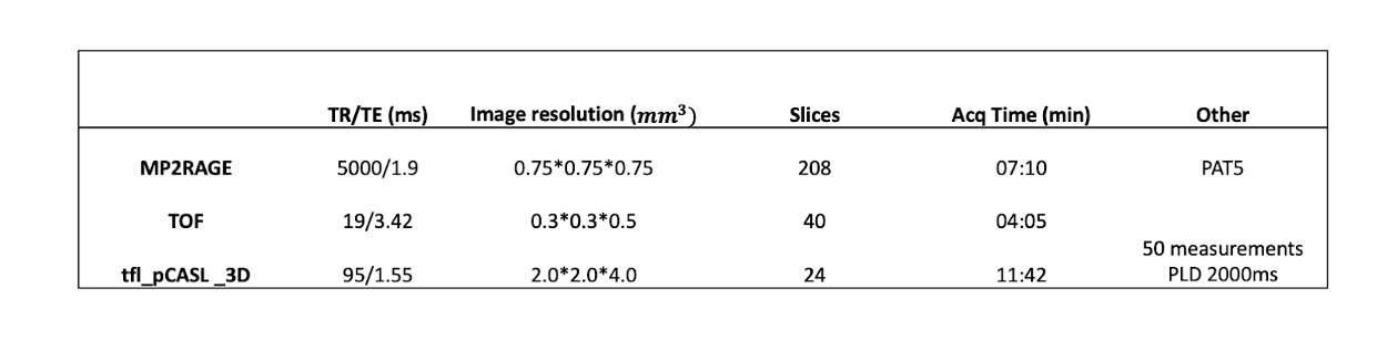
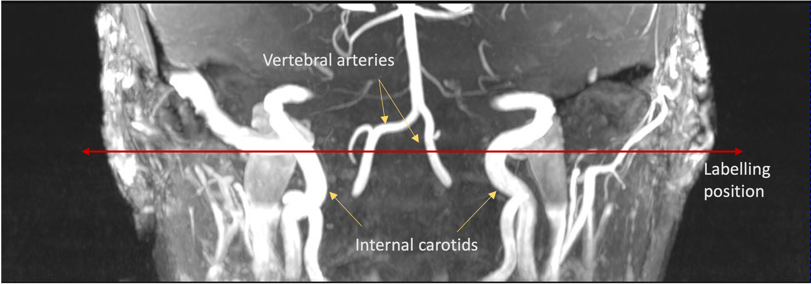
Methods: Healthy volunteers were scanned on a 7T scanner (MAGNETOM Terra pTx, Siemens Healthcare, Erlangen, Germany) with local ethical approval. The scanning protocol outlined in Figure 2 remained consistent for the coils. A Time-of-Flight MR angiogram (ToF-MRA) was acquired for calculating the label offset, ensuring that the labeling plane intersected the four major vessels (the right and left branches of the vertebral arteries and the right and left branches of the internal carotid arteries, Figure 3). B0 and B1+ maps were acquired followed by a calibration scan for GRAPPA. The pCASL sequence was run in circularly polarized (CP) mode. Raw data from the scanner were reconstructed using a MATLAB application (LOFT_TFL_PCASL_3D) provided by the University of Southern California as part of the pCASL package. Image registration to Montreal Neurological Institute 152 (MNI152) space was performed using FLIRT in FSL via the T1w MP2RAGE image. Voxel-wise temporal SNR (tSNR) of each scan was calculated by taking the temporal mean of the control images divided by the temporal standard deviation. Voxel-wise percentage difference in tSNR (tSNR) was then calculated astSNR = (tSNR(NV) -tSNR(HC)) / (tSNR(NV) +tSNR(HC)) 200%The two coils used in this experiment have different coverage; thus, we would expect the labelling and inversion efficiency of the two coils to be different. Since CBF calculations are highly sensitive to these values, we decided to compare the CBF ratios between grey and white matter to reduce any confounding effects arising from the differing spatial B1+ distribution between the coils.
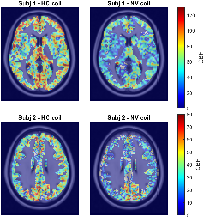
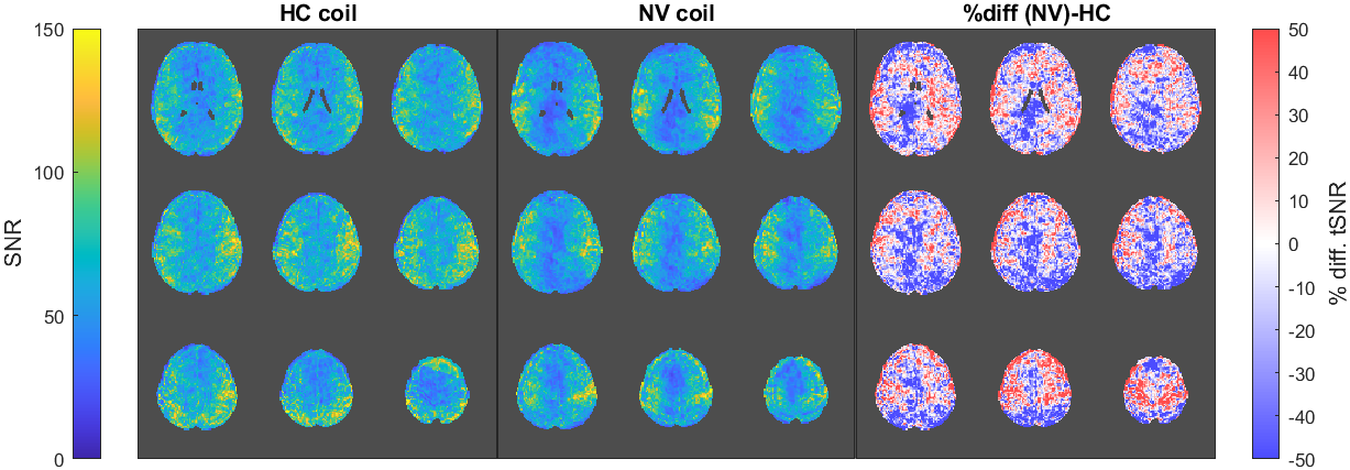
Results and Discussion: Figure 4 displays the GM CBF maps in MNI152 space for two subjects on each of the coils. The average GM/WM CBF ratios are 1.95 and 1.51 for the HC and NV coil respectively. Future work to determine the labelling and inversion efficiency of the sequence in the novel NV coil will allow for the quantification of CBF values. There is a slight decrease in tSNR, especially in the occipital lobes, with the NV coil compared to the conventional HC (Figure 5). This is likely to be a combination of two factors. First, the NV coil might have a lower B1+ value in the occipital lobe than HC, resulting in a lower flip angle. Additionally, the receive profiles of both coils are different with the NV coil having a smaller number of receive loops covering a larger area. In this initial feasibility study, we have not exploited the full parallel transmission (pTx) capabilities of the NV coil. We believe that the combination of region-specific pTx pulse design on the labelling plane and the extended coil coverage will allow us to achieve more consistent labelling, and improve the performance of pCASL at 7T.
Conclusion: This study has shown the feasibility of pCASL in healthy volunteers with a novel 8TxRx56Rx neurovascular coil at 7T.
Acknowledgements
No acknowledgement found.References
References: 1. Detre JA, Leigh JS, Williams DS, Koretsky AP. PERFUSION IMAGING. Magnetic Resonance in Medicine. Jan 1992;23(1):37-45. doi:10.1002/mrm.19102301062. Pohmann R, Speck O, Scheffler K. Signal-to-Noise Ratio and MR Tissue Parameters in Human Brain Imaging at 3, 7, and 9.4 Tesla Using Current Receive Coil Arrays. Magnetic Resonance in Medicine. Feb 2016;75(2):801-809. doi:10.1002/mrm.256773. Zuo ZT, Wang R, Zhuo Y, Xue R, St Lawrence KS, Wang DJJ. Turbo-FLASH Based Arterial Spin Labeled Perfusion MRI at 7 T. Plos One. Jun 2013;8(6)e66612. doi:10.1371/journal.pone.00666124. Meixner CR, Eisen CK, Schmitter S, et al. Hybrid B1+-shimming and gradient adaptions for improved pseudo-continuous arterial spin labeling at 7 Tesla. Magnetic Resonance in Medicine. Jan 2022;87(1):207-219. doi:10.1002/mrm.289825. Williams SN, Allwood-Spiers S, McElhinney P, et al. A Nested Eight-Channel Transmit Array With Open-Face Concept for Human Brain Imaging at 7 Tesla. Frontiers in Physics. Jul 2021;9701330. doi:10.3389/fphy.2021.7013306. Gunamony S, Feinberg D. An 8-channel transmit 64-channel receive compact head coil for Next Gen 7T scanner with head gradient insert. ISMRM; 2022; London.7. Baskaran D, McElhinney P, Williams S, Allwood-Spiers S, Porter D, Gunamomy S. Eight-channel transceiver array for combined head and neck imaging at 7Tesla. presented at: ISMRM 2023 Annual Meeting and Exhibition; 2022; Toronto, ON, Canada.Figures