1448
Assessing RF-induced heating safety of MRI scans with shoulder implants at 7T1Diagnostic Radiology, Cleveland Clinic, Cleveland, OH, United States, 2MRI Research, Cleveland Clinic, Cleveland, OH, United States, 3Orthopedic Surgery, Cleveland Clinic, Cleveland, OH, United States
Synopsis
Keywords: Safety, Safety
Motivation: Brain MRI with shoulder implants are often contraindicated at 7T with head coils due to possibility of radiofrequency (RF)-induced heating. Such implants are not uncommon in older adults; contraindication results in increasing proportion of elderly to be denied high-field scans. Assessing heating safety of MRI at 7T with implants is of significance.
Goal(s): To measure shoulder implant heating during MRI at 7T with pTx head coils and assess safety thereof.
Approach: Temperature of a reverse total shoulder implant was measured with fluoroptic sesnors during MRI at Siemens 7T Terra with pTX head coil.
Results: No significant heating (>1°C) of the implant was observed.
Impact: Findings from this study may enable brain scanning individuals with shoulder implant at 7T, thus making improved image quality a possibility.
Introduction
Annually more than 50000 people get shoulder replacement surgery in the United States (orthoinfo.aaos.org), and the number is on the rise.[1] However, radiofrequency (RF)-induced heating is of concern in MRI scans with metallic/conducting implants. Some of these implants have been cleared to be scanned at 1.5 or 3T under certain conditions.[2] However, the heating related safety of these implants is largely unknown at higher field strengths along with parallel transmit (pTx) technology, which often results in implanted patients not getting the benefit of MRI with high signal-to-noise and contrast-to-noise ratios[3] with reduced RF inhomogeneity.[4] We have measured shoulder implant heating at 7T with a pTx head coil to assess safety of performing neuro MRI with these implants.Methods
All MRI scans were performed in a 7T Terra scanner (Siemens, Erlangen, Germany) with a pTx (Nova 8Tx/32Rx) head coil. A reverse total shoulder implant (Enovis, Austin, TX, stem length 7.5 cm) was positioned in an anthromorphic ASTM phantom was filled up with polyacryclic gel (ASTM F2182 – 11a standard) having similar conductivity and dielectric constant as human tissue. Several MRI scans routinely used for clinical and/or research purpose at our center were performed (Table 1). The specific absorption rates (SAR) of the sequences are shown in Table 2. Temperatures during the MRI scan at the screw, head (CoCrMo alloy) and stem (Ti6Al4V alloy + CP Ti porous coating) of the implant was measured using fluoroptic temperature sensor (model m3300, Luxtron (Lumasense Technologies), Santa Clara, CA, USA) (Fig. 1). Temperatures during the MR scans were also monitored at a reference point located about 30 cm away from the implant. A finite-difference time-domain (FDTD) method[5] based software (XFdtd 7.4; Remcom Inc., State College, PA, USA) was used to calculate induced RF fields both with and without the presence of a conductor positioned as the implant in an RF transmit coil, which was modeled as 16-rung high-pass quadrature birdcage coil with 17.5 cm radius, 33 cm length and tuned to 297 MHz.Results and Discussion
No significant heating (>1°C) was observed during any of the scans. A turbo spin echo (TSE) was the highest SAR generating sequence used in this study. The changes in temperature at different locations during this TSE scan are shown in Fig. 2. The B-field simulated distribution along YZ plane (containing the conductor) shows no significant changes in presence of the conductor (Fig. 3) assuring no degradation of image quality.This study suggests that scanning a reverse total shoulder implant at 7T with a pTx coil may be considered safe as long the exact protocol used in these tests are followed. However, users should re-evaluate the safety at their centers because of certain possible differences in hardware configuration and pulse sequences.
Conclusion
A reverse total shoulder implant under the configuration tested in this study can be scanned at 7T with a pTx head coil with time averaged RF power of up to 5.0 W without any significant heating of the implant.Acknowledgements
No acknowledgement found.References
[1] Farley KX, Wilson JM, Kumar A, Gottschalk MB, Daly C, Sanchez-Sotelo J, et al. Prevalence of Shoulder Arthroplasty in the United States and the Increasing Burden of Revision Shoulder Arthroplasty. JB JS Open Access 2021;6.
[2] Clementi V, Zanovello U, Arduino A, Ancarani C, Baruffaldi F, Bordini B, et al. Classification Scheme of Heating Risk during MRI Scans on Patients with Orthopaedic Prostheses. Diagnostics (Basel) 2022;12.
[3] Trattnig S, Springer E, Bogner W, Hangel G, Strasser B, Dymerska B, et al. Key clinical benefits of neuroimaging at 7T. Neuroimage 2018;168:477-89.
[4] Zhang Z, Yip CY, Grissom W, Noll DC, Boada FE, Stenger VA. Reduction of transmitter B1 inhomogeneity with transmit SENSE slice-select pulses. Magn Reson Med 2007;57:842-7.
[5] Taflove A, Hagness SC. Computational Electrodynamics: the Finite-Difference Time-Domain Method. Artech House, Norwood, MA, USA 2005.
Figures
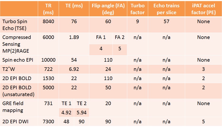
Table 1. MRI scans performed with relevant parameters. iPAT: integrated parallel acquisition techniques, PE: phase encode, n/a: not applicable.
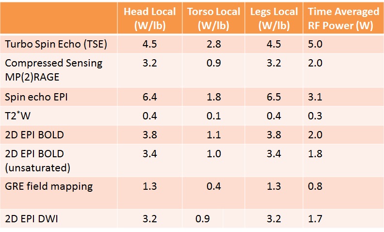
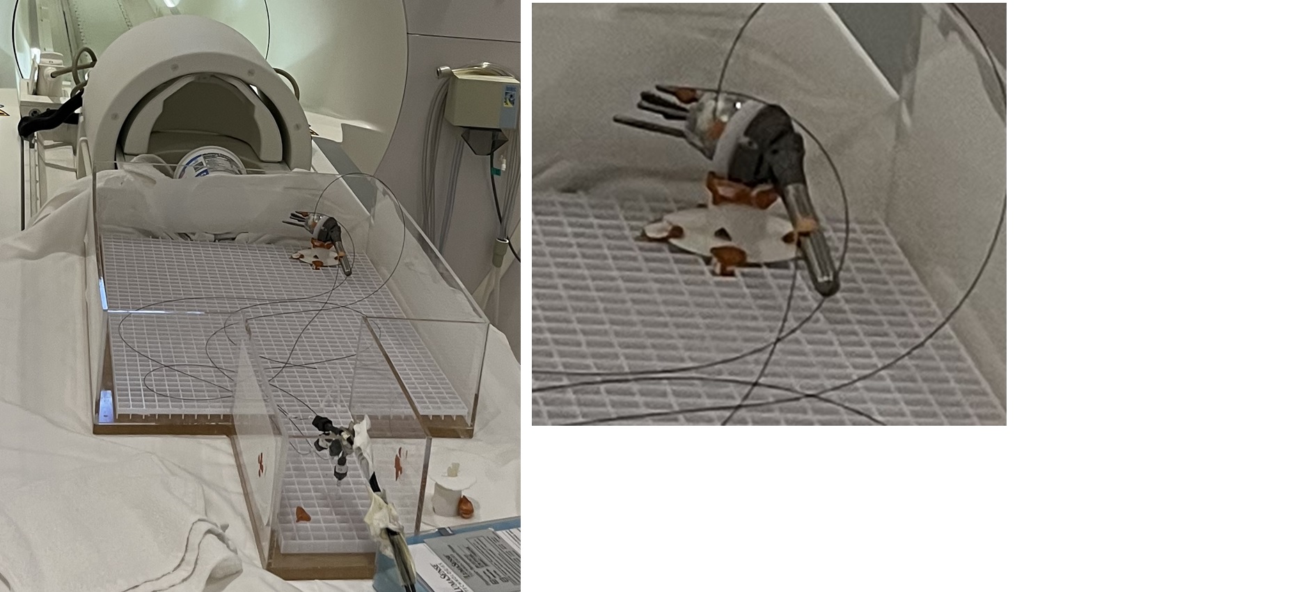
Fig. 1. Experimental setup with the thermal probes.
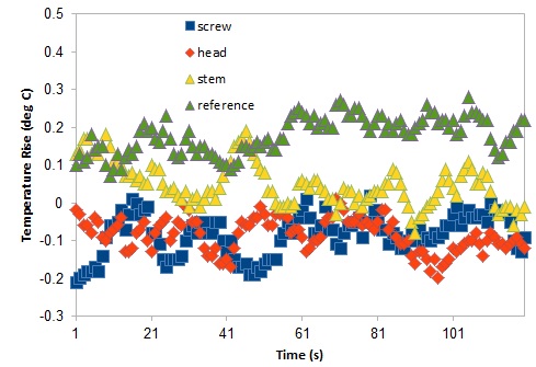
Fig. 2. Temperature change during TSE scan. No significant heating was observed at screw, head and stem of the implant.
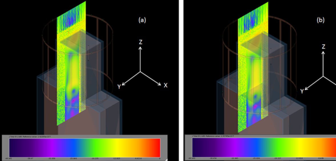
Fig. 3. B-field along YZ plane (a) without and (b) with conductor on the plane. No significant difference is observed.