1446
Evaluating the Performance of a Visual Coil in Physiological and Thermal Noise Dominated Regimes at Ultra-High Magnetic Field1Western University, Robarts research Institute, Center for Functional and Metabolic Mapping, London, ON, Canada
Synopsis
Keywords: RF Arrays & Systems, New Devices
Motivation: Advancements in array coil technology may not yield proportional gains in temporal SNR when physiological fluctuations are the primary noise source.
Goal(s): We constructed a highly dense array covering the visual cortex and evaluated its noise characteristics and performance as a function of resolution.
Approach: We evaluated image SNR and temporal SNR in vivo at 7 Tesla and produced BOLD activation maps using a visual paradigm.
Results: The improvement in image SNR with decreased resolution was significantly higher than the improvement in temporal SNR. This suggests physiological noise dominance at the lower resolution.
Impact: The image and temporal SNR offered by the visual coil allows for sub-millimeter resolution. Insight into the transition between thermal and physiologically noise dominated regimes will aid in reducing the risk of false positives in fMRI using this dense array.
Introduction
The noise in the fMRI time series comprises both a thermal component and a physiological component with thermal noise unaffected by MR signal levels while physiological noise scales linearly with the MR signal [1]. Efforts to enhance image SNR by improving the array coil may therefore not lead to a commensurate improvement in time-series (temporal) SNR if physiological fluctuations are the dominant source of noise [2]. Surface coil arrays, designed to enhance SNR in a targeted brain region, often feature smaller coil elements to increase image SNR. Increasing the array coil count (or decreasing coil size) increases the MR signal, but the impact of reducing coil element size on temporal SNR is complex. Smaller coil elements will produce a higher signal, yet they also have a lower ratio of sample-to-coil noise (i.e., a lower Q-ratio due to being loaded by a smaller volume of tissue), and the higher signal levels equate to proportionally higher physiological noise. It is therefore important to evaluate the noise characteristics of highly dense arrays to determine their performance gains as a function of resolution. In this study, we have developed a dense, 32-channel coil array covering the visual cortex, designed to extend the fMRI resolution into the sub-millimeter range—this is particularly relevant for laminar/columnar studies. We evaluate its performance at two common fMRI isotropic resolutions: 0.8 mm and 2 mm.Method
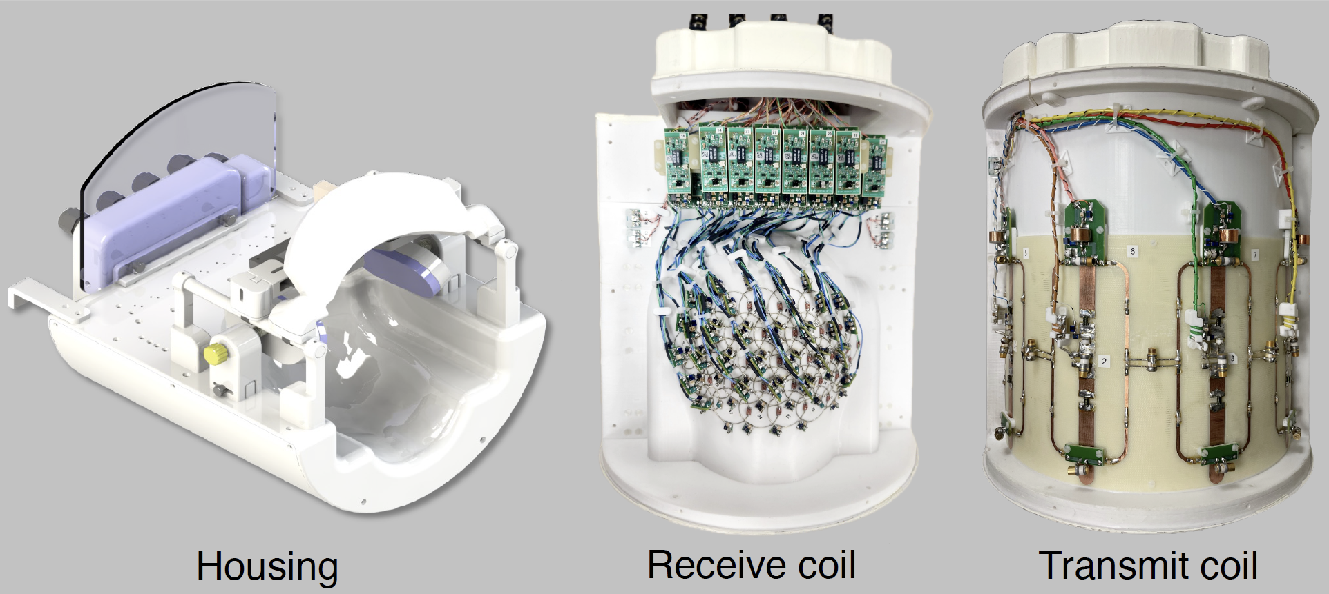
Visual coil: Figure 1 shows the 7T visual coil designed to image the occipital lobe. The inner surface of the visual coil housing follows the contour of an average adult head size, including being tapered to the nape, to minimize the distance between receive elements and the head. The receive coil is comprised of 32 overlapped circular loops, covering an oval-shaped region centered on the primary visual cortex. Each element included active and passive detuning and was connected to a low-input-impedance preamplifier. The transmit coil features four pairs of overlapping loops and dipoles with capacitive decoupling between adjacent loops.Image acquisition: Imaging was performed on a head-only 7T magnet. Rigorous safety testing was performed to ensure robust operation and subject safety before scanning human participants. One participant provided written consent and underwent multiple scanning sessions. The imaging slab was positioned parallel to the calcarine sulcus, and 15 slices were obtained using a multi-band 2D EPI sequence with TR/TE = 1250/20 ms, GRAPPA = 3, partial Fourier = 6/8. Time series were acquired with 0.8- and 2-mm isotropic resolution. Noise scans were acquired with no RF excitation to calculate image SNR. Functional scans were conducted in separate sessions with the same imaging parameters as the signal and noise scans, and the HCP movie clips were used as a visual stimulus.
Temporal and image SNR: Temporal and image SNR were evaluated for each of the datasets with different voxel volumes. All time-series were initially motion-corrected using AFNI. Temporal SNR maps were then calculated for each EPI time series by calculating the ratio of the mean signal of each pixel through the linearly de-trended time course to the standard deviation of that pixel through the time course. Image SNR maps were then converted to absolute SNR units using the method presented by Kellman et al [3], and both image and temporal SNR maps were corrected for bias in magnitude measurements. SPM12 was used for motion correction of the functional data and producing BOLD activation maps.
Results and Conclusion
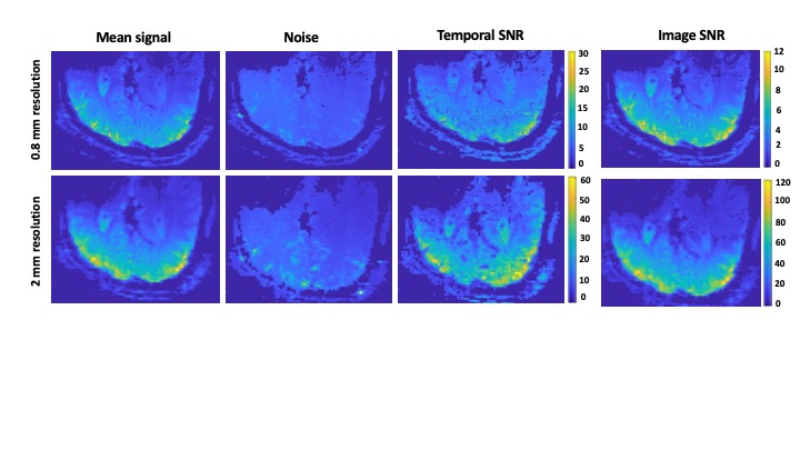
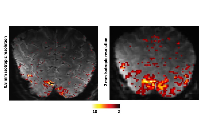
Figure 2 illustrates the signal, noise, and tSNR maps from each time series (0.8-mm and 2-mm isotropic resolution) and the corresponding image SNR maps. Within a gray matter region of interest, we observed an image SNR ratio of 9.4 between the 2-mm and 0.8-mm resolutions, with a corresponding tSNR ratio of 2.3. This suggests a predominant influence of physiological noise in the low-resolution data, as evident by the low-resolution noise maps aligning more closely with the gray matter structure. It is essential to consider that the higher signal produced at ultra-high field, combined with the high sensitivity of small coil elements in a densely packed array, comes with increased physiological noise, thus increasing the resolution until thermal noise predominates is advantageous for the densely packed UHF array. This information can be used to guide the choice of resolution for fMRI studies performed with this coil to help mitigate false positives. In general, the introduced visual coil provides high SNR and shows promise for performing laminar/columnar studies at high resolution [4]. An example of the BOLD activation maps obtained with the visual coil at the two mentioned resolutions is also shown in Figure 3.Acknowledgements
No acknowledgement found.References
1. Krüger G, Glover GH. Physiological noise in oxygenation‐sensitive magnetic resonance imaging. Magn Reson Med. 2001;46:631–7.
2. Triantafyllou C, Polimeni JR, Wald LL. Physiological noise and signal-to-noise ratio in fMRI with multi-channel array coils. Neuroimage. 2011;55:597–606.
3. Kellman P, McVeigh ER. Image reconstruction in SNR units: A general method for SNR measurement†. Magn Reson Med. 2005;54:1439–47.
4. Bollmann S, Barth M. New acquisition techniques and their prospects for the achievable resolution of fMRI. Prog Neurobiol. 2020;207:101936.
Figures