1444
Sodium and Proton Knee MRI using a 32 Channel Transmit and Receive Modular Array at 7.0 Tesla1Radiology, University of Melbourne, Parkville, Australia, 2Biomedical Engineering & Graeme Clark Institute, University of Melbourne, Parkville, Australia, 3MRITools GmbH, Berlin, Germany
Synopsis
Keywords: RF Arrays & Systems, MSK, Sodium, Ultra-High Field, Knee
Motivation: Overcome the limitations of current sodium/proton knee coil configurations using a flexible RF coil array system
Goal(s): Acceptance testing of a 32 channel (16 proton and 16 sodium) RF coil array and demonstrate applicability for sodium and proton MRI of the knee.
Approach: 8 hexagonal modules (4 coils each) were assembled in a volumetric configuration, with sodium and proton MRI quality assessed for phantom and human knee imaging.
Results: High quality sodium volumetric images of the knee were obtained while anatomical proton images were achieved using a standard TSE sequence with additional GRAPPA acceleration and without repositioning
Impact: A 32 channel modular array has been demonstrated to be an effective and flexible solution for both proton and sodium MRI of the knee. Sodium sensitivity was optimised while maintaining proton performance sufficient to acquire accelerated high quality anatomical images.
Introduction
Sodium MRI has the potential to have an impact on research and treatment of musculoskeletal (MSK) conditions. It can detect changes in cartilage and muscle morphology associated with dysregulation of sodium homeostasis that cannot be detected by proton MRI or other imaging methods. However, the low sensitivity, spatial resolution, and the lack of overall anatomical detail in sodium MSK images necessitates the acquisition of proton images while maintaining consistent knee flexion. Previous dual sodium/proton coils have been reported for dedicated knee imaging.1,2 However, these coils are bespoke and unavailable for commercial purchase. In addition, the small number of channels limit the proton imaging acceleration capabilities such that a significant amount of additional time is spent on proton imaging that could be dedicated to improving the quality and quantification of the sodium images. The previous rigid cylindrical construction also limits the use to knee, hand or elbow imaging unlike the trend in modern flexible proton MSK coil arrays that can be rearranged for imaging other MSK targets such as hips, ankles or shoulders. To address these limitations, we have investigated the use of a commercially available (MRITools GmbH, Berlin Germany) flexible modular coil system to obtain quality volumetric sodium images of the knee while ensuring efficient proton anatomical imaging. We hypothesised that an inner array of sodium coils would give optimal sodium performance with an outer proton array facilitating efficient imaging of knee anatomy despite a reduction in coil quality caused by the arrangement. A proton version of the modular coil system has previously been reported3 to be effective for proton imaging of the spine and shoulder.Methods
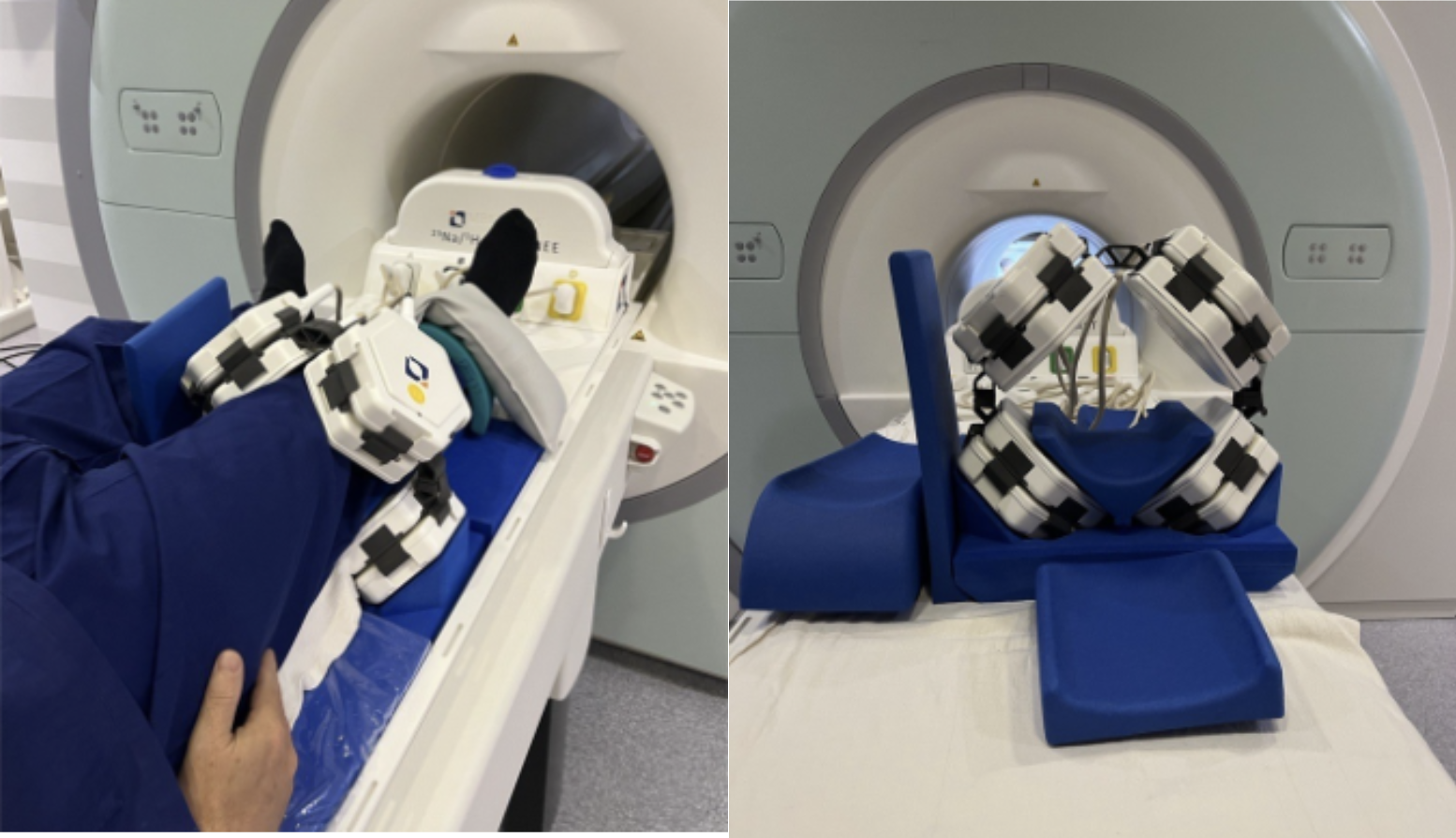
The 32 channel array (4 modules of proton and 4 of sodium) with appropriate connectors for a knee configuration (Figure 1) and a compact interface box to allow for linear transmission of the array was provided by MRI.TOOLS GmbH (Berlin, Germany). Evaluation and validation was performed in EMF simulations and on cylindrical phantom containing saline and five sodium standards (in agar), using a 2D GRE sequence (TR/TE = 500/20, GRAPPA = 3, NEX =2, FOV = 210x210 mm, matrix = 444 x 333 and Acq = 4min) and a 2D TSE sequence (TR/TE = 3200/49ms, ETL =11, GRAPPA = 3, NEX =2, FOV = 210x210 mm, matrix = 576 x 432 and Acq = 3min). The array was then tested on a human volunteer (50 years) with a protocol consisting of proton and sodium imaging. Proton axial 2D TSE sequence (210 mm FOV, 144x144 matrix, 11 echoes, TR/TE = 6000/49ms, GRAPPA =3, NEX =4, Acq. = 1min52sec).Sodium MRI was performed using a MERINA sequence4 (TE=0.4ms, FA=90deg, TR=150ms, 4000proj, TRO=4ms, FOV=300mm, resolution = 3.1mm isotropic, TA=10:00min). Sodium images were manually co-registered to the proton images using ITK-Snap (http://www.itksnap.org).
Results
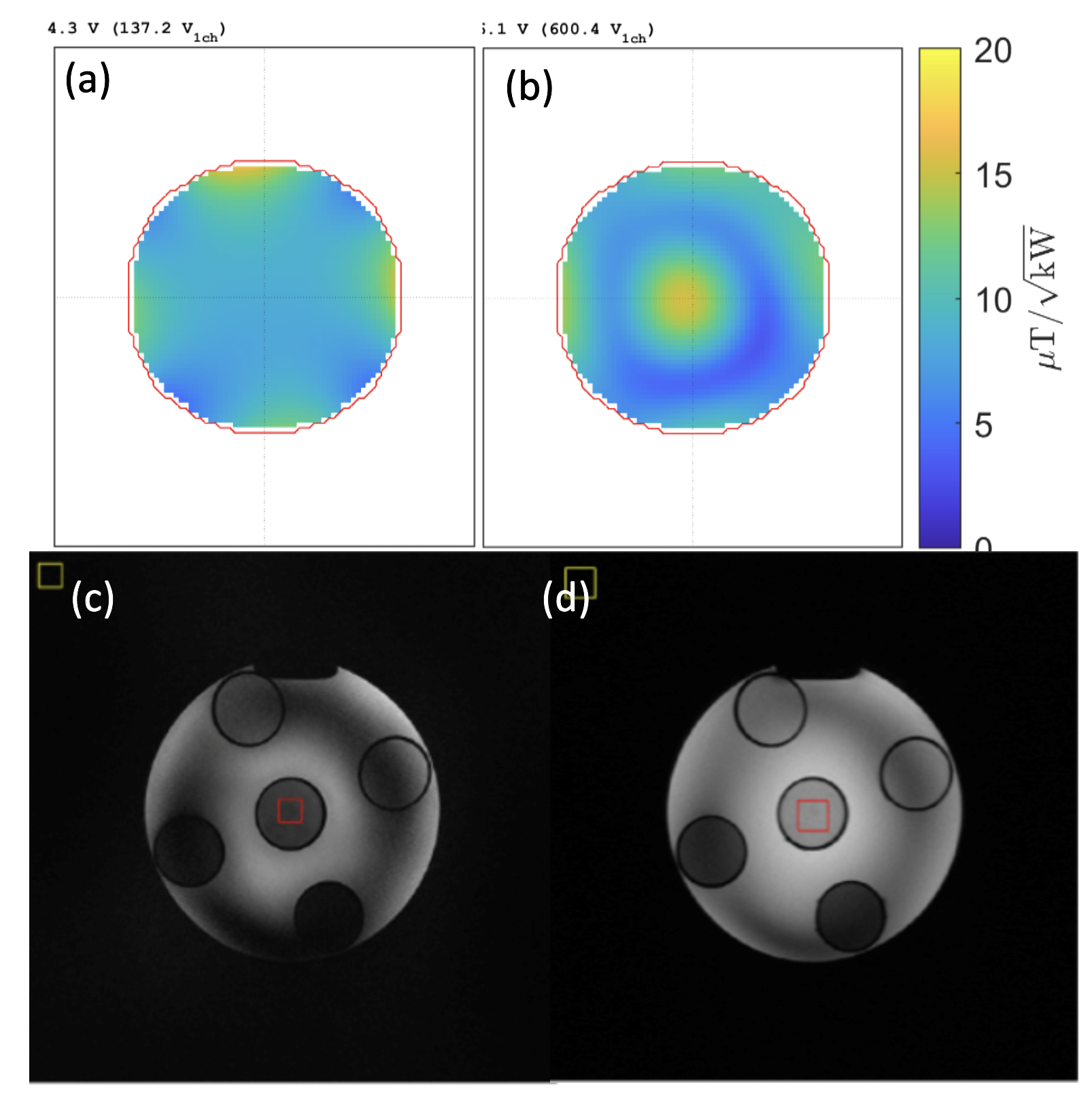
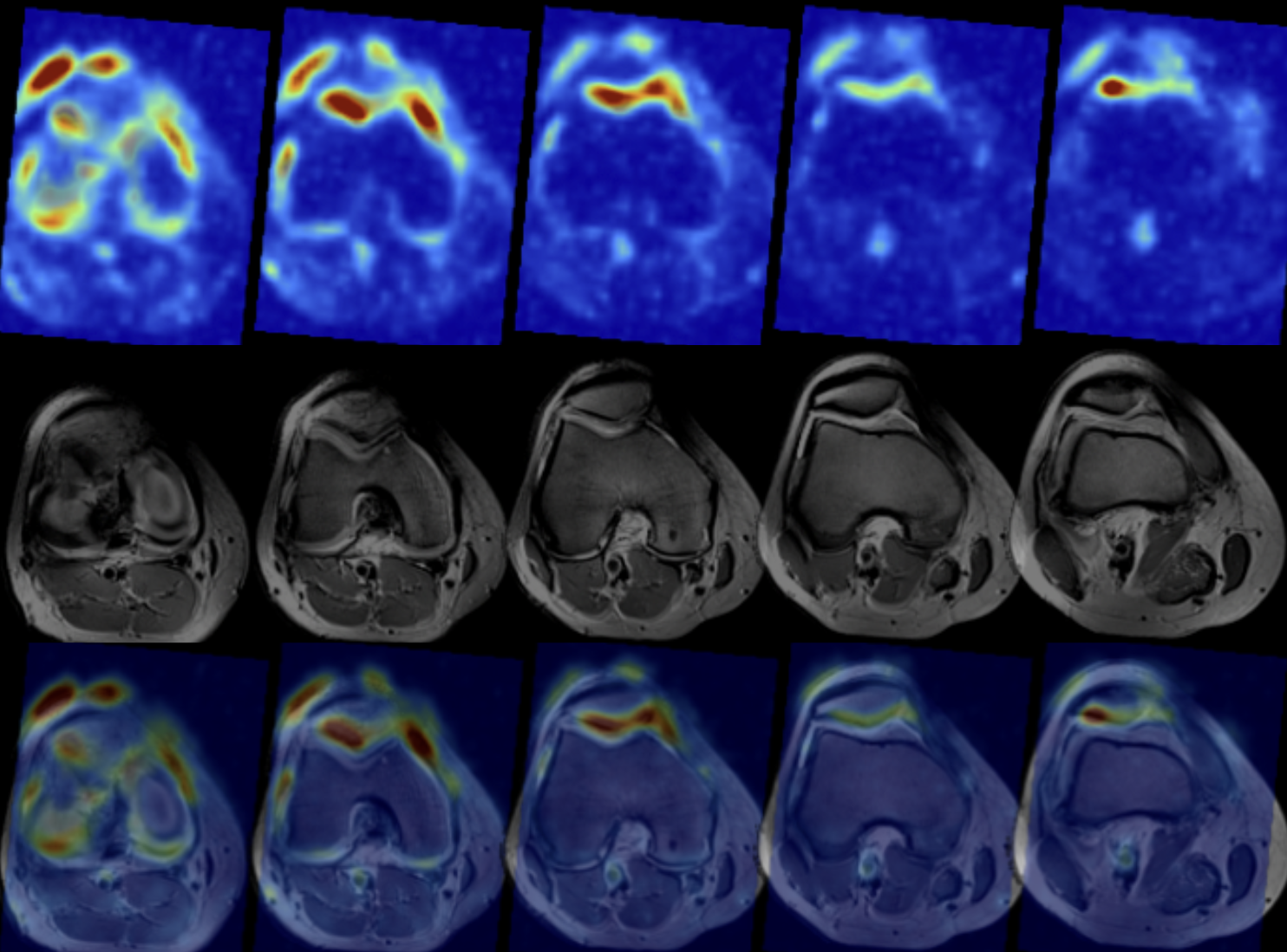
2D GRE and TSE phantom imaging produced sufficiently high quality (Figure 2c and d) with the high conductivity saline phantom producing the usual B1+ signal dropout at 7T and correlating with the B1+ simulation plots (Figure 2a and b). The in vivo imaging (Figure 3) demonstrated this coil array could achieve excellent sodium imaging with clinical quality proton MRI in an imaging protocol suitable for studying clinical cohorts. The sodium signal SNR was estimated to be 16+/-4 in the cartilage compared to 140 +/- 31 for the 2D TSE imaging.Discussion and Conclusion
This 32 channel RF coil array setup allows for flexible sodium and proton imaging for MSK applications. The construction prioritises the SNR of the sodium imaging however the flexible proton array still allows for sufficient quality imaging to be achieved whilst taking advantage of scan accelerations made possible by the phased array receive elements. The flexible and modular nature of the arrays facilitates rearrangement of the hexagonal building blocks for optimised imaging of other body regions and MSK targets.Acknowledgements
The authors acknowledge the facilities and scientific and technical assistance of the National Imaging Facility, a National Collaborative Research Infrastructure Strategy (NCRIS) capability, at the Melbourne Brain Centre Imaging Unit, University of Melbourne.References
- Brown R, et al. A Flexible Nested Sodium and Proton Coil Array with Wideband Matching for Knee Cartilage MRI at 3 Tesla. Magn Reson Med. 2016 Oct;76(4):1325–34.
- Brown, R. et al., Design of a Nested Eight-Channel Sodium and Four-Channel Proton Coil for 7T Knee Imaging. Magn. Reson. Med. 2013, 70, 259–268.
- Andreas Graessl et al., En route to clinical ultrahigh field musculoskeletal MR using multi-purpose transceiver RF modules for spine und shoulder imaging at 7.0 T., Proc. Intl. Soc. Mag. Reson. Med. 22, 2014
- Blunck, Yasmin, et al. "3D‐multi‐echo radial imaging of 23Na (3D‐MERINA) for time‐efficient multi‐parameter tissue compartment mapping." Magnetic resonance in medicine 79.4 (2018): 1950-1961.
Figures