1442
A self-decoupled 20/32-channel Tx/Rx array for simultaneous brain and spinal cord MRI at 7T1Department of Electrical and Computer Engineering, Vanderbilt University, nashville, TN, United States, 2Vanderbilt University Institution of Imaging and Science, Nashville, TN, United States, 3Department of Radiology and Radiological Science, Vanderbilt University Medical Center, Nashville, TN, United States
Synopsis
Keywords: RF Arrays & Systems, RF Arrays & Systems
Motivation: Simultaneous functional imaging of the brain and spinal cord can offer valuable insights into the interactions and processing pathways between these organs
Goal(s): The goal is to build an transmit array and receive array for 7T the brain and spinal cord MRI.
Approach: The approach uses corner-fed self-decoupling for transmit array and center-fed self-decoupling for receive array.
Results: A 20-channel modular self-decoupled transmit array and 32-channel overlapped receive array were fabricated. Both arrays exhibit excellent matching performance, acceptable decoupling, and high detuning ability. Thanks to the self-decoupling technology, both arrays are splitable which simplifies the setup and allows for easier access by subjects.
Impact: The proposed device will solve the hardware issue and permit studies of healthy subjects to improve our understanding of the resting-state connectivity and interaction between the brain and spinal cord, compensating impaired sensory, motor, or autonomic functions in neurological disorders.
Introduction
fMRI studies have provided new insights into the functional architecture of the brain [1-4]. In addition to brain fMRI, spinal cord fMRI has recently emerged as a powerful tool for studying motor and sensory/pain pathways in the spinal cord [5,6]. Simultaneous functional imaging of the brain and spinal cord can offer valuable insights into the interactions and processing pathways between these organs in both normal and abnormal states, such as spinal cord injury, chronic pain, and motor diseases. Unlike RF coils used in brain imaging, only a few RF coil designs have been proposed for simultaneous brain and spinal cord imaging [7,8]. Due to the extended longitudinal coverage required, the number of transmit coils may need to be eight. Additionally, it is preferable to keep the coils separate to ensure easy patient access. In this work, we have developed a 20/32-channel Tx/Rx array for simultaneous brain and spinal cord MRI. We employed a self-decoupling method to simplify coil fabrication and enable modular designs.Method
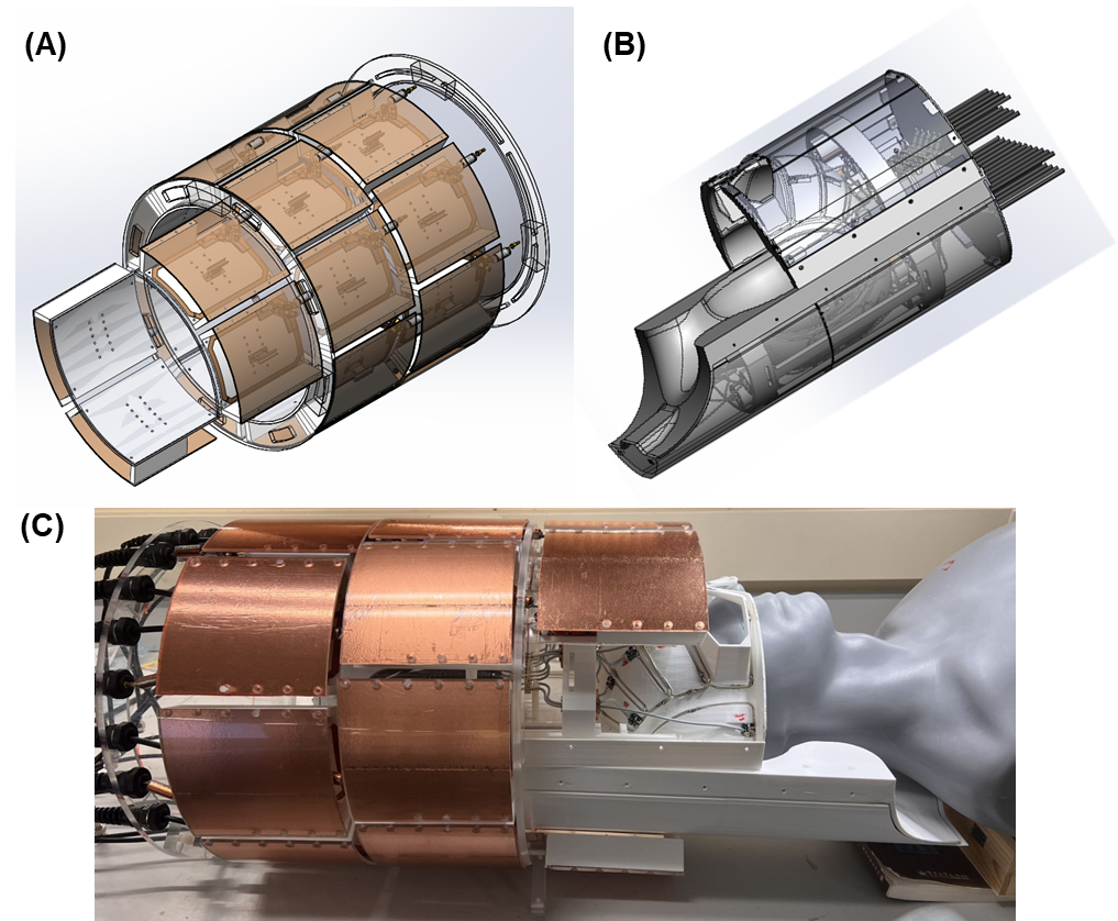
Self-decoupled Tx array:The transmit array consists of 20 self-decoupled modular coils (Figure 1A and Figure 1C). Each coil has dimensions of 10x10 cm² and is equipped with its own cable trap and RF shield. Each coil was fed at the corner to enable decoupling capability in both horizontal and vertical directions [9]. The gap between the elements in the same row and the elements in adjacent rows is approximately 1.5 cm. The 20 transmit coils arranged in three rows (8 coils, 8 coils, 4 coils) as shown in Figure 1b. The third row extends to cover a portion of the chest and back areas to ensure the entire cervical spine is included.
Self-decoupled Rx coil:
The receive array is composed of center-fed self-decoupled coils (Figure 1B and Figure 1C) [9]. Self-decoupling was employed to ensure isolation between coils in the same row, while geometrical decoupling was implemented to ensure separation among different rows. The top part consists of 10 coils, while the bottom part comprises 22 coils, covering the entire back of the head and the cervical spine.
Bench test:
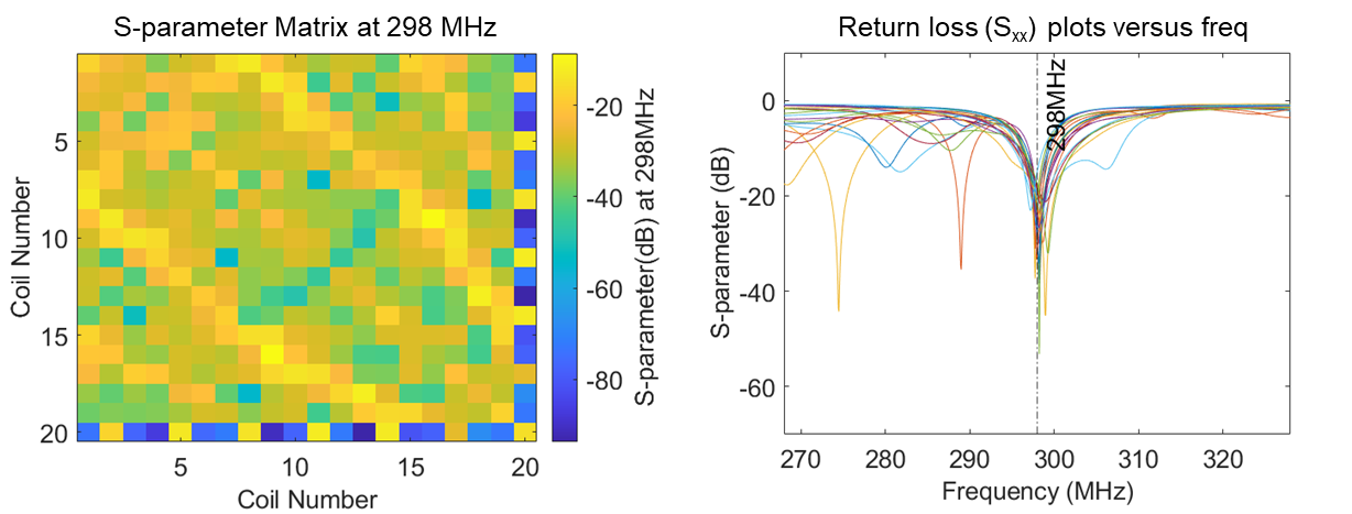
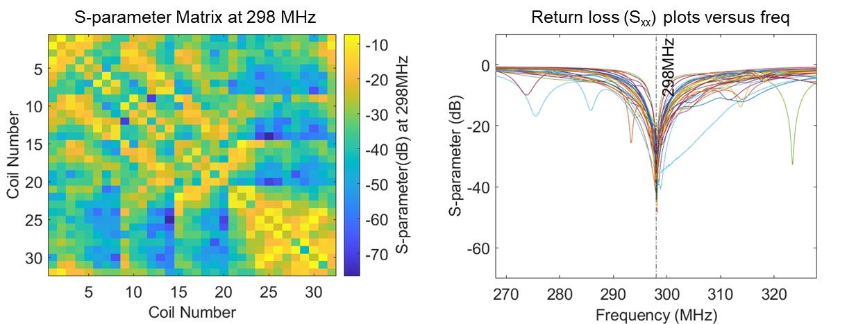
Both the transmit array and receive array were tested on the workbench. The S-parameters, including matching and decoupling, were recorded with a human head and shoulder phantom loaded. When measuring the S-parameters of the transmit array, the receive array was detuned, and vice versa. In addition to tuning, matching, and decoupling measurements, we also assessed the detuning performance for both the transmit array and receive array. Furthermore, for the receive array, we measured the preamplifier decoupling performance.
Results and Discussions
Figure 2 displays the bench test results of the Tx array. All coils were well-tuned to 298 MHz and matched to 50 ohms, with Sxx below -20 dB. The inter-element isolation among transmit coils is below -12 dB, indicating less than 10% power crosstalk. Although it is not as ideal as the original two-channel self-decoupled array (with isolation at -30 dB), it is still acceptable, especially considering the lightweight loading of the transmit coils and the complexity of the multi-row design.Figure 3 presents the bench test results of the Rx array. Similar to the Tx coils, the return loss of the receive coils is negligible when loaded with the phantom (< -25 dB). The coil isolation is better than that of the Tx array, with an average value of up to -21 dB. Both the Tx coils and Rx coils exhibit excellent detuning performance of <-20 dB. By adjusting the cable length, the Rx coils achieve a preamplifier decoupling of <-25 dB.
Unlike head-only imaging, multi-row Tx arrays are preferred for simultaneous brain and spinal cord imaging because they can independently control the transmit field and local SAR in these two areas. Currently, most 7T scanners are equipped with only 8 transmit channels. To operate twenty Tx coils, we recommend using either the Butler Matrix circuit or the recently proposed array-compared circuits. We found that the modular design in the Tx array significantly simplifies coil fabrication and allows for easy patient access. We did not employ such a modular design for the Rx array, as it might lead to imaging nulls in z-direction gaps. We are currently working towards implementation in the 7T scanner and the safety considerations for inside the bore. Both the mechanical and the circuit designs will be made open source in the future.
Acknowledgements
This work was supported by NIH grants R01 EB031078. The content is solely the responsibility of the authors and does not necessarily represent the official views of the National Institutes of Health.References
1. Ogawa S, Lee TM, Kay AR, Tank DW. Brain magnetic resonance imaging with contrast dependent on blood oxygenation. Proc Natl Acad Sci U S A. 1990;87(24):9868-72. Epub 1990/12/01. doi: 10.1073/pnas.87.24.9868. PubMed PMID: 2124706; PubMed Central PMCID: PMCPMC55275.
2. Bandettini PA, Wong EC, Hinks RS, Tikofsky RS, Hyde JS. Time course EPI of human brain function during task activation. Magn Reson Med. 1992;25(2):390-7. Epub 1992/06/01. doi: 10.1002/mrm.1910250220. PubMed PMID: 1614324.
3. Kwong KK, Belliveau JW, Chesler DA, Goldberg IE, Weisskoff RM, Poncelet BP, et al. Dynamic magnetic resonance imaging of human brain activity during primary sensory stimulation. Proc Natl Acad Sci U S A. 1992;89(12):5675-9. Epub 1992/06/15. doi: 10.1073/pnas.89.12.5675. PubMed PMID: 1608978; PubMed Central PMCID: PMCPMC49355.
4. Conrad BN, Barry RL, Rogers BP, Maki S, Mishra A, Thukral S, et al. Multiple sclerosis lesions affect intrinsic functional connectivity of the spinal cord. Brain. 2018;141(6):1650-64. Epub 2018/04/13. doi: 10.1093/brain/awy083. PubMed PMID: 29648581; PubMed Central PMCID: PMCPMC5972586.
5. Cohen-Adad J, Gauthier CJ, Brooks JC, Slessarev M, Han J, Fisher JA, et al. BOLD signal responses to controlled hypercapnia in human spinal cord. Neuroimage. 2010;50(3):1074-84. Epub 2010/01/12. doi: 10.1016/j.neuroimage.2009.12.122. PubMed PMID: 20060914.
6. Robert L. Barry, S. Johanna Vannesjo, Samantha By, John C. Gore, Seth A. Smith, “Spinal cord MRI at 7T”, in NeuroImage, Volume 168, Pages 437-451, March 2018
7. Zhang, B., Seifert, A. C., Kim, J. W., Borrello, J., & Xu, J. (2017). 7 Tesla 22‐channel wrap‐around coil array for cervical spinal cord and brainstem imaging. Magnetic resonance in medicine, 78(4), 1623-1634.
8. May, M. W., Hansen, S. L. J., Mahmutovic, M., Scholz, A., Kutscha, N., Guerin, B., ... & Keil, B. (2022). A patient‐friendly 16‐channel transmit/64‐channel receive coil array for combined head–neck MRI at 7 Tesla. Magnetic Resonance in Medicine, 88(3), 1419-1433.
9. Yan, X., Gore, J.C. & Grissom, W.A. “Self-decoupled radiofrequency coils for magnetic resonance imaging”. Nat Commun 9, 3481 (2018). https://doi.org/10.1038/s41467-018-05585-
Figures