1441
Improvement in Receive Sensitivity at Ultra-High Field in Brain via Metasurfaces1University of Pennsylvania, Philadelphia, PA, United States, 2Center for Advanced Metabolic Imaging in Precision Medicine, Department of Radiology, University of Pennsylvania, Philadelphia, PA, United States
Synopsis
Keywords: New Devices, Spectroscopy
Motivation: Quantification of magnetic resonance spectroscopy (MRS) spectra requires sufficient SNR to detect changes in metabolic state. At ultra-high field, low concentration metabolite require length scan times to obtain sufficient SNR.
Goal(s): To use a novel metasurface design to enhance the B1- receive sensitivity in phantom and in vivo brain proton spectroscopy data at 7T, resulting in increased SNR without additional scan time.
Approach: Receive sensitivity enhancement was measured in phantom and in vivo experiments with and without the metasurface present.
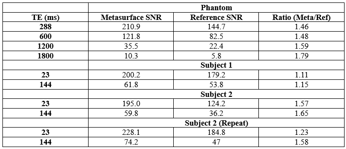
Results: The metasurfaces enhanced phantom spectral SNR by an average of 58%, while in vivo spectral SNR was improved on average by 38%.
Impact: The work impacts 7T spectroscopy techniques by improving the inherent receive sensitivity, yielding higher SNR spectra. Future work will focus on demonstrating this application in patent populations to reduce scan times and enhance the signal from low concentration metabolites.
Introduction
Ultra-high field (≥7T) magnetic resonance spectroscopy (MRS) has allowed for more detailed studies if multiple pathologies such as high-grade gliomas1 and Alzheimer's Disease2 y benefitting from the increased signal-to-noise ratio (SNR), increased chemical dispersion, and reduction in J-coupling for strongly coupled systems3. However, a limitation that is still present at these field strengths is the inherently inhomogeneous B1- field, restricting spectral SNR of low concentration metabolites. Current methods for enhancing spectral signal include building higher quality receive arrays with more channel or acquiring more signal averages. However, few methods exist to enhance the receive field during acquisition. Studies have been performed using high permittivity dielectric materials claiming to increase spectral SNR by increasing coupling between the coil array and body4,5. However, dielectric pads are limited by their permittivity and have a relatively short shelf life6. Recently, other groups have found success using metasurfaces to enhance the B1- field at 1.5T for wrist and knee imaging7,8. Therefore, the goal of this work was to enhance phantom and in vivo proton spectroscopy SNR at 7T using a novel metasurface design by increasing the B1- field.Methods
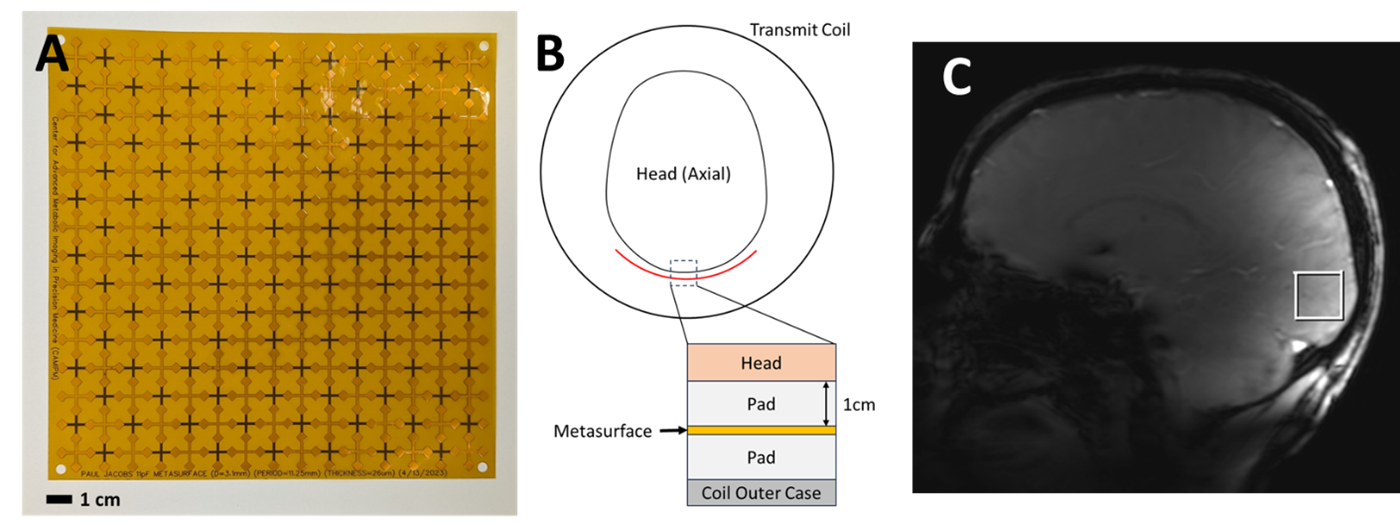
All data was acquired on a 7T system (MAGNETOM Terra, Siemens Healthcare, Erlangen, Germany) using a single-channel transmit/32-channel phased array proton head coil (Nova Medical, Wilmington, MA, USA) with all experiments being performed with and without a prototype metasurface (Figure 1a). Phantom MRS spectra and a noise-only (transmit voltage set to 0V) signal were acquired via a PRESS localized 20×20×20mm single voxel spectroscopy (SVS) sequence with the following parameters: TR=3000ms, TE=288, 600, 1200, 1800ms, and 32 averages, on a 1H MRS phantom (BRAINO, GE Healthcare). Noise estimates were computed as the standard deviation of the signal-free trailing points of the FIDs. Scans were also acquired at various transmitter reference voltages (100-400V), including the calibrate voltage for the voxel location. In vivo MRS brain data was acquired on two subjects with written informed consent under an approved institutional regulatory board protocol. The same sequence parameters were used except for echo times of 23ms and 144ms, with the voxel placed in the occipital lobe adjacent to the metasurface (Figure 1b and1c) with calibrated transmitter reference voltages to account for B1+ differences. Spectral SNR was computed as the peak height of the phased NAA resonance, divided by the standard deviation of the noise. Care was taken to obtain the same shim and resulting water spectral line width for the metasurface and control scans.Results
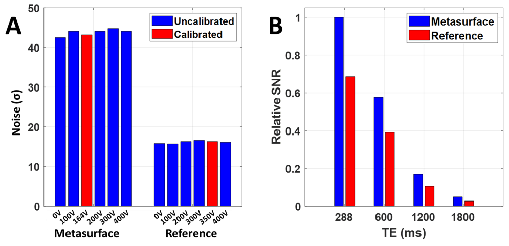
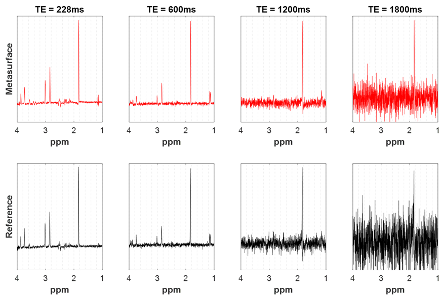
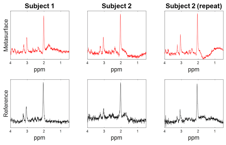
Figure 2a shows the quantified phantom noise measurements across transmitter voltages, with the metasurface showing consistently higher noise than the reference. Noise within either experimental group did not show a dependence on transmitter voltage. Figure 2b shows the quantified relative SNR of each of the phantom spectra, seen in Figure 3, which increased in the presence of the metasurface by an average of 58%. In vivo data seen in Figure 4, also qualitatively shows an increase in SNR when the metasurface was used in comparison to the reference case. This is consistently seen across both subjects scanned and in the same subject repeat acquisition. Table 1 shows the fully quantified results for both the phantom and in vivo datasets, in which the in vivo data saw an average increase of 38% when the metasurface was used.Discussion and Conclusion
In this work we showed thar receive field sensitivity could be improved for MR spectroscopy via a novel metasurface design at 7T. We were able to demonstrate this for both phantom and in vivo experiments where the average enhancement was 58% and 38% respectively. These comparisons are made with the transmitter voltage calibrated so that the increase in B1+ efficiency is negated. Metasurfaces have conventionally been used to enhance the B1+ field via induced conduction currents creating a secondary magnetic field9. This can also be thought of during the receive portion of the scan, where the B1- field also interacts with the metasurface causing secondary currents to form and produce an additional receive field7. Limitations of this study include visible variation in the spectral lipid content, most likely due to slight voxel placement differences near the skull. This contrast with the phantom data which allowed for the voxel to be placed closer to the edge and therefore more in the metasurface enhancing region. In the future, additional in vivo data will be acquired, and SNR will be quantified via image-based methods.Acknowledgements
Research reported in this work was supported by the National Institutes of Health via the National Institute of Biomedical Imaging and Bioengineering under award number P41EB029460.References
1. McCarthy L, Verma G, Hangel G, et al. Application of 7T MRS to High-Grade Gliomas. AJNR Am J Neuroradiol. Oct 2022;43(10):1378-1395. doi:10.3174/ajnr.A7502
2. Marjanska M, McCarten JR, Hodges JS, Hemmy LS, Terpstra M. Distinctive Neurochemistry in Alzheimer's Disease via 7 T In Vivo Magnetic Resonance Spectroscopy. J Alzheimers Dis. 2019;68(2):559-569. doi:10.3233/JAD-180861
3. Henning A. Proton and multinuclear magnetic resonance spectroscopy in the human brain at ultra-high field strength: A review. Neuroimage. Mar 2018;168:181-198. doi:10.1016/j.neuroimage.2017.07.017
4. de Heer P, Bizino MB, Versluis MJ, Webb AG, Lamb HJ. Improved Cardiac Proton Magnetic Resonance Spectroscopy at 3 T Using High Permittivity Pads. Invest Radiol. Feb 2016;51(2):134-8. doi:10.1097/RLI.0000000000000214
5. Lee BY, Zhu XH, Rupprecht S, Lanagan MT, Yang QX, Chen W. Large improvement of RF transmission efficiency and reception sensitivity for human in vivo(31)P MRS imaging using ultrahigh dielectric constant materials at 7T. Magn Reson Imaging. Oct 2017;42:158-163. doi:10.1016/j.mri.2017.07.019
6. Teeuwisse WM, Brink WM, Webb AG. Quantitative assessment of the effects of high-permittivity pads in 7 Tesla MRI of the brain. Magn Reson Med. May 2012;67(5):1285-93. doi:10.1002/mrm.23108 7. Chi Z, Yi Y, Wang Y, et al. Adaptive Cylindrical Wireless Metasurfaces in Clinical Magnetic Resonance Imaging. Adv Mater. Oct 2021;33(40):e2102469. doi:10.1002/adma.202102469
8. Yi Y, Chi Z, Wang Y, et al. In vivo MRI of knee using a metasurface-inspired wireless coil. Magn Reson Med. Oct 10 2023;doi:10.1002/mrm.29870
9. Vorobyev V, Shchelokova A, Efimtcev A, et al. Improving B1+ homogeneity in abdominal imaging at 3 T with light, flexible, and compact metasurface. Magn Reson Med. Jan 2022;87(1):496-508. doi:10.1002/mrm.28946
Figures