1436
In-Ovo MRI: design of a large-scale RF coil model1Neuroscience Research Institute, Gachon University, Incheon, Korea, Republic of, 2Department of Health Sciences and Technology, GAIHST, Gachon University, Incheon, Korea, Republic of, 3Department of Biomedical Engineering, Gachon University, Incheon, Korea, Republic of
Synopsis
Keywords: RF Arrays & Systems, RF Arrays & Systems
Motivation: The poultry industry utilizes MRI to assess egg quality, especially to identify fertilized eggs. Image quality across eggs should be similar and with efficient filling factor.
Goal(s): The goal is to design a specialized coil for MRI that can efficiently and accurately scan multiple eggs , providing the same image quality for each egg.
Approach: The proposed coil has a multiloop structure. We used electromagnetic simulations to compute the magnetic field and compared the proposed coil with a volume and individual loop coils.
Results: Validated through simulations and MR images, the coil displayed the scalability and, enabling the imaging of multiple eggs
Impact: The proposed coil design shows better egg per area filling factor efficiency and uniform image quality for each egg and also scalability for high-throughput egg screening. This design promises to enhance multiple egg imaging quality control in the poultry industry.
Introduction
The utilization of MRI technology within the poultry industry holds significant importance in optimizing egg production processes and ensuring product quality. MRI provides producers with the capability to make decisions about egg quality through image analysis. A recent development in the poultry sector involves the use of MRI for identifying fertilized eggs, with the potential to scan approximately 9000 eggs per hour [1]. This advancement presents a promising tool for elevating efficiency and precision in egg production and quality control. While MRI is traditionally associated with human and small animal imaging, recent studies have ventured into the realm of avian egg imaging [2]. These studies focus on comprehending the development and structural growth of embryos, including the analysis of egg fertilization without the need to breach the eggshell [3].Methods
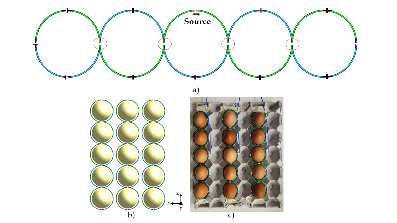
Our research introduces a coil design capable of generating a uniform |B1|-field for multiple eggs while utilizing a single channel. This innovative coil design takes the form of a multiple loop structure, that can be scaled for multiple loops, as illustrated in Fig. 1a. Comprising two distinct layers – the upper (displayed in green) and lower (displayed in blue) – with a small vertical separation, this coil's conductor lines adhere to a half-circumference geometry, with the other half following a continuous semicircular wave pattern. From a top view, the coil may appear as multiple loop coils; however, the unique feature lies in the fact that the loop is not fully closed and connected for each turn as shown by the red circle, except at the extremes. To ensure symmetrical |B1|-field performance, the source is strategically positioned at the coil's center, as indicated in Fig. 1a. The coil consists of a radius of 25mm and 5 turn, with a length of 255 mm and capacitors are placed for tuning as shown in Fig. 1a. We compared the performance of the proposed coil with three other coil configurations: an 8-channel volume coil with, a 5 channel individual loop array coil of 25 mm radius each, and a 3-channel multi loop coil array with 15 eggs, as illustrated in Fig. 1b. Electromagnetic simulations were performed for each coil, utilizing Sim4life software from Zurich ZMT. Every coil was tuned and matched to the operating frequency of a 7T MRI scanner, corresponding to 297.4 MHz. The coil was constructed, tuned, and matched to 297MHz and 50 Ohms, featuring a radius of 25mm, with each channel accommodating 5 eggs. As depicted in Fig. 1c, we constructed three channels for image acquisition, allowing us to acquire images of 15 eggs.Results
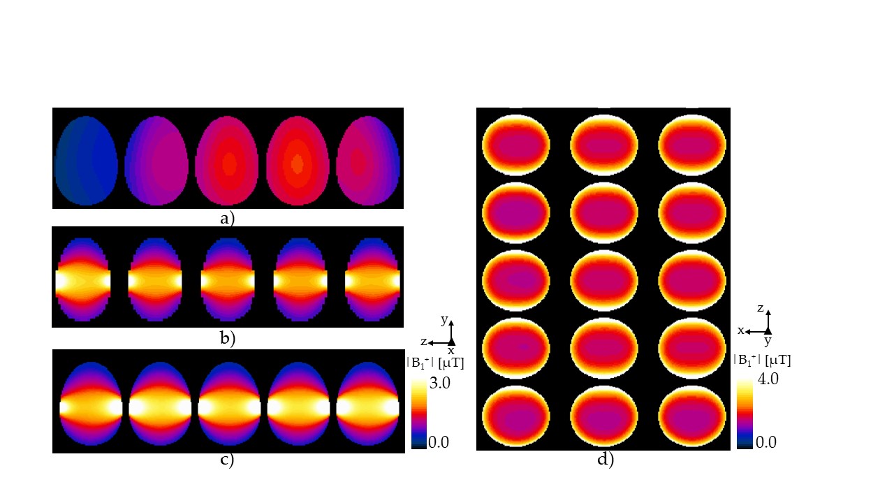
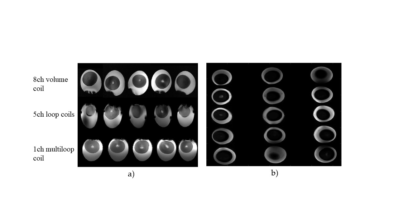
Figure 2a displays the |B1|-field of the 5 eggs acquired using the 8-channel array coil, while Figure 2b shows the field for the individual loop coils. Notably, the proposed coil, presented in Figure 2c with the 5 eggs in the z-y axis, provides a |B1|-field of similar intensity to that of the single loop coils while reducing the space between eggs. The x-z view shown in Fig. 2d displays the magnetic field for the 15 eggs with the 3ch of the proposed coil. it should be noted that the uniformity and a similar field intensity and patterns is acquired for each egg. Images of the three types of coils were acquired utilizing a 7T MRI scanner from Siemens (Magnetom) and a Turbo Spin Echo pulse sequence. Figure 3a offers a comparison of egg images obtained using a clinical head 8-channel volume coil (top), individual 5-channel array coil (middle), and the 1-channel braid-coil (bottom). In Figure 3b, the image from the 3-channel braided coil array reveals the acquisition of images of 15 eggs.Discussions
This study introduces a remarkable coil capable of capturing MR images of multiple eggs with a minimal number of transmission and reception channels, all while maintaining high image intensity and uniformity for each egg. We anticipate that the proposed coil will find utility in advancing in-Ovo MRI applications and research endeavors.Acknowledgements
This work was supported by the Institute for Information & Communications Technology Promotion (IITP) grant funded by the Korean government (MSIP) (No. 2021-0-00490)References
1) M. Johnson, (2021, November). MRI technology can help identify which eggs are fertile. [Online]. Available: https://www.wattagnet.com/latest-news/article/15534623/mri-technology-can-help-identify-which-eggs-are-fertile
2) R. Klose, F. Streckenbach, S. Hadlich, T. Stahnke, R. Guthoff, A. Wree, M. Frank, S. Langner, O. Stachs and T. Lindner. “Ultra-high-field MRI in the Chicken Embryo in Ovo-a Model for Experimental Ophthalmology,” Klin. Monbl. Augenheilkd., vol. 234 no. 12, pp. 1458-1462, 2017.
3) B. Shojaei, H. Tavakoli, M.J. Aghasizade and H.R. Sayyah Badkhor. “Morphologic evaluation of the fertilized and unfertilized ostrich egg during the early stages of development (MRI analysis),” Anat. Sci. J., vol. 11 no.4, pp. 169-174, 2014.
Figures