1435
A Novel 8-Channel 31P/1H DODO Transceiver Array for Human Brain Imaging and Performance Comparison with 31P/1H TEM Volume Coil at 7T1Center for Magnetic Resonance Research, University of Minnesota, Minneapolis, MN, United States, 2Department of Biomedical Engineering, State University of New York at Buffalo, Buffalo, NY, United States
Synopsis
Keywords: RF Arrays & Systems, RF Arrays & Systems, X-nuclear array coil
Motivation: 31P MRSI provides a valuable tool for metabolic imaging of human brain, and is crucial for studying neuroenergetics and neurological diseases, however, faces the low SNR challenge.
Goal(s): To construct a dual-tuned 8-channel 31P/1H transceiver array head coil with excellent performance for both proton structural MRI and 31P MRSI at 7T.
Approach: We constructed an 8-ch 31P/1H transceiver array coil based on the novel double tuned and double matched (DODO) coil element, and compared its performance with the 31P/1H TEM volume coil.
Results: The 8-ch 31P/1H transceiver array coil shows significantly higher performance compared to 31P/1H TEM volume coil.
Impact: We developed an 8-ch 31P/1H transceiver array coil with excellent performance based on the DODO coil design. This technology has broad applications for in vivo human brain 31P MRSI studies and can be adapted to other multinuclear array coil designs.
Introduction
31P MRS imaging (31P-MRSI) is valuable for non-invasive study of human brain phosphorus metabolites and metabolism, including the NAD metabolites and NAD+/NADH redox state, which regulates glucose metabolism. It has been frequently applied to study neurological disorders such as Alzheimer's and Parkinson’s diseases 1-3. However, 31P-MRSI suffers from low signal-to-noise ratio (SNR). From the RF coil perspective, a RF coil should operate at not only 31P but also 1H Larmor frequency, which is necessary for brain anatomical imaging and B0 shimming to achieve high-quality 31P-MRSI. Traditional dual frequency coil design typically uses an LC trap circuit in the resonant loop to generate an additional resonant frequency 4. Additionally, several novel methods including interleaved array coils, nested loops and dual tuned TEM volume coil, have been used to construct dual-tuned 31P/1H coils for whole-brain imaging at UHF 5-7. However, all these techniques typically result in a tradeoff between the performance of either 31P or 1H. In this work, we propose a novel double-tuned and double-matched loop coil element (DODO coil, 8) operating at both 31P and 1H frequencies. This proposed DODO coil element demonstrates similar performance to single-tuned control coils for either 31P or 1H frequencies. Consequently, we have developed an 8-channel 31P/1H array human head coil based on the DODO coil design, which exhibits excellent imaging performance compared to a 31P/1H TEM volume coil 5.Methods
The 31P/1H dual-tuned DODO coil and single-tuned 31P and 1H (control) coils with the same geometry and size were constructed (Fig. 1A) and evaluated by the S-parameters measured using a VNC (Rohde & Schwarz ZNBT8 16-port Vector Network Analyzer 9 kHz – 8.5 GHz) (Fig. 1B). The 8-ch 31P/1H DODO coil and the TEM volume coil were loaded with a head-shaped inorganic phosphate (Pi) phantom for acquiring proton density image and 31P chemical shift imaging (CSI) on a Siemens MAGNETOM 7T scanner. The 31P CSI was acquired with: 18x18x15 cm FOV; 19x19x15 matrix under fully relaxed condition. The 31P B1+ (RF magnetic field) maps were quantified based on the CSI spectrum integrals using the double flip angle method, and the 31P SNR maps were quantified based on the CSI spectrum integral divided by spectrum noise.Results
A 31P/1H DODO loop coil (15cm by 8cm) was designed and constructed, then compared with the imaging performance of the single-tuned 31P and 1H loop control coils, loaded with the Pi phantom (Fig. 1A). The DODO loop coil, which relies on the inductors (40nH) to block high frequency currents, and small capacitors (~1pF) to block low frequency currents, demonstrated excellent S11 parameters for both 31P and 1H (better than -27dB) and excellent isolation between the two resonances (S12 ≤ -17 dB) under loaded condition as shown in Fig.1B. Figure 2 demonstrates similar performance in terms of the B1+ strength for proton and 31P, 31P SNR, and CSI maps at nominal 90 degree pulse flip angle between the 31P/1H DODO loop coil with single-tuned control loop coils. Noticeably, 31P MRSI noise level for both DODO and control coils were similar.The prototypes of the 8-ch 31P/1H DODO array head coil and the 31P/1H TEM volume coil are shown in Fig. 3A. The 31P B1+ maps of the 8-ch 31P/1H DODO array coil show ~16% higher in central regions and ~72% higher in peripheral regions as compared to the 31P/1H TEM volume coil (Fig. 3B). In Fig.4, the 8-ch 31P/1H DODO array coil (CP mode) demonstrates high quality proton density images, and superior 31P SNR with 60% gain in the central regions and 370% gain in the peripheral regions as compared to the 31P/1H TEM volume coil loaded with head-shaped Pi phantom.
Discussion
The 31P/1H DODO array head coil avoids the LC trap circuits 4 in the main resonant loop structure, thus achieves high SNR and performance for both 31P and 1H imaging. The coil structure is simple since each loop element can operate at two resonant frequencies. Noticeably, it also demonstrates near 0.2 uT/volt B1+ strength in the center of the Pi phantom, which is significantly higher than 0.12-0.13 uT/volt as reported in the literature 6, 7.Conclusion
We constructed and demonstrated a novel 8-ch 31P/1H dual-frequency transceiver array head coil based on the DODO coil concept, which can be applied to design and construct different multinuclear array coils. More importantly, the new coil shows superior performance and should be promising for in vivo human brain 31P MRSI studies at 7T.Acknowledgements
This work was supported, in part, by NIH grants: U01 EB026978, R01CA240953, R01NS133006 and P41EB027061.References
1. Lu, M., X.H. Zhu, and W. Chen, In vivo 31P MRS assessment of intracellular NAD metabolites and NAD+/NADH redox state in human brain at 4 T. NMR Biomed, 2016. 29(7): p. 1010-7.
2. Lei, H., et al., In vivo 31P magnetic resonance spectroscopy of human brain at 7 T: an initial experience. Magn Reson Med, 2003. 49(2): p. 199-205.
3. Zhu, X.H., et al., In vivo NAD assay reveals the intracellular NAD contents and redox state in healthy human brain and their age dependences. Proc Natl Acad Sci U S A, 2015. 112(9): p. 2876-81.
4. Shen, G.X., F.E. Boada, and K.R. Thulborn, Dual-frequency, dual-quadrature, birdcage RF coil design with identical B1 pattern for sodium and proton imaging of the human brain at 1.5 T. Magnetic Resonance in Medicine, 1997. 38(5): p. 717-725.
5. Zhang, X., A circular-polarized double-tuned (31P and 1H) TEM coil for human head MRI/MRS at 7T, in Proceeding ISMRM 2003. p.423.
6. Brown, R., et al., A nested phosphorus and proton coil array for brain magnetic resonance imaging and spectroscopy. NeuroImage, 2016. 124: p. 602-611.
7. Avdievich, N.I., et al., Double‐tuned 31P/1H human head array with high performance at both frequencies for spectroscopic imaging at 9.4T. Magnetic Resonance in Medicine, 2020. 84(2): p. 1076-1089.
8. Li, X., et al. DOuble tuned and DOuble matched large-size loop coil (DODO) design and evaluation for 17O MRSI and 1H MRI application at 7T. in proceeding ISMRM 2022, p.5077.
Figures
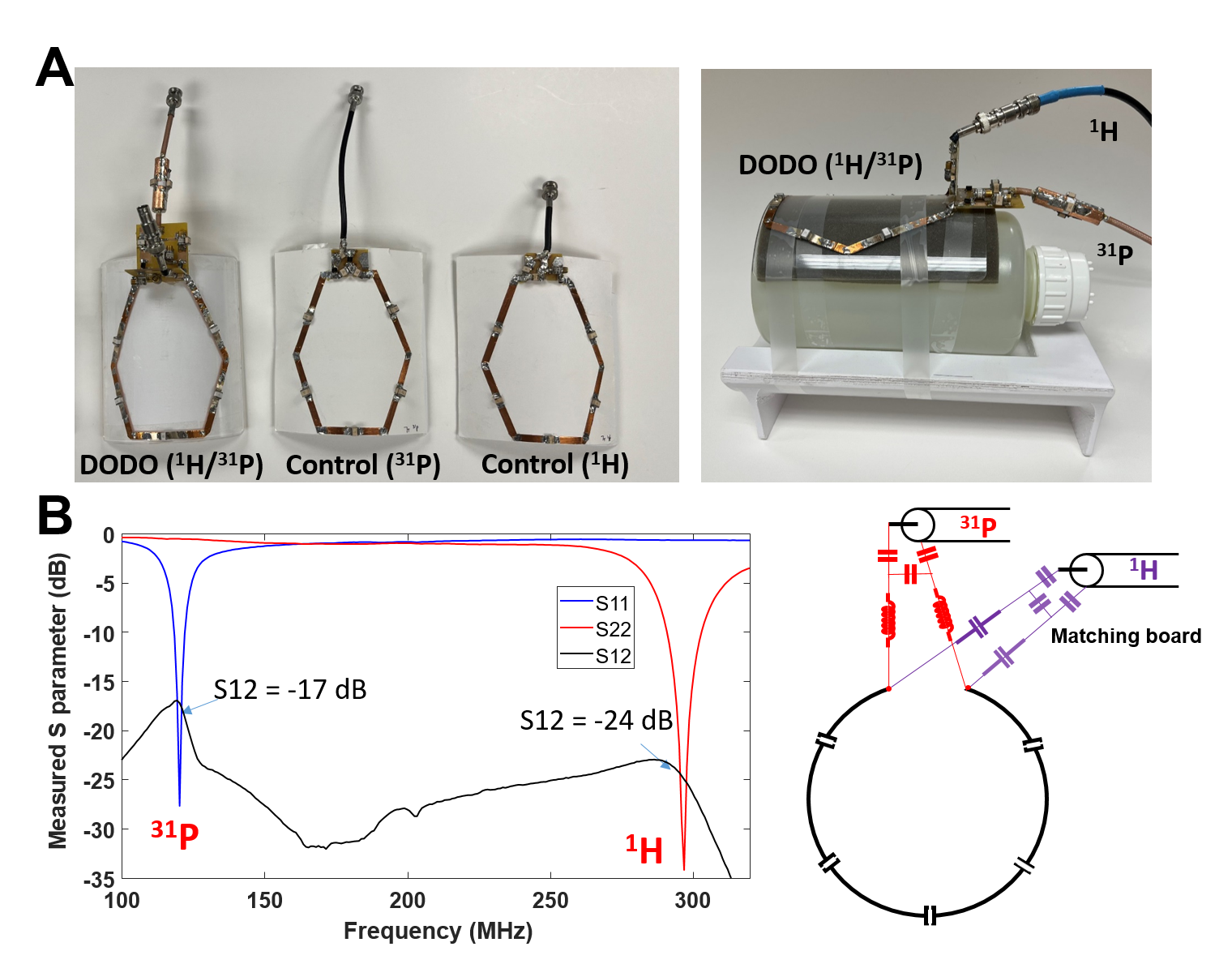
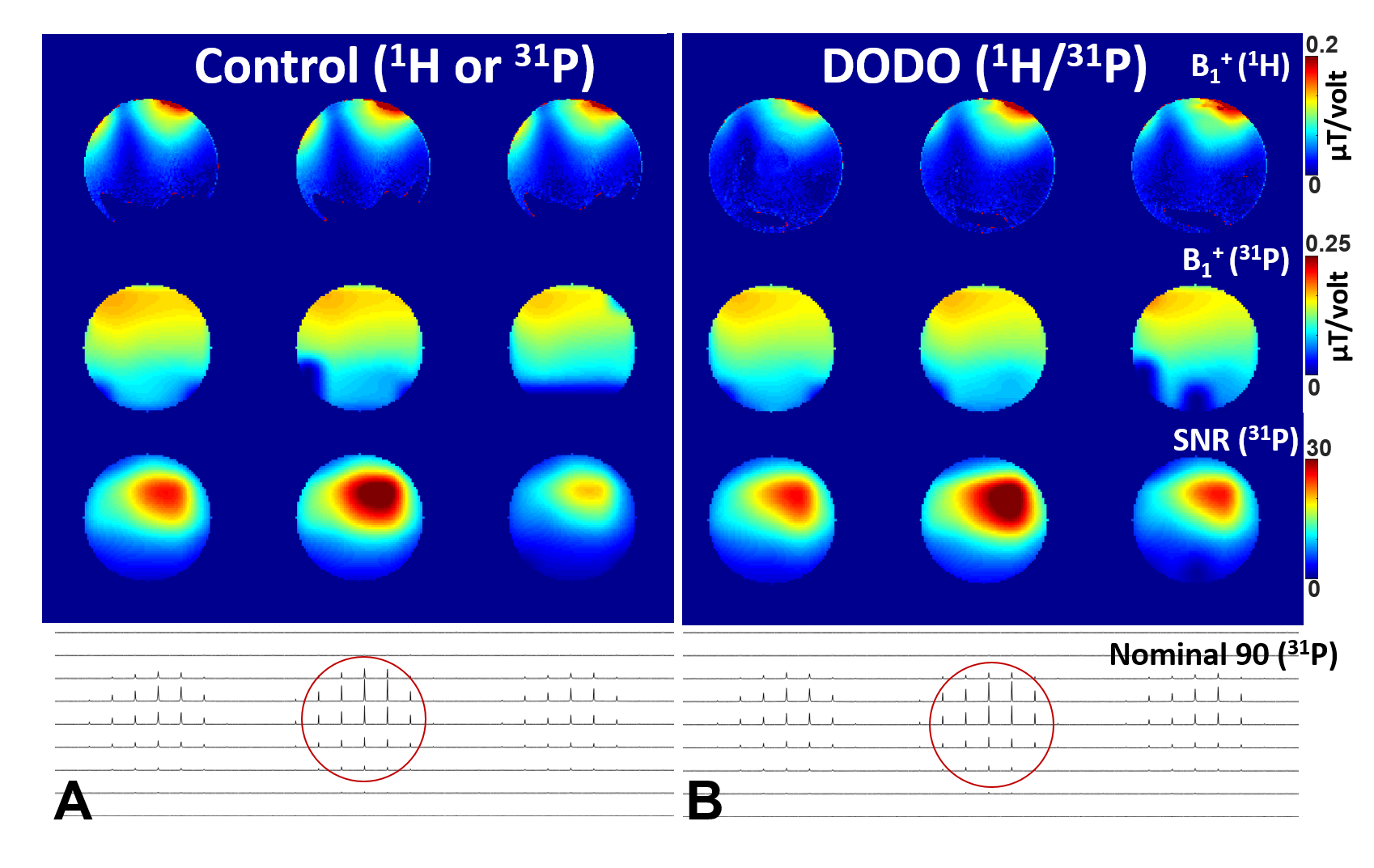
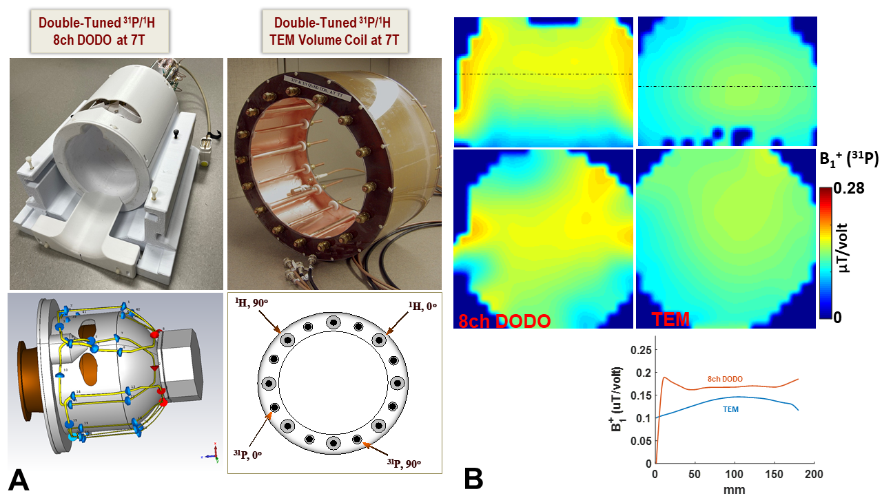
Figure 3. (A) The coil assembly and layout for the 8-ch 1H/31P DODO transceiver array coil and 1H/31P TEM volume coil for human brain imaging at 7T. (B) The B1+ maps (in coronal and axial orientation) measured using the 3D 31P MRSI, the 8-ch 1H/31P DODO transceiver array coil (left panel) and 1H/31P TEM volume coil (right panel) respectively, and a head-shaped Pi phantom.
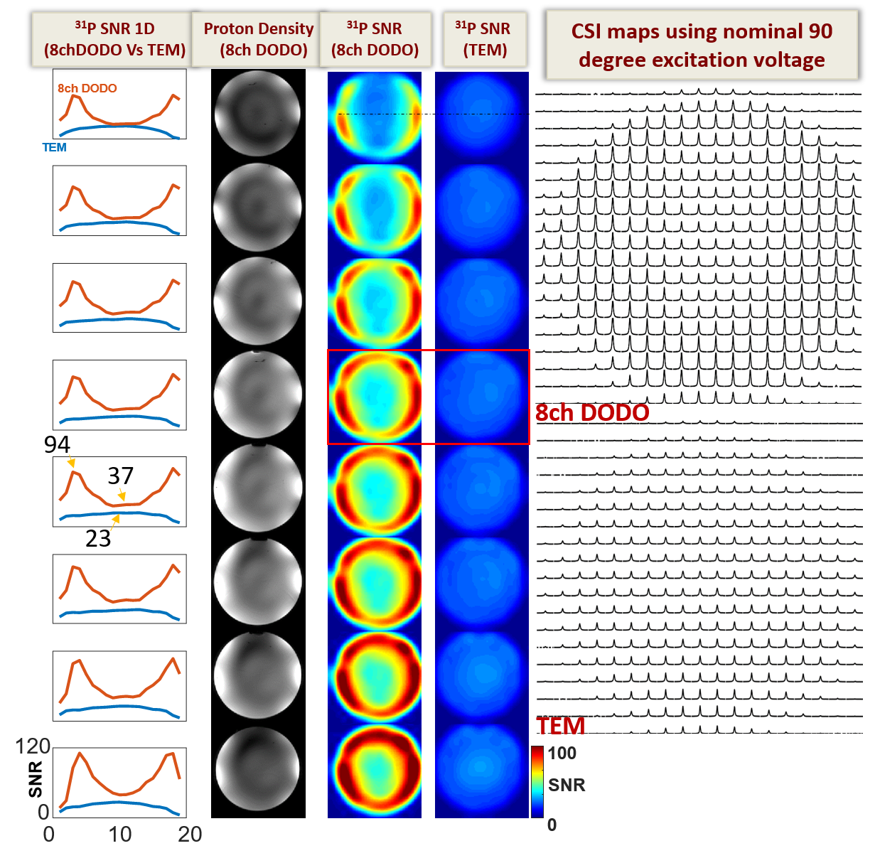
Figure 4. From left to right are the 1D SNR profiles through the center of the SNR maps in axial orientation for the 8-ch 1H/31P DODO transceiver array coil vs. 1H/31P TEM volume coil; proton density maps acquired using the DODO coil in the CP mode; 31P SNR of selected slices (in axial orientation) for the two coils; and representative 31P CSI for the center axial slices (red box) for the two coils at nominal 90-degree RF excitation pulse voltage.