1425
An Integrated/Radiofrequency Wireless Neonatal Coil Array with Global Navigation Satellite System (GNSS) for Clock Synchronization in MRI1Duke University Medical Physics Graduate Program, Durham, NC, United States, 2Duke-UNC Brain Imaging and Analysis Center, Durham, NC, United States, 3GE Healthcare, Aurora, OH, United States
Synopsis
Keywords: RF Arrays & Systems, RF Arrays & Systems
Motivation: Wireless clock synchronization must be implemented to achieve a wireless MRI receive coil architecture.
Goal(s): Our goal was to implement atomic clock timing via global navigation satellites signals (GNSS) to the receive coil in the scanner for high precision clock correction and synchronization.
Approach: We performed benchtop measurements to measure the precision of clock correction achievable with GNSS and precision time protocol (PTP), as well as modified an iRFW coil to receive these GNSS signal from within the scanner bore.
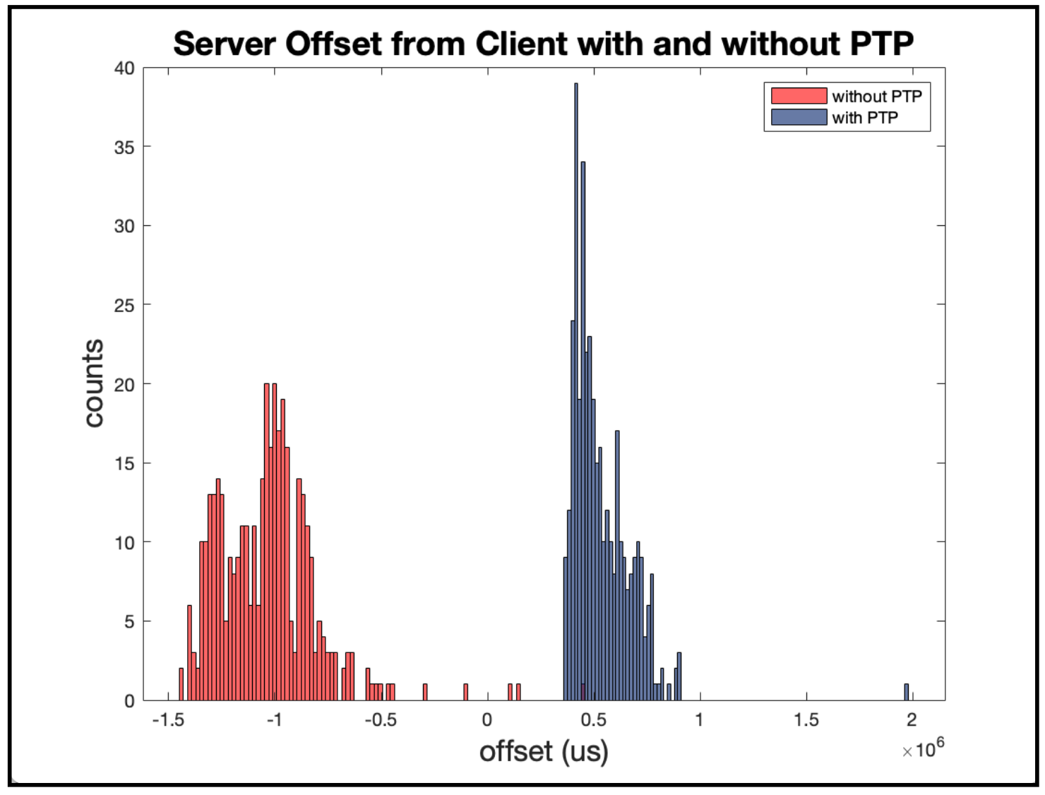
Results: Bench-top clock measurements showed nanosecond precision time-synchronization using PTP-GNSS, and the iRFW-GNSS successfully acquired atomic clock time signals within the scanner bore.
Impact: The iRFW-GNSS coil design can perform the wireless transfer MRI data and clock-syntonization regardless of scanner platform allowing for wide spread adoption of wireless MRI for new and existing scanners.
Introduction
MRI with adult-sized radio-frequency (RF) coil arrays to assess brain anomalies in neonatal patients1,2,3 can produce a non-uniform signal-to-noise ratio (SNR) in the brain. A neonatal-specific coil array can provide a more uniform SNR, but routing of its heavy cable assembly and accompanying wired physiological monitoring devices required to maintain image quality and safety significantly increases setup time and therefore anesthetization dose4. Ideally, a neonate-specific array would be form-fitting and able to wirelessly transmit data outside the bore for a uniform SNR in the brain and to decrease burn risk, setup time, and anesthetization by removing the cabling. Recently, integrated RF/wireless (iRFW) coil array simulations showed a uniform 3T MRI (e.g.,127.7 MHz) SNR in the neonatal head and a radiated signal sufficient for high-throughput WIFI-6E (e.g.,5.5 GHz) wireless MRI data transfer outside the bore by allowing these RF-currents to flow on the same coil conductor5. However, MR data acquired and wirelessly transmitted from an iRFW array to the console must also be wirelessly time-synchronized with the pulse sequence to generate an image6. Fortunately, our novel GNSS-enabled iRFW coil design (iRFW-GNSS) that can perform simultaneous MR and GNSS (e.g.,1.575 GHz) signal reception can be integrated into the array for precision-time-protocol (PTP) wireless time-synchronization within the bore. Here, iRFW-GNSS coil array simulations will be performed to determine the optimal design that provides a uniform SNR in the brain and radiated signals sufficient for both WIFI data transfer and GNSS signal reception in the bore. Proof-of-concept MRI experiments with a four-channel iRFW-GNSS array will be performed to evaluate its ability to image and acquire GNSS signals in the scanner. Finally, benchtop measurements will be performed to assess PTP time-synchronization for MRI.Methods
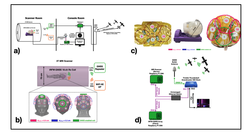
16-channel iRFW-GNSS soccer-ball coil array simulations using two coil element diameters (i.e., 5.2 and 6.9 cm) were performed on a 13 cm phantom inside a 70 cm bore at the MRI, WIFI-6E, and GNSS frequencies to determine the array’s SNR and its radiated power from the WIFI/GNSS gain patterns coupled to access point dipoles (AP) located 3m away (Fig. 1a). Specifically, six 5.2 and ten 6.9 cm diameter coil elements were arranged in a soccerball geometry to provide a uniform SNR Coefficient-of-Variance in the principal planes. Additionally, the WIFI/GNSS gain patterns for all coil elements were simulated and the two WIFI and one GNSS coil elements that provided the most radiated signal (i.e., S21) to the APs were selected as iRFW and iRFW-GNSS coil elements, respectively. Next, a four-channel soccerball coil array with three 6.9 and one 5.2 cm diameter coil elements was constructed on a semi-rigid enclosure near a 13 cm spherical phantom (Fig. 1b). Each coil element was tuned to the Larmor frequency and SNR maps were acquired using a FSPGR sequence on a GE Premier scanner. Next, the coil element with the best GNSS gain pattern from simulations was modified into an iRFW-GNSS coil by inserting high-impedance filters between the coil and the MRI and GNSS ports, which was tuned to the resonate at the GNSS frequency, for RF-isolation. New SNR maps were then acquired with the array and the iRFW-GNSS satellite carrier-to-noise ratio (C\N0) received in the bore from a patch antenna on the scanner room wall was measured to evaluate its wireless GNSS performance. The patch was connected to a separate GNSS antenna located in an adjacent room that supplied it and a Grandmaster Clock (GMC) with satellite C\N0 and timing data (Fig. 1c). Lastly, benchtop PTP measurements between the GMC and Raspberry Pi (Fig. 1d) were performed for a 12-hour period to determine satellite availability and evaluate PTP over half a satellite orbit.Results
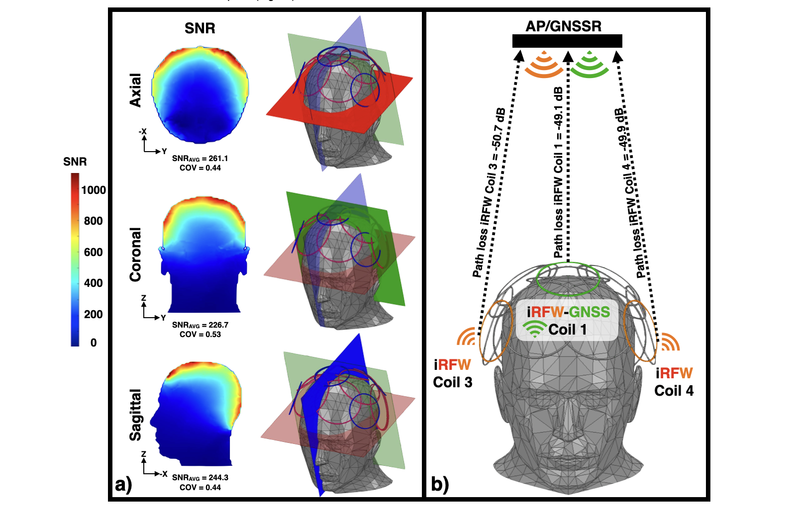
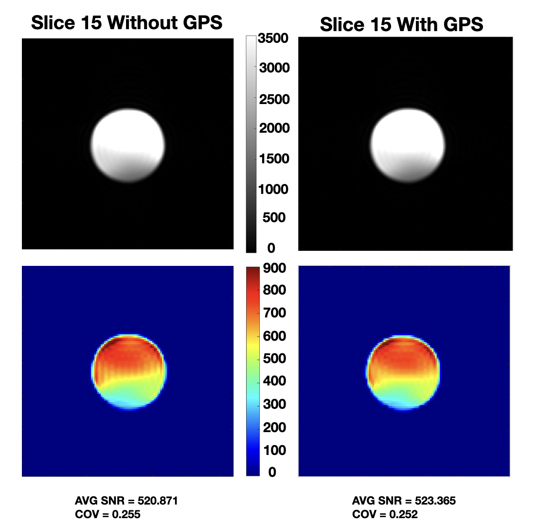
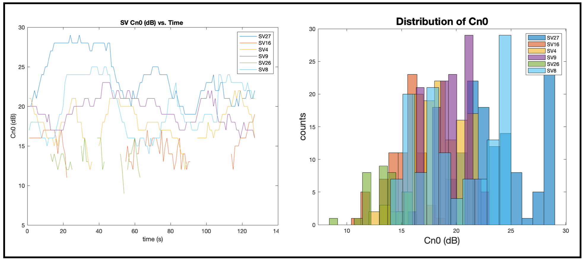
Simulations showed a uniform SNR COV in the axial, coronal, and sagittal planes of 0.46, 0.53, and 0.44, respectively and WIFI/GNSS gain patterns that provide a minimum S21 of -49.9 and -49.1 dB to the AP, which is sufficient for WIFI-6E and GNSS wireless reception (Fig. 2). The four-channel MRI and iRFW-GNSS coil arrays showed little difference in phantom SNR uniformity with COVs of 0.774 and 0.779, respectively, over the entire volume (Fig. 3) Further, the iRFW-GNSS coil element was able to continuously receive data from six satellites (Fig. 4) with an average C\NO of 18.2 dB for wireless time-synchronization. Lastly, benchtop PTP measurements showed nanosecond timing precisions between the GMC and RPi (Fig. 5).Discussion
This work shows that the iRFW-GNSS design can reliably receive satellite data in the bore, which can be used to time-synchronize the array to the scanner with nanosecond precision for image reconstruction without degrading image quality.Acknowledgements
No acknowledgement found.References
1) Rutherford MA, Pennock JM, Dubowitz LM. Cranial ultrasound and magnetic resonance imaging in hypoxic-ischaemic encephalopathy: a comparison with outcome. Dev Med Child Neurol. 1994 Sep;36(9):813-25. doi: 10.1111/j.1469-8749.1994.tb08191.x. PMID: 7926331.
2) Rademaker KJ, Uiterwaal CS, Beek FJ, van Haastert IC, Lieftink AF, Groenendaal F, Grobbee DE, de Vries LS. Neonatal cranial ultrasound versus MRI and neurodevelopmental outcome at school age in children born preterm. Arch Dis Child Fetal Neonatal Ed. 2005 Nov;90(6):F489-93. doi: 10.1136/adc.2005.073908. Epub 2005 Jun 14. PMID: 15956095; PMCID: PMC1721983.
3) Cawley P, Padormo F, Cromb D, Almablis J, Marenzana M, Teixeira, R, et al. Development of neonatal-specific sequences for portable ultralow field magnetic resonance brain imaging: a prospective, single-centre, cohort study. https://doi.org/10.1016/j.eclinm.2023.102253
4) Hughes EJ, Winchman T, Padormo F, Teixeira R, Wurie J, Sharma M, Fox M, Hutter J, Cordero-Grande L, Price AN, Allsop J, Bueno-Conde J, Tusor N, Arichi T, Edwards AD, Rutherford MA, Counsell SJ, Hajnal JV. A dedicated neonatal brain imaging system. Magn Reson Med. 2017 Aug;78(2):794-804. doi: 10.1002/mrm.26462. Epub 2016 Sep 19. PMID: 27643791; PMCID: PMC5516134.
5) Dickinson OJ, Taracila V, Oversonn DK, Lynch S, Reed B, Robb F, Song A, Truong TK, Darnell D. Simulations of an integrated RF/wirelss neonatal head coil array for multiple-innput multiple output (MIMO) wireless MRI data transmission. Proc. Intl. Soc. Mag. Reson. Med. 31 (2022)
6) Scott G. Pilot Tone Software Synchronization for Wireless MRI Receivers. Greig Scott1, Proc. Intl. Soc. Mag. Reson. Med. 26 (2018)
Figures