1424
Inexpensive Easy-To-Use Commercial MRI Readable Thermometer1NIST, Boulder, CO, United States, 2University of Colorado, Boulder, CO, United States, 3Calimetrix, Madison, WI, United States, 4Cleveland Clinic, Cleveland, OH, United States
Synopsis
Keywords: Phantoms, Thermometry
Motivation: To demonstrate the utility of inexpensive easy-to-use commercial MRI-readable thermometers for monitoring phantom temperatures during imaging and enable temperature corrections in an MSK relaxometry phantom developed for inter-site relaxation time (T1, T2, T1ρ) comparisons.
Goal(s): Provide convenient thermometry for temperature corrections for phantom based intersite comparisons of relaxation time and diffusion measurements.
Approach: Commercial incubator thermometers with a temperature range of 15°C to 50°C were characterized for phantom thermometry.
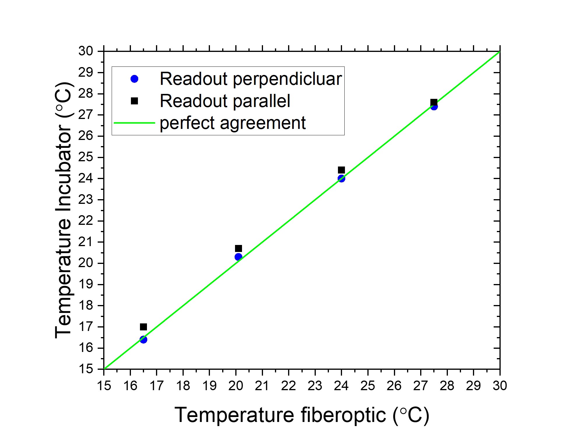
Results: Commercial incubator thermometers demonstrated to be MRI readable with an accuracy of ±0.2°C using standard imaging protocols, as long as the thermometer axis is perpendicular to the readout direction.
Impact: Easy to use MRI-readable thermometry is available to enable temperature corrections to minimize confounding factors in inter-site comparisons.
Introduction
Determining the accuracy of MRI-based measurements requires the use of calibrated phantoms. Many parameters, including relaxation times and diffusion-based biomarkers, are temperature dependent. T1, T2, and water diffusivity can vary by 1%/°C to 3%/°C(1, 2). Accurate measurements require accurate temperature measurement and correction. Measurement of temperature using externally introduced thermometers can lead to error since these measurements are usually made before or after scanning and do not account for temperature variation during scan time. Further, these temperature measurements are hard to include in the imaging data set and are often lost. MRI-readable thermometers are preferred since the temperature is stored within the image data. Fiber optic, liquid crystal thermometers(3), chemical shift thermometers(4) are available for incorporation within phantoms, but these are expensive and complex. Here we look at the utility and accuracy of inexpensive commercial incubator thermometers for MRI-readable thermometry. The immediate application was for incorporation into a musculoskeletal (MSK) relaxometry phantom (Calimetrix, Madison, WI) to allow temperature corrected inter-site comparisons.Methods
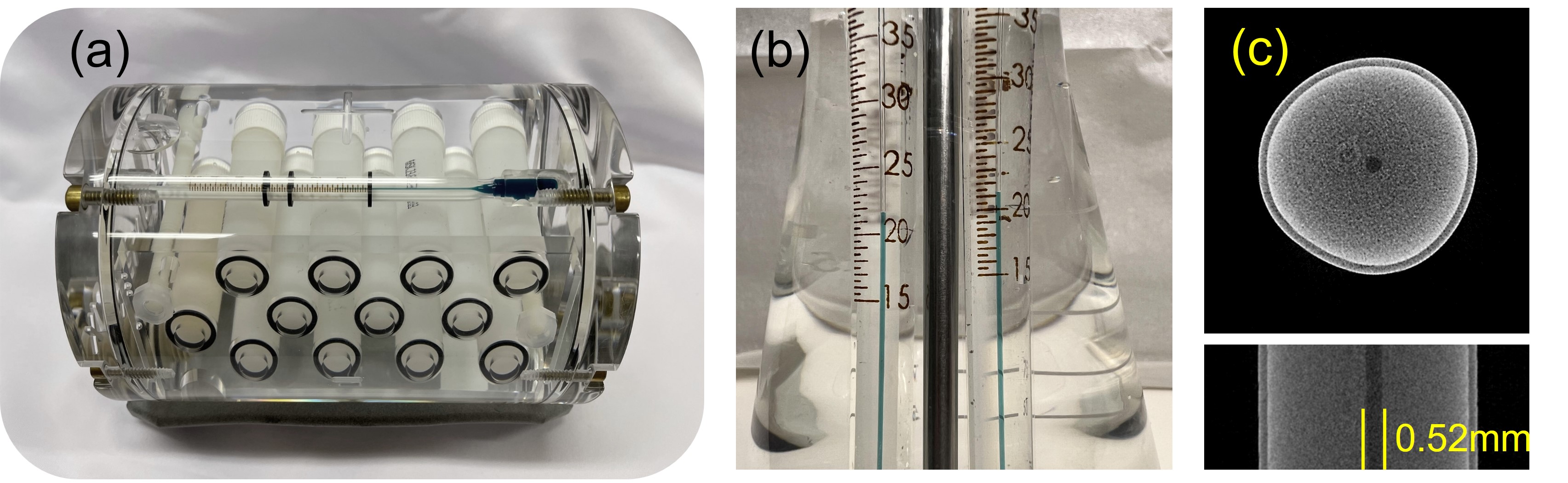
Commercial incubator thermometers with a temperature range of 15°C to 50°C were purchased with cost <$90.00 each. The thermometers were 135mm long, 7mm diameters (Fig.1), nontoxic and robust. The thermometer fill was Enviro-Safe® Green Liquid, which is a citrus oil formulation with a green dye. The stated accuracy, ±0.5°C, was confirmed by comparison to a NIST-calibrated thermometer (Fig.1b). The thermometer column diameter measured 0.52mm using microCT (Fig.1c). The fill solution is clearly visible on most MRI scans, with the thermometer column visible on higher resolutions scans with voxel sizes ≤1mm (Fig.2). Rectangular O-rings (6.5mm inner-diameter, 1.25mm thickness) were added to the outside of the thermometer to delineate the 15°C and 30°C temperature marks (Fig.1a) and allow MRI readability. The O-rings were positioned under a magnifier using an insertion tube to ensure the O-rings were straight and positioned accurately. The spacing between the O-rings was 23.0 mm, giving a spatial sensitivity of 0.64°C/mm. The thermometer was inserted into various phantoms including a temperature-controlled phantom with a NIST-traceable fiber optic thermometer. The imaging was performed on a 3T Siemens Prisma Fit using a 15-channel knee coil and a 3T preclinical scanner, using a bird cage coil and a temperature control system. The temperature was determined using T(°C)=15+15(l ⁄ L) , where L is the measured length between the top of the lower O-ring and the bottom of the top O-ring, and l is the distance height of the thermometer column from the top of the bottom O-ring (Fig 2b). Since the measurement is relative, the distances can be measured in any units by any method, the spatial calibration of the image is not relevant. The thermometer fill had a T1=1400ms at 21°C and a visible chemical shift along the readout direction of 0.5 to 1mm (Fig.3c).Results and Discussion
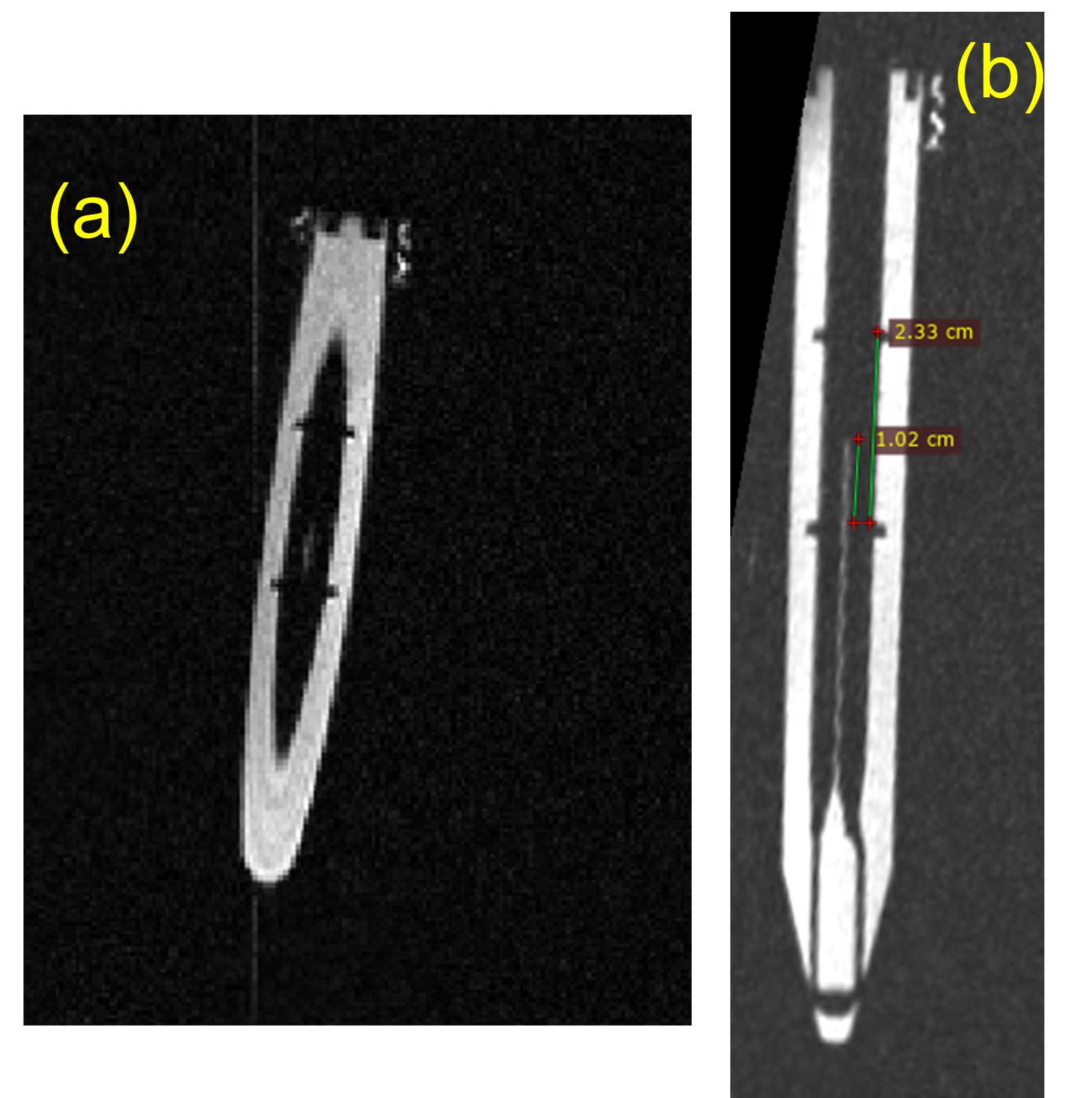
Thermometer images with phantom temperature 16.5±0.2°C are shown in Fig.3 with the thermometer axis along the phase encode(PE) and readout(RO) directions. The MRI determination of the incubator thermometer temperature was 16.4°C and 17.0°C with the thermometer axis along the PE and RO directions, respectively. When the RO direction is aligned with the thermometer axis, the chemical shift is also along the thermometer axis and leads to an error of approximately 0.5°C. When the thermometer is aligned along the PE direction, the chemical shift is perpendicular to the thermometer column and does not contribute to the measurement error, which was 0.1°C. The incubator temperature vs the NIST traceable thermometer temperature is shown in Fig.4. The standard deviation of the error is 0.14°C and 0.44°C for scans with RO perpendicular and parallel to the thermometer axis, respectively.When the thermometer is not well aligned with a principal scanner axis, the thermometer can be read with a standard volume scan (Fig.5). Figure5(a) shows a coronal thermometer image, (b) shows a software generated oblique slice aligned with the thermometer axis, clearly showing the column and sufficient for temperature determination (albeit with lower accuracy due to possible chemical shift artifact).To obtain a measurement accuracy of <0.15°C, some thermometer prescreening and imaging with the RO perpendicular to the thermometer axis are required. The chemical shift artifact can be measured using images such as Fig.3(c) and calibrated out if the optimal orientation cannot be achieved. For an accuracy of <1°C, simple inclusion and readout of the thermometer is all that is needed.Conclusions
Inclusion of inexpensive commercial incubator thermometers into phantoms allows for MRI readable temperature without the use of special pulse sequences. It can be read with navigator, volume, and spin-echo scans and can be used for temperature corrections for inter-site quantitative imaging comparisons.Acknowledgements
We Acknowledge funding support from NIH/NIAMS R01 AR077452 and the Arthritis Foundation.References
1. Stupic KF, Ainslie M, Boss MA, Charles C, Dienstfrey AM, Evelhoch JL, Finn P, Gimbutas Z, Gunter JL, Hill DLG, Jack CR, Jackson EF, Karaulanov T, Keenan KE, Liu G, Martin MN, Prasad PV, Rentz NS, Yuan C, Russek SE. A standard system phantom for magnetic resonance imaging. Magnetic Resonance in Medicine. 2021;n/a(n/a). doi: https://doi.org/10.1002/mrm.28779.
2. Holz M, Heil SR, Sacco A. Temperature-dependent self-diffusion coefficients of water and six selected molecular liquids for calibration in accurate 1H NMR PFG measurements. Physical Chemistry Chemical Physics. 2000;2(20):4740-2. doi: https://doi.org/10.1039/b005319h.
3. Keenan KE, Stupic KF, Russek SE, Mirowski E. MRI-visible liquid crystal thermometer. Magnetic Resonance in Medicine . 2020;84(3):1552-63. doi: https://10.1002/mrm.28224.
4. Swanson S, Malyarenko D, Chenevert T. MR Thermometry in Phantoms Using Bulk Magnetic Susceptibility. Proc Intl Soc Mag Reson Med 2018;26:5648.
Figures
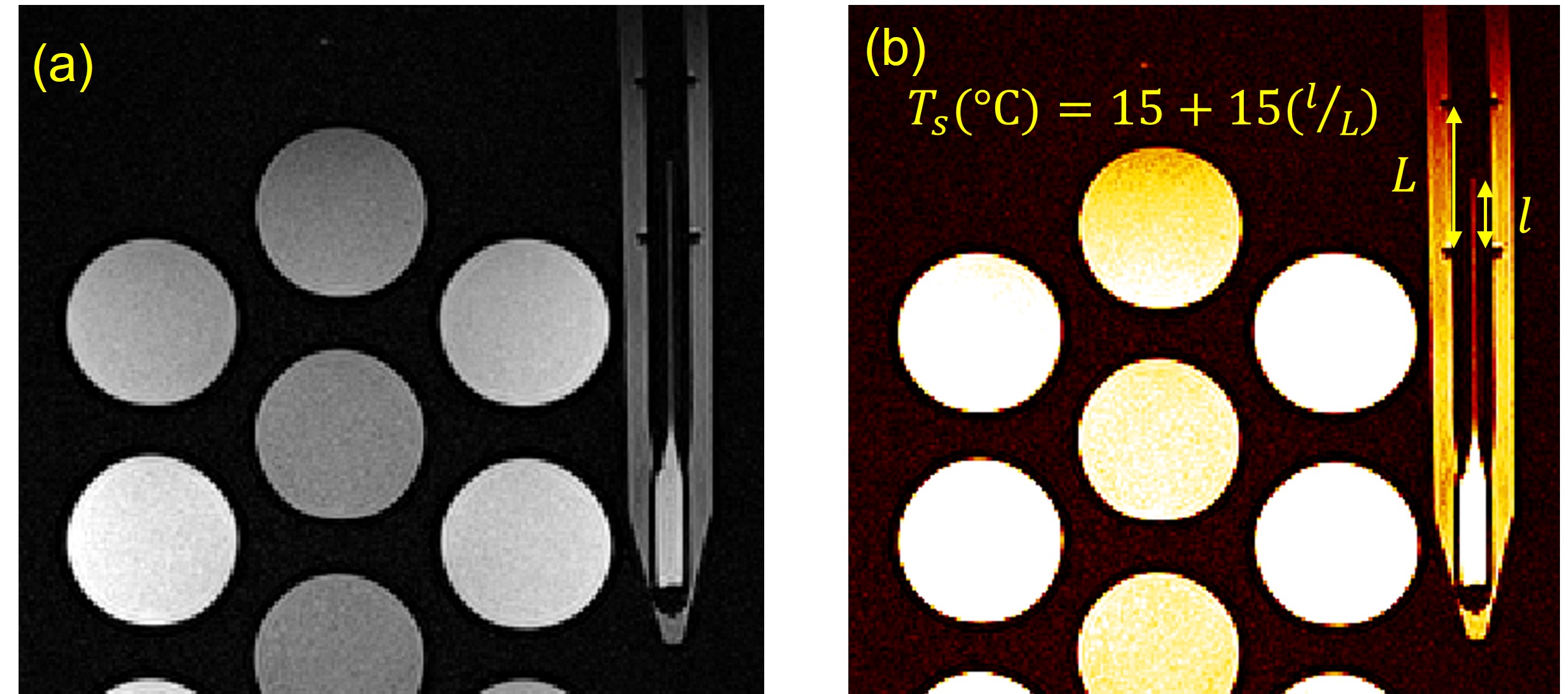
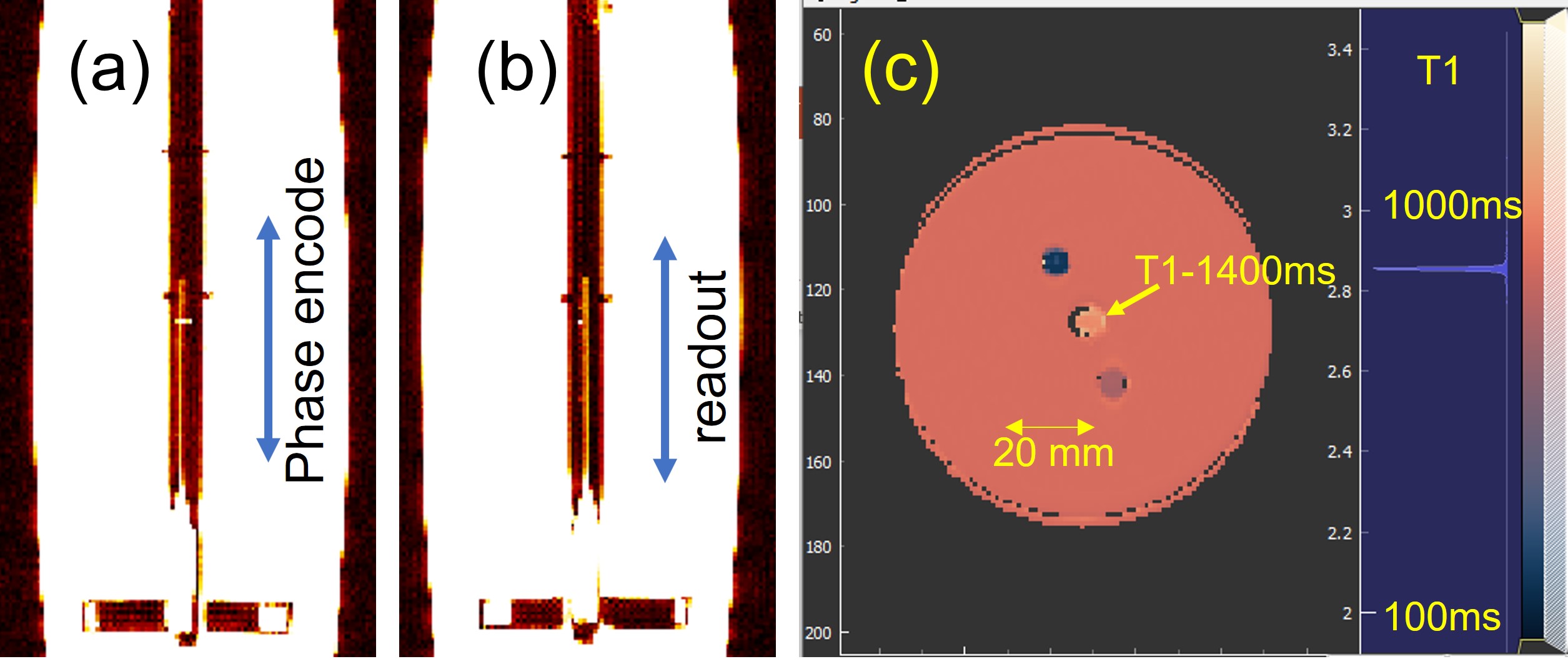
(a), (b) Coronal images (0.63mm in-plane, 2mm slice thickness) with thermometer axis along phase encode and readout directions, respectively. The chemical shift is visible in (a) with the thermometer column off the central axis. The phantom temperature is 16.5±0.2°C, while the incubator thermometer reads 16.4°C. In (b), the chemical shift is along the thermometer column and not obviously visible. The incubator thermometer measures 17.0°C. (c) Axial T1 map (0.63mm in-plane, 6mm slice thickness) showing incubator thermometer bulb in center with a T1=1400ms with a chemical shift.