1422
Continuous In-line Monitoring of Perfusion Culture Viability with Contact-Free Magnetic Resonance Relaxometry1Electrical Engineering and Computer Science, Massachusetts Institute of Technology, Cambridge, MA, United States, 2CAMP IRG, SMART Centre, CREATE, Singapore, 3Biological Engineering, Massachusetts Institute of Technology, Cambridge, MA, United States
Synopsis
Keywords: New Devices, Relaxometry
Motivation: Bioreactor cell density measurements currently require removing samples from the culture. This limits how many samples can be taken in a day and precludes real-time culture monitoring.
Goal(s): We sought to take nondestructive cell density measurements every ten minutes without removing cells from the bioreactor.
Approach: We built a system that takes T2 relaxometry measurements through the bioreactor’s silicone tubing every ten minutes and compared these to measurements taken by a commercial cytometry system.
Results: The T2 relaxation data closely tracks the true cell density. Changes made to the culture are detected much more quickly by the relaxometer.
Impact: Magnetic resonance relaxometry can be used to track bioreactor culture growth in real time, and makes it possible to identify and correct problems with the culture before the culture fails.
Introduction
Frequent, low-latency measurements of bioreactor culture growth are critical for achieving maximum culture efficiency and viability over a multi-week culture. Traditional cell density and viability measurements are disruptive to the culture because they require samples to be removed from the bioreactor and eventually discarded. This makes it difficult to take measurements frequently enough for feedback control and rapid detection of culture problems. In this work, we show that T2 relaxation measurements taken through the walls of the bioreactor tubing can be used to monitor the cell density in real time.Methods
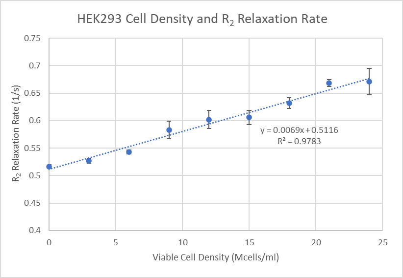
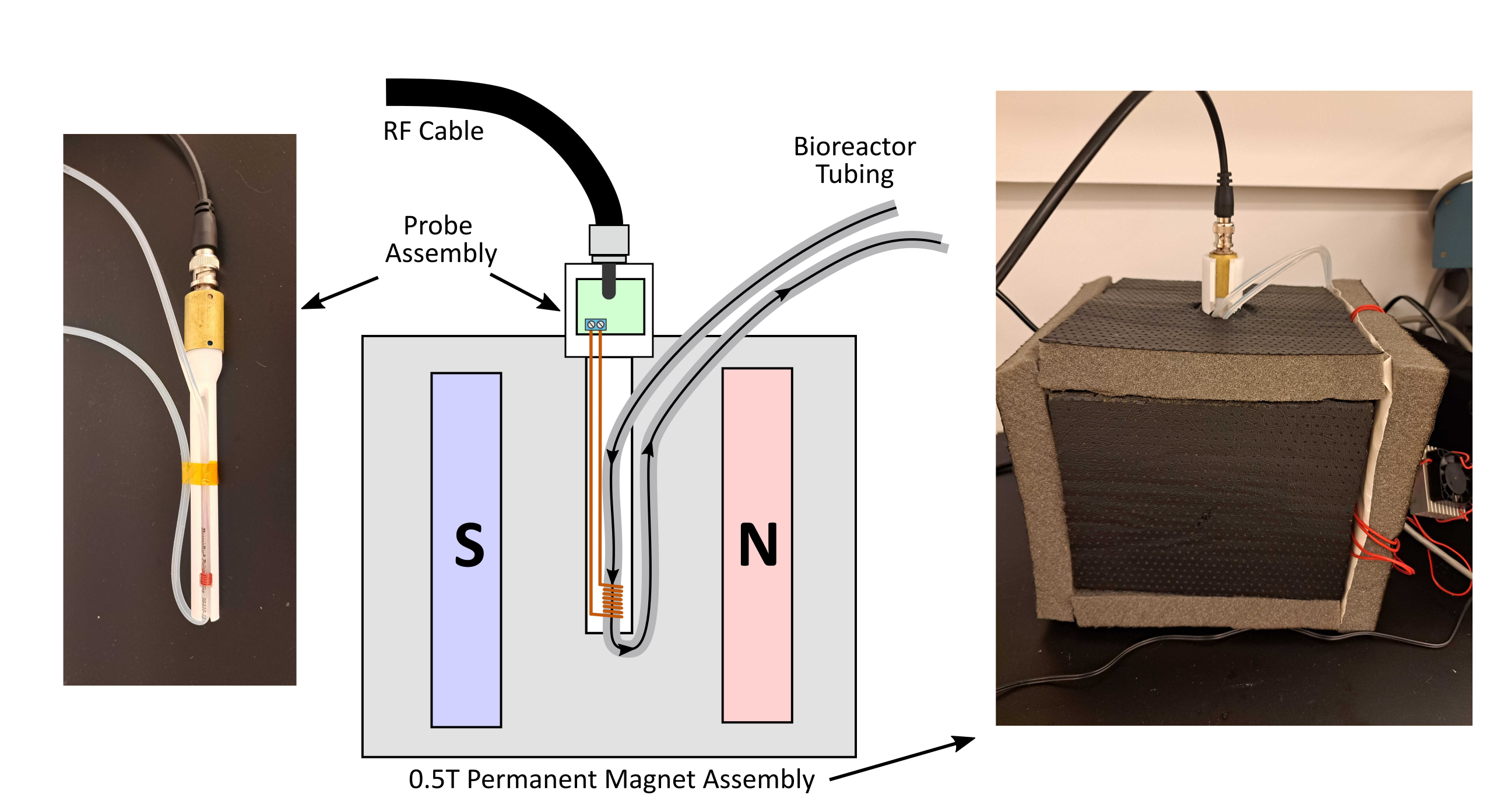
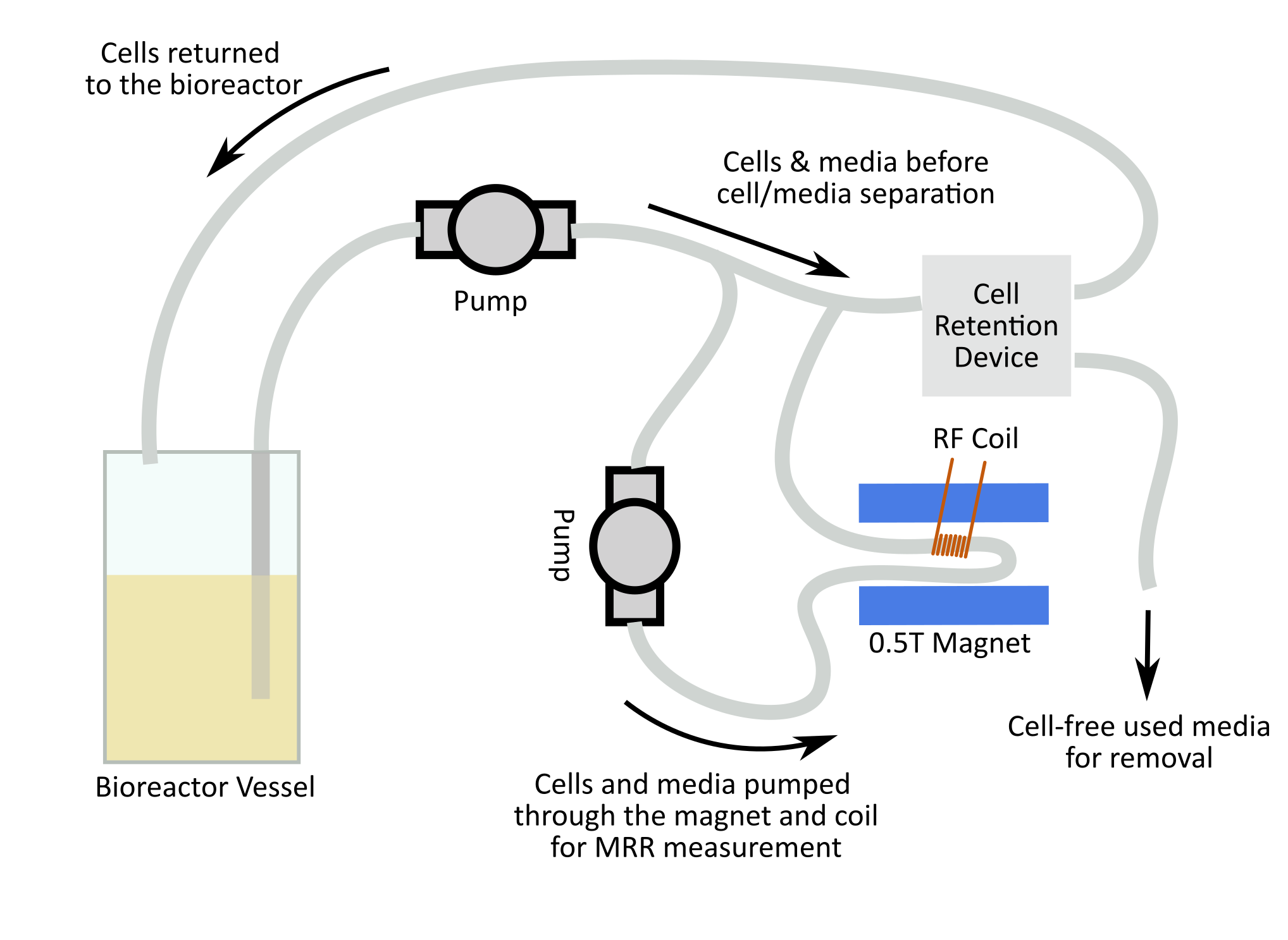
First, we established the relationship between the T2 relaxation time and cell viable density by measuring samples with controlled cell densities in a commercial magnetic resonance relaxometer (MRR). To monitor the cell density in the culture in real time, we wound a solenoidal coil around the silicone tubing of a mini bioreactor used for continuous culture of HEK293 cells. We connected this coil to a tuning circuit and inserted it into a half-Tesla permanent magnet assembly that stabilizes the magnet temperature and eliminates the need for frequency calibration during the culture. Finally, we sealed and autoclaved the bioreactor system before starting the culture, and at no point during the setup of the relaxometry system did we open or disconnect the tubing.We used a low-cost relaxometer built around an off-the-shelf software-defined radio to take 6 measurements of the T2 relaxation time per hour using a CPMG experiment with 500µs echo spacing and 4000 echoes. The first 500 echoes in each experiment were discarded as they are contaminated with signal from the silicone tubing. We fit an exponential to the echo amplitudes with a non-linear least squares approach before lowpass filtering the raw T2 measurements to improve the accuracy of the cell density estimate.
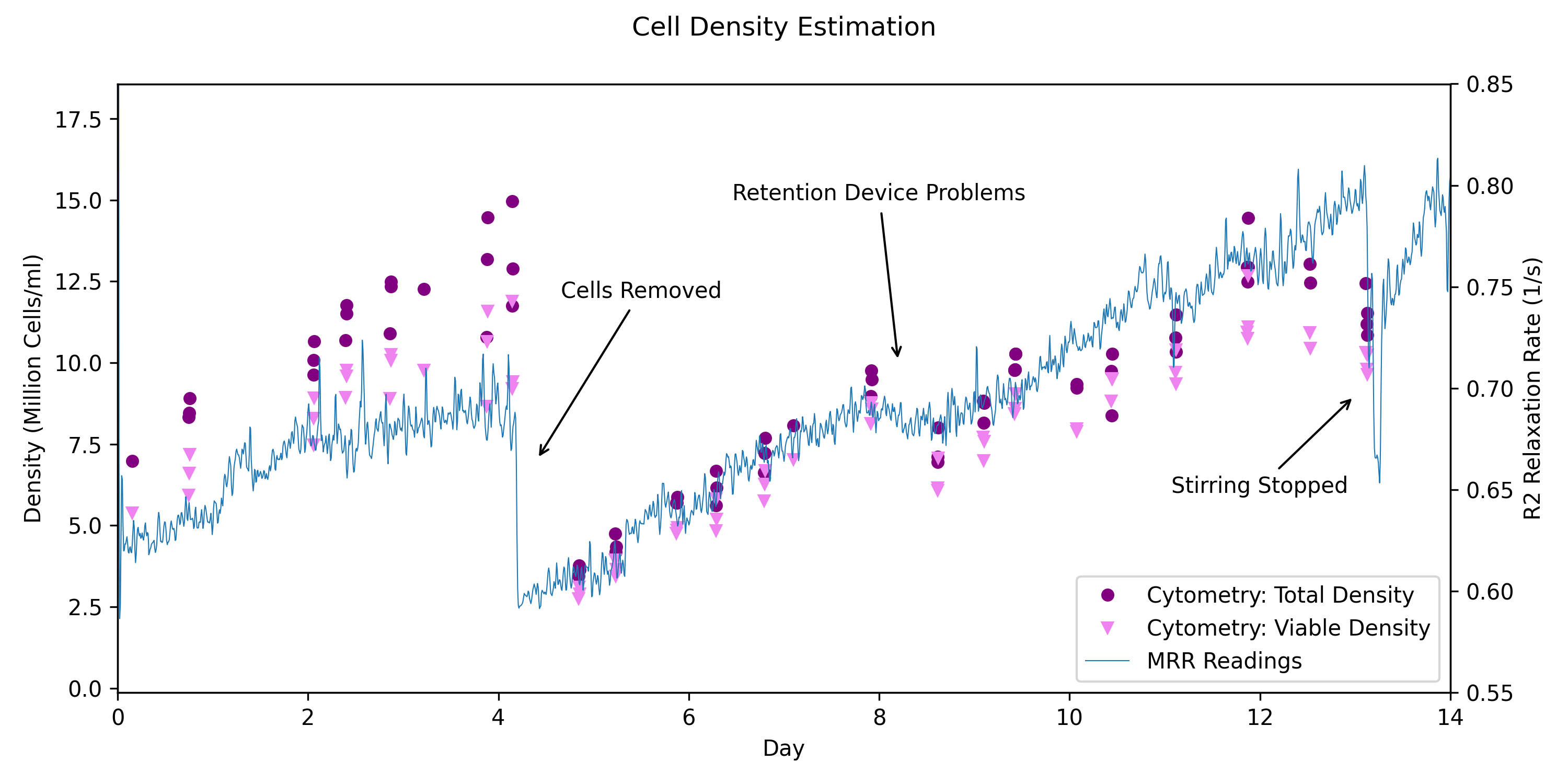
After 4 days, we removed most of the cells from the bioreactor, and on day 8 problems with the cell retention device caused a drop in the cell density. Finally, the stirrer in the bioreactor was turned off for an hour on day 13 before the culture is terminated. The cell density predicted by the MRR and the response of the MRR-based estimates to these disturbances were compared to measurements of the total cell density and viability taken daily with a commercial cytometry-based cell analyzer.
In addition to the cytometry measurements, we regularly removed and centrifuged samples from the culture to measure the T2 relaxation time of the cell pellet. This allows us to determine the extent to which variability in cell morphology influences the relaxometry measurements of the cell/media suspension.
Results
A chart showing the relaxometry and cytometry data is given in Figure 4. The relaxation time measurements have a temporal resolution of 1 sample every 10 minutes over 2 weeks, compared to one or two samples per day from the cytometry measurements. The raw relaxometry estimates over a period of 1 day have a standard deviation of 1.2Mcells/ml, compared to 0.8Mcells/ml for the cytometry measurements. Before filtering, the drop in cell density following the removal of the cells is apparent after 10 minutes. With causal lowpass filtering to reduce the standard deviation of cell density measurements over one day to 0.7Mcells/ml, this drop is detectable after 40 minutes, 36 times faster than daily cytometry measurements. The T2 relaxation times of pelleted cell samples did not change significantly during the culture.Discussion
There is good agreement between the cytometry data and the relaxation time measurements, which suggests that T2 relaxometry is an effective technique for estimating the cell density of a HEK cell culture. The relaxometer is able to detect the drops in cell density caused by the removal of cells from the culture and problems with the bioreactor much more quickly than the cytometry system. Consistent differences between the cytometry and MRR measurements at the beginning and end of the culture warrant further study and may be caused by differences in how the MRR and cytometer are connected to the bioreactor, or the addition of an anti-clumping agent after the removal of cells on day 4.Conclusion
Bioreactors are used to produce a wide array of critically important therapeutic products, from insulin to the viral vectors produced in the culture described here. Frequent, nondestructive MR-based measurements of a cell culture make it possible to detect and address drops in the cell density before the culture fails, and since the technique described here does not require direct access to the cells, it eliminates the complexity and risk of contamination associated with additional wetted components.Acknowledgements
No acknowledgement found.References
No reference found.Figures