1416
Proof of concept for the integration of a low-Tc SQUID in MRI detection at 1 mT1Chipiron, Paris, France
Synopsis
Keywords: Low-Field MRI, Low-Field MRI
Motivation: Ultra-low MRI systems provide a reduced SNR.
Goal(s): Our goal is to improve the SNR by incorporating SQUID-based RF detection into the ULF MRI system.
Approach: We build a custom-made SQUID MRI system and acquire the first 2D image of a phantom as proof of concept.
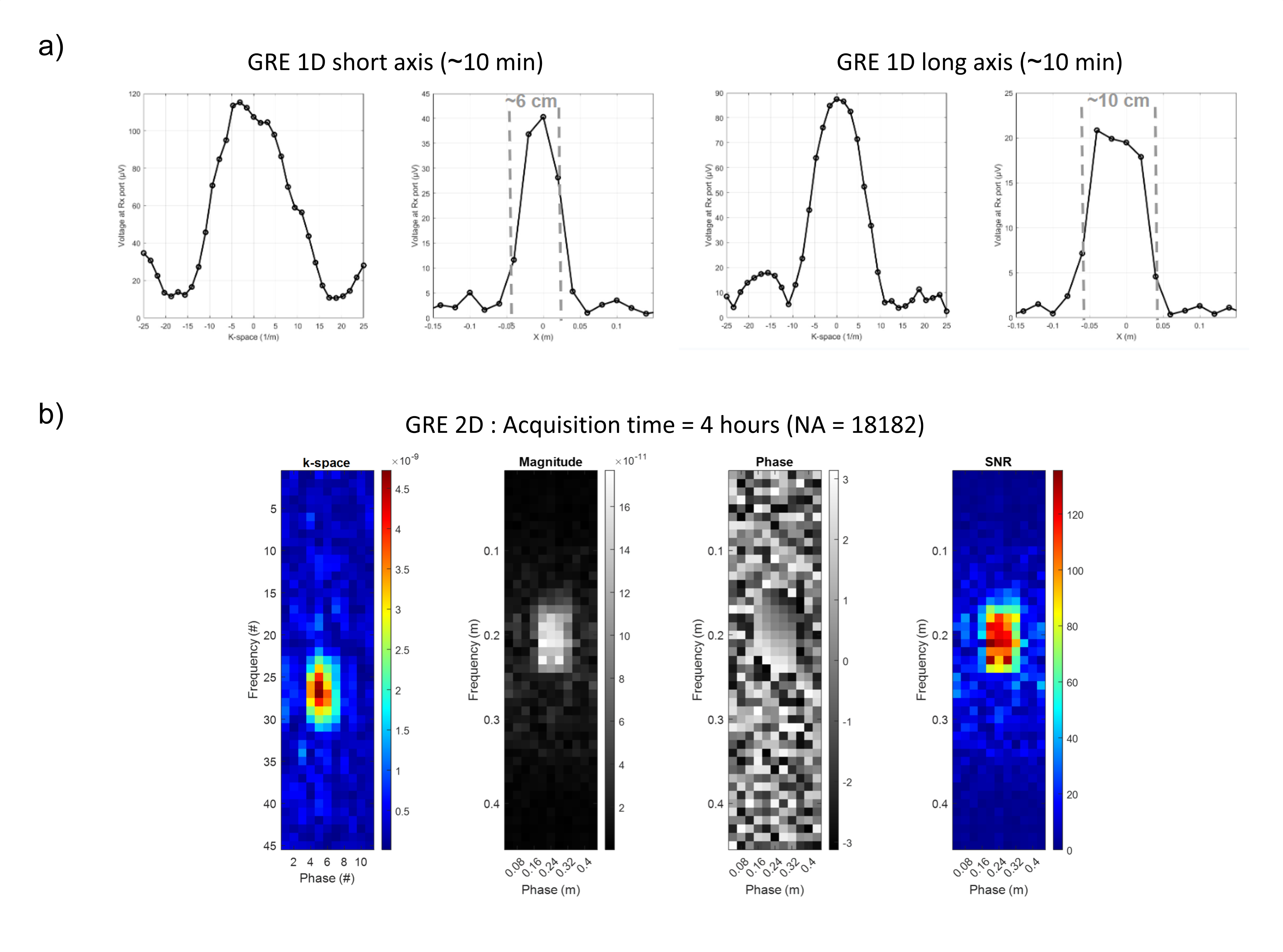
Results: we successfully acquired a 2D image of a phantom in 4 hours with a resolution of 15×12.5 mm²
Impact: Striving for MRI accessibility, we develop a portable ultra-low field scanner using highly sensitive SQUID detection. Our first 2D image marks the initial stride toward achieving clinically competitive image quality.
Introduction
Over the past decade, a new approach has emerged in the field of MRI, which involves the use of ultra-low field (ULF<10mT) scanners, diverging from the conventional trend of utilizing higher magnetic fields as the cutting-edge technology. Besides the cost-effectiveness and portability of ULF MRI, operating at these low magnetic fields has shown improvements in T1 contrast in some tissues1,2, leading to more efficient diagnostics of various medical conditions, including cancer. The main problem of ULF MRI lies in a detected signal typically orders of magnitude lower compared to clinical-field MRI, which impacts the signal-to-noise ratio (SNR) in the images. Several methods3,4 are being explored to address this limitation and improve the sensitivity of ULF MRI. The inherent challenge of poor sensitivity in ULF MRI can be mitigated by choosing a superconducting quantum interference device (SQUID) for signal detection. Various groups2,5 used SQUIDs and multi-channel surface coils at 4.2K employing liquid Helium (LHe) cooled cryostats. The use of cryogenic liquid and room-temperature samples incompatibility with 4.2K SQUID and coils are detrimental to achieving portable and low-cost MRI systems. Here, we propose our first implementation of a cryogen-free SQUID detector inductively coupled to a customized volume gradiometer-based RF coil operating at room temperature6,7. We further present our first 2D image acquired using a fully custom-made SQUID-MRI scanner at 1mT.Methods
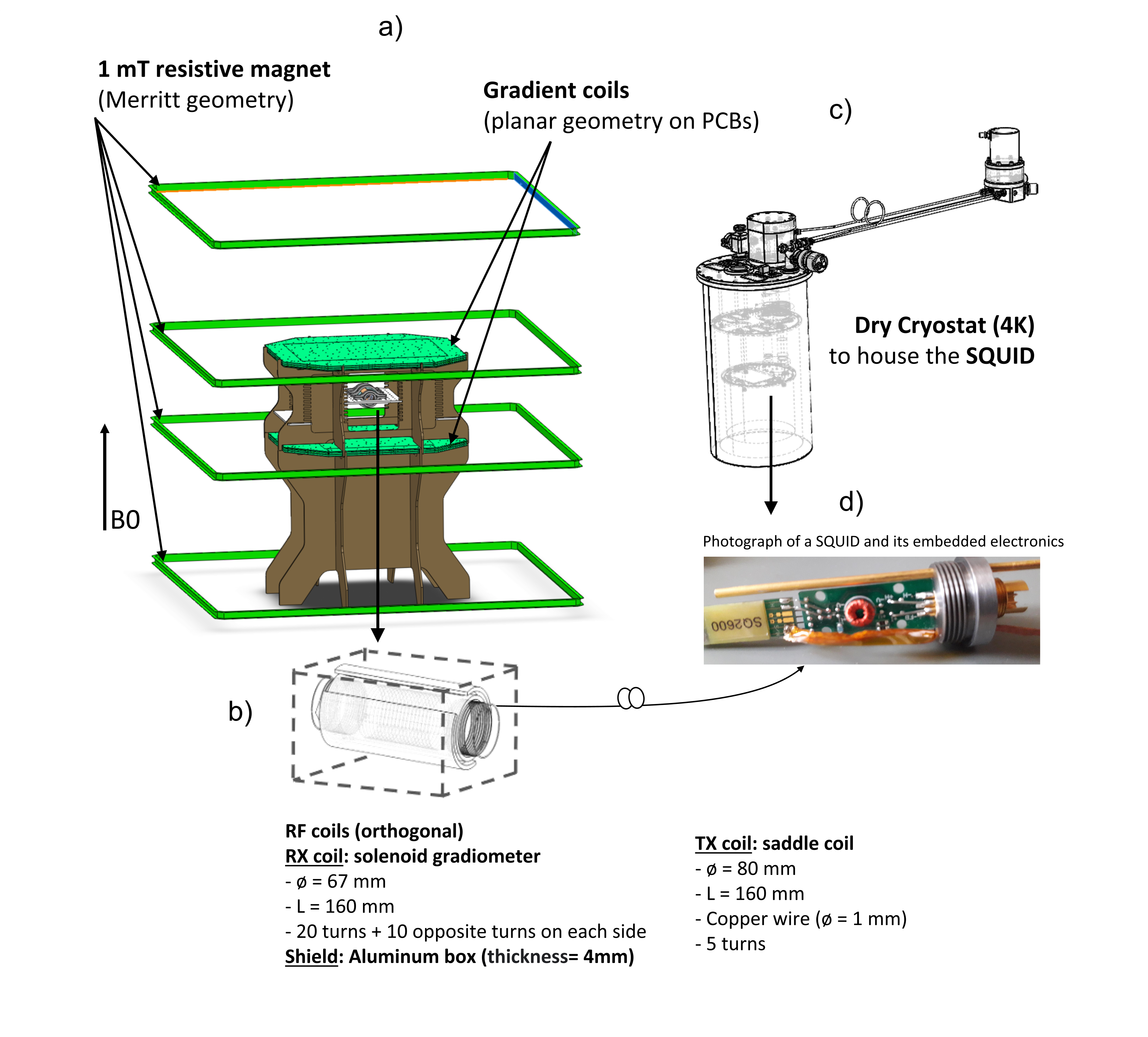
SQUID sensorA micrometer-sized low critical temperature Niobium-based SQUID is coupled to a larger flux transformer in a current-sensing configuration. This transformer comprises a 300K RF receive coil and a 4.2K superconducting input coil positioned in close proximity to the SQUID in a washer design. Fig.1 illustrates the cryogen-free cryostat, which relies on a pulse tube cryocooler and is used to house the SQUID at a temperature of 4.2K. The magnetic flux seen by the SQUID is directly proportional to the one passing through a volume gradiometer used as RF pickup coil in inductive conditions.
MRI hardware
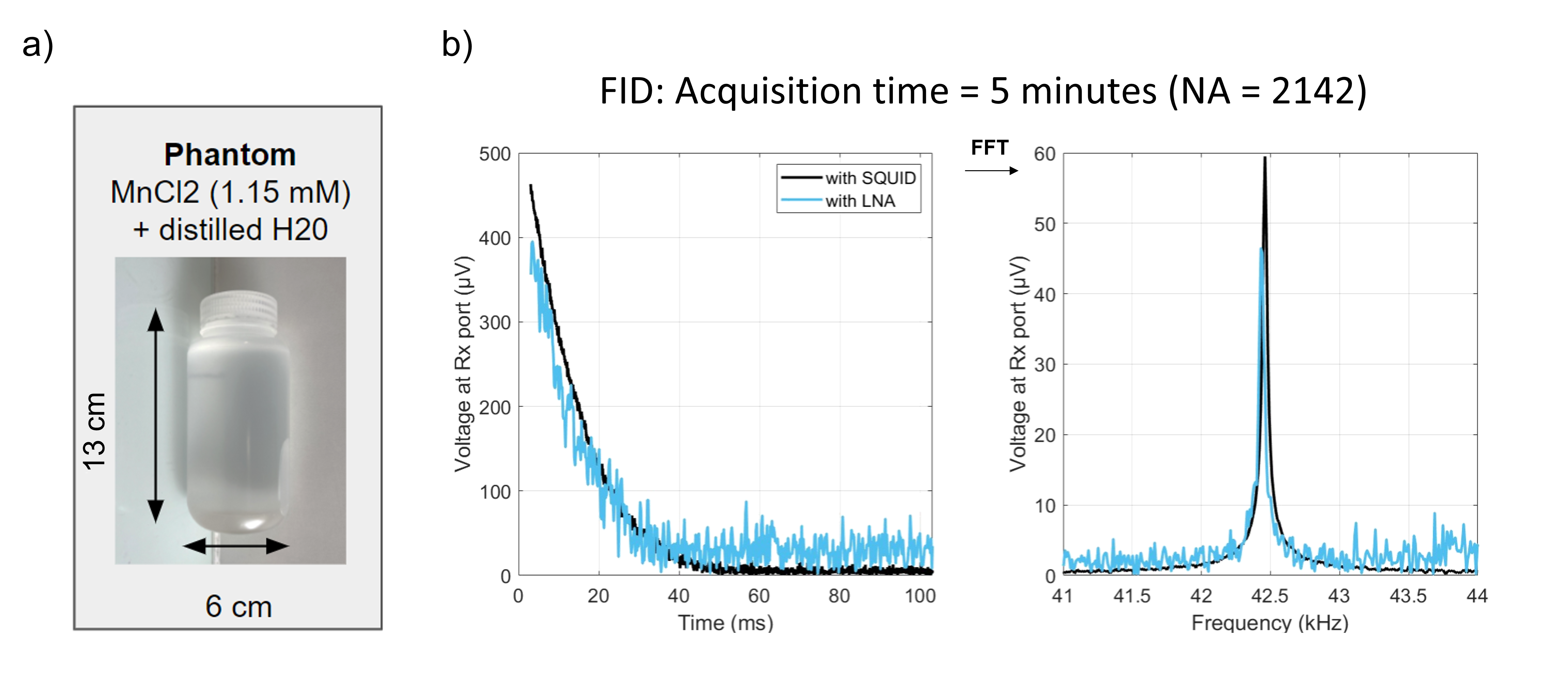
A Merrit coil electromagnet fed by a current source generates a polarization field (B0) of 1mT (Fig.1a). The RF field transmission is performed using an 80mm-diameter saddle coil with 5 turns, while the RF reception is achieved using a solenoid-based volume gradiometer that is linked to the SQUID (Fig.1b). Both RF coils are tuned to 42.5kHz. A volume gradiometer is used for effective filtering of far-field noise6. The phantom consists of a 1.15mMol MnCl2-based solution (T1=11±3ms) contained in a bottle (Fig. 2a). An FID signal, a 1D profile and a 2D image using gradient echo sequences were acquired. Sequence parameters are shown in the legend of Figure 3.
Results
Figure 2b shows the average demodulated received signal and its spectrum using the RF receive coil connected to the SQUID. The peak in the signal is observed at the expected proton Larmor frequency at 1mT. For comparison, an FID was obtained replacing the SQUID by a commercial low-noise preamplifier (LNA) with 80 dB gain. The use of the SQUID yields a seven-fold enhancement in signal-to-noise ratio (SNR) compared to the LNA (Fig.2b). Figures 3a and 3b display 1D profiles along the phantom short and long axes, as well as a 2D image of the phantom. These profiles and the 2D image correspond to the dimensions of the bottle.Discussion
The experimental results show that the SQUID sensor combined with the room temperature solenoid gradiometer can effectively detect the NMR signal. With equivalent gain, the noise introduced by the preamplifier (2nA/√Hz) is four orders of magnitude greater than the SQUID’s equivalent current noise (0.5pA/√Hz). Therefore, we confirmed experimentally that with our customized ULF MRI setup, the SNR obtained with the solenoid gradiometer and the SQUID is seven times higher than that obtained with the LNA. The 1D profile and 2D image confirm that the dimensions match those of the bottle, and that the resolution is 15×12.5 mm².Conclusion
ULF MRI is an emerging and promising technology that has yet to be fully explored. The integration of the SQUID technology into our ULF scanner promises to dramatically improve signal sensitivity, hence envisioning clinical employment of MRI at such field regime. We expect to achieve a substantial improvement in the SNR of at least 100 times. To achieve this goal, we are actively refining the connection between the 300K RF coil and the SQUID. Furthermore, we are investigating strategies such as receiver coil cooling to reduce Johnson noise in the pickup coil, which scales linearly with the temperature and the integration of active noise cancellation techniques to further minimize unwanted noise.Acknowledgements
No acknowledgement found.References
1. Busch S. et al. MRM, 67:1138–1145, 2012
2. Clarke J. et al., Annu. Rev. Biomed. Eng., 9:389–412, 2007
3. Zotev VS et al., JMR, 207:78-88, 2010
4. Liu Y. et al., Nature Communications, 12, 7238, 2021
5. Seton, HC et al., Cryogenics, 45:348-355
6. Saniour et al., ESMRMB 2023
7. Fiorito et al., ISMRM-BIC 2023
Figures