1413
Calibration-Free pTx of the Cervical, Thoracic, and Lumbar Spinal Cord at 7T1Physikalisch-Technische Bundesanstalt (PTB), Berlin, Germany, 2Charité – Universitätsmedizin Berlin, Berlin, Germany, 3NeuroPoly Lab, Institute of Biomedical Engineering, Polytechnique Montréal, Montreal, QC, Canada, 4Centre de recherche du CHU Sainte-Justine, Université de Montréal, Montréal, QC, Canada, 5Functional Neuroimaging Unit, CRIUGM, Université de Montréal, Montréal, QC, Canada, 6Mila-Quebec AI Institute, Montréal, QC, Canada, 7Medical Physics in Radiology, German Cancer Research Center (DKFZ), Heidelberg, Germany, 8Center for Magnetic Resonance Research, University of Minnesota, Minneapolis, MN, United States
Synopsis
Keywords: Parallel Transmit & Multiband, Parallel Transmit & Multiband, 7 Tesla, Spinal Cord, Universal Shim
Motivation: Addressing the issue of lengthy parallel transmit (pTx) adjustment times caused by the absence of a dedicated spinal cord (SC) coil setting, which limits SC imaging at ultra-high field.
Goal(s): Enhance imaging efficiency by universal shim modes that can be applied without the need for additional adjustment time for different SC regions and coils.
Approach: We have built a library of channel-wise B1+ maps and optimized universal RF shims to optimize transmit homogeneity and efficiency.
Results: The proposed universal shims significantly improve B1+ efficiency, achieving a 50% enhancement compared to the default shim mode, while eliminating the need for subject-specific pTx adjustments.
Impact: The development of universal shims not only enhances SC imaging efficiency at ultra-high field but also streamlines the process by eliminating lengthy subject-specific pTx adjustments, expecting SC imaging to become more usable for non-pTx experts.
Introduction
Ultrahigh-field (UHF) MRI has shown to improve the diagnosis of neurological disorders within the human brain.1 However, extending it to the spinal cord (SC) still faces challenges like spatial flip-angle (FA) variations, limited transmit efficiency, and motion interference.2 To address spatial FA variations, tailored and universal parallel transmission (pTx) techniques have been proposed.3-6 However, dynamic pTx is more susceptible to ΔB0 variations and exhibit reduced lower temporal B1+ efficiency than static pTx (RF shimming). These challenges are amplified in SC imaging due to the lower B1+ efficiency and more pronounced ΔB0 variations compared to the human brain. Therefore, universal RF shim solutions have recently been proposed for the brain and cervical spine in a simulation study.7In this in-vivo study, we investigate the feasibility and benefits of using universal RF shims (US) to optimize FA distributions in the cervical, thoracic, and lumbar spine. The US solutions were successfully validated at 7T in three previously unexamined volunteers, covering the cervical and the thoracolumbar SC, respectively.
Methods
MRI scans were performed on 21 healthy volunteers (9M/12F, 21-56years, 18-35kg/m2) at 7 Tesla (Magnetom 7T, Siemens, Germany) using pTx (step2.3) and two certified RF coils (MRI.Tools GmbH, Germany). One coil was tailored for carotid imaging (8TX/8RX), while the other was optimized for heart imaging (8TX/32RX).Relative B1+ maps for US calculations were acquired differently for the cervical spine (C-Spine) and thoracolumbar spine (T/L-Spine) under free-breathing. For C-Spine, 2D B1+ maps were obtained with a Cartesian gradient-echo (GRE) sequence8 (nominalFA=15°, TE/TR=1.78/4ms, FOV=384x224mm2, voxel-size=2x2x4mm3). In T/L-Spine, 3D B1+ maps were collected for each volunteer using a radial phase-encoding GRE scan9 (nominalFA=20°, TE/TR=2.02/40ms, FOV=250x312x312mm3, voxel=2x2x2mm3, TA=205 seconds).
Universal phase-only RF shims were individually optimized for the C- and T/L-Spine using a training library of 6 and 9 volunteers, respectively. The US design aimed to balance homogeneity using the coefficient of variation (CV): $$CV=\frac{std(\left|\sum_{ch=1}^{N_c}{B_{1,ch}^+b_{ch}}\right|_{ROI})}{mean(\left|\sum_{ch=1}^{N_c}{B_{1,ch}^+b_{ch}}\right|_{ROI})},$$ and the transmit efficiency (η): $$\eta=\frac{\left|\sum_{ch=1}^{N_c}{B_{1,ch}^+b_{ch}}\right|_{ROI}}{\sum_{ch=1}^{N_c}\left|B_{1,ch}^+\right|_{ROI}},$$ based on channel-wise B1+ maps in the library combined with one global set of complex RF phase factors for manually selected SC regions of interest (ROIs).
Three unseen test-cases underwent additional, vendor-provided absolute B1+ mapping10 w/wo the US (nomFA=8°, TE/TR=2.18/5000ms, FOV=384x384mm2, slice-thickness=5mm).
2D GRE-based ΔB0 maps were obtained in the C-Spine (nomFA=20°, TE1/TE2/TR=3.06/4.08/818ms, FOV=256x256x256mm3, voxel=2x2x2mm3) for subject-tailored B0 shimming (up to the 2nd order) using SC Shimming-Toolbox.11 The regularized least-squares B0 shimming process involved the following pre-processing steps: manual selection of the C-Spine centerline, automated cylindrical mask generation (d=20mm), and phase unwrapping (scikit-image; threshold=0.05).
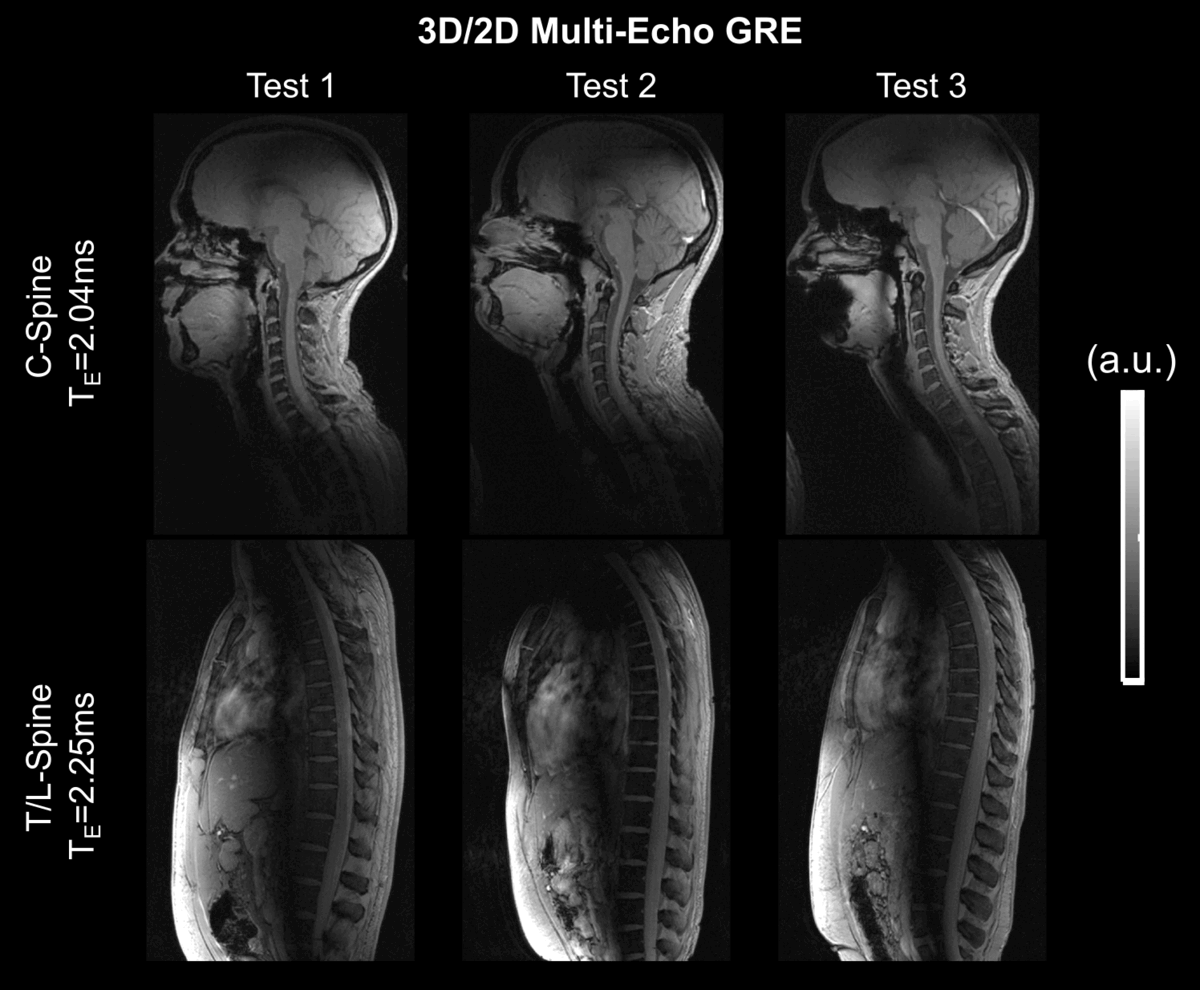
3D high-resolution Cartesian multi-echo GRE scans were obtained for C-Spine under free-breathing (nominalFA=10°, TE1/TE2/TE3/TR=2.04/4.08/6.12/27.69ms, FOV=352x212x192mm3, voxel=1x1x1mm3), and during breath-hold in 2D for T/L-Spine (nominalFA=30°, TE1/TE2/.../TE5/TR=2.25/5.25/.../14.25/40ms, FOV=384x244mm2, voxel=1x1x2mm3).
Results and Discussion
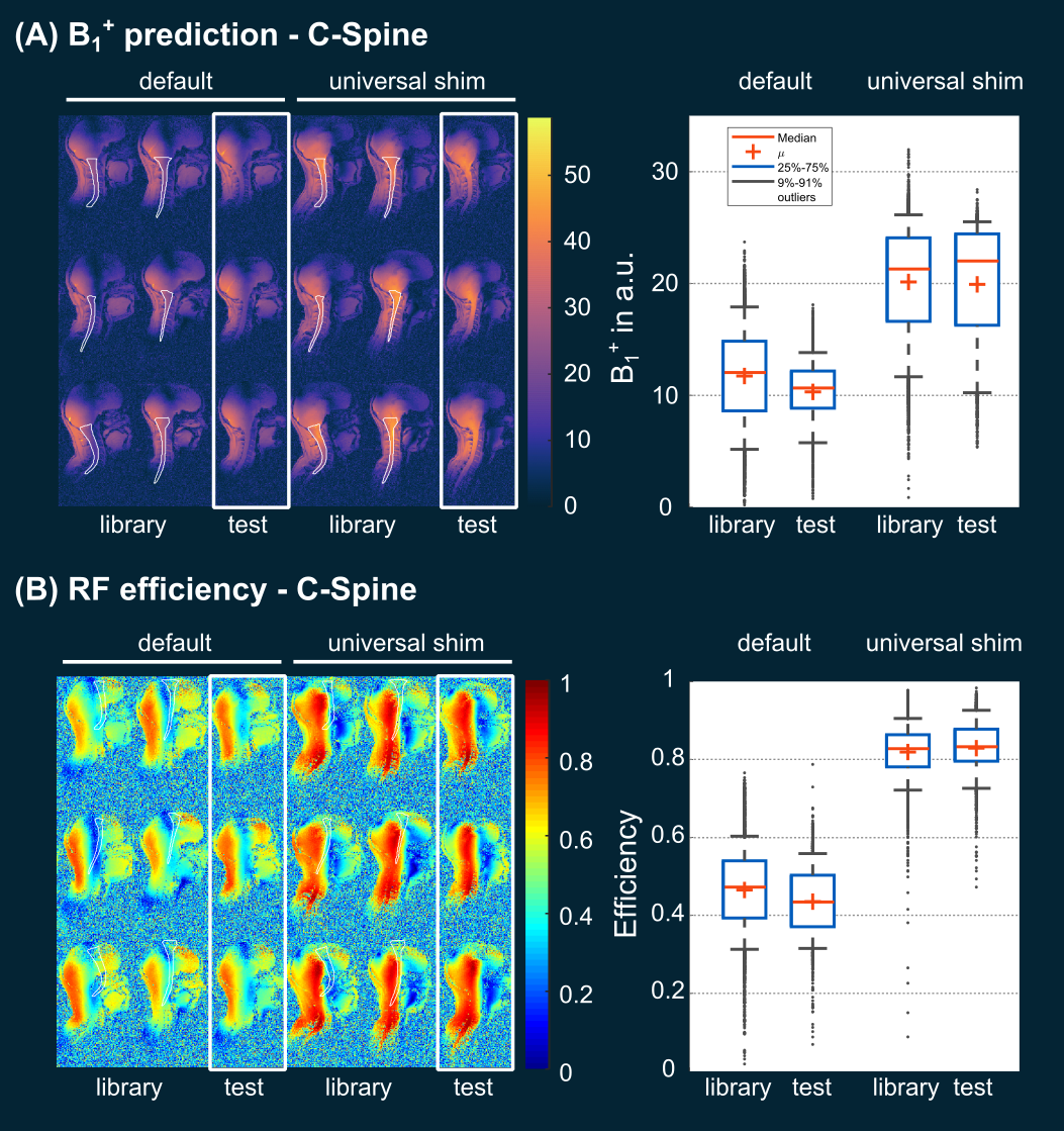
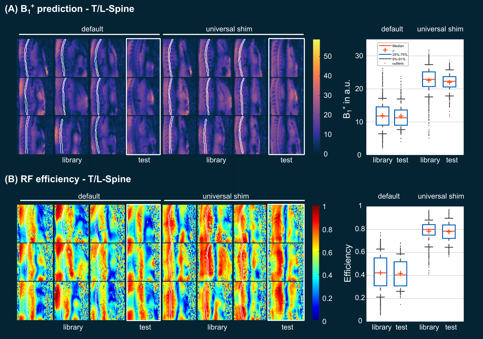
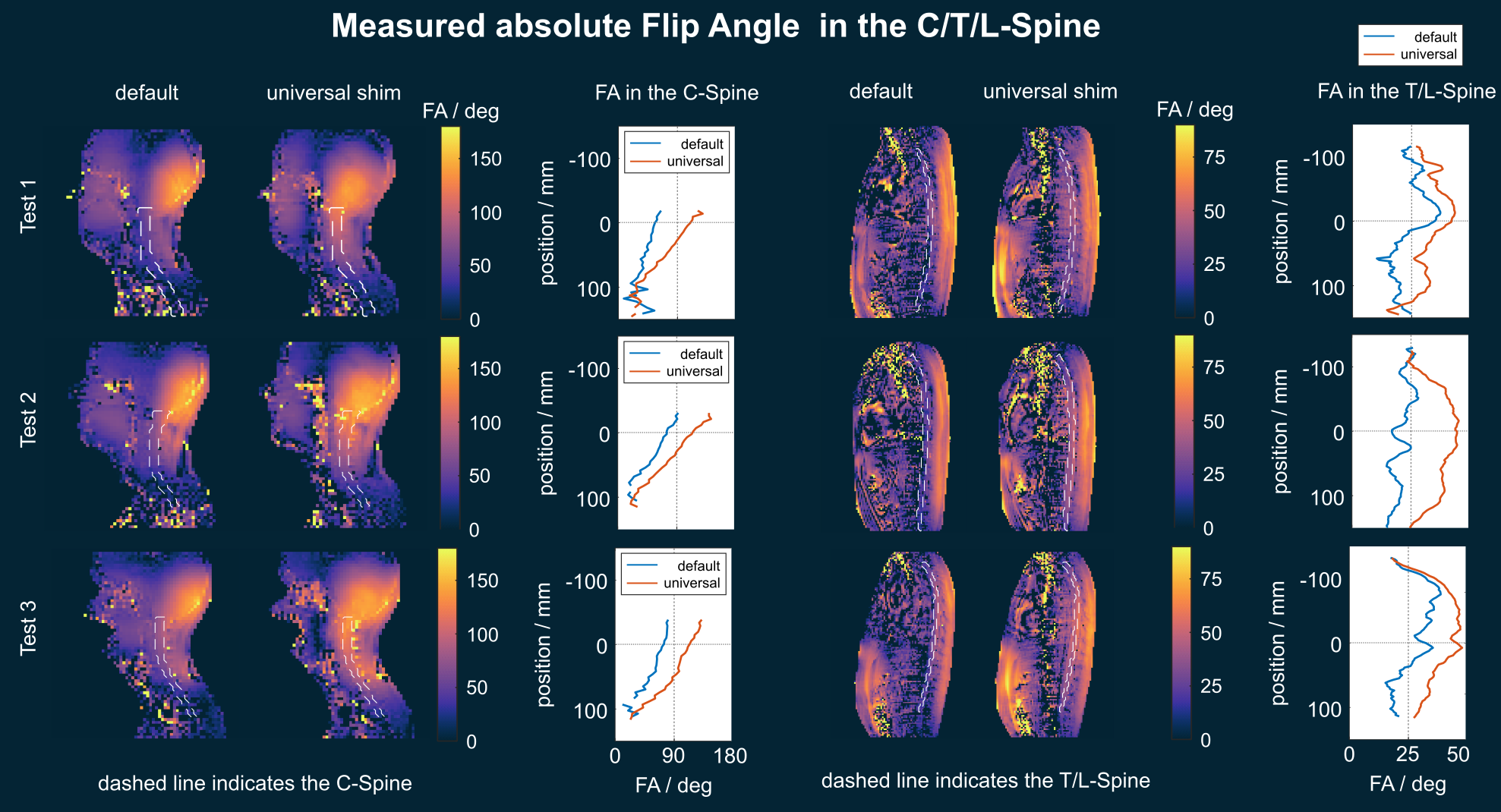
Figs. 1 and 2 compare the default shim (not optimized for the SC) with C- and T/L-Spine universal shims (US). In C-Spine test cases, the US showed a slight CV reduction (29% to 28%) and a two-fold η increase (0.44 to 0.83). Similar trends were observed in the T/L-Spine. Across all test cases, US reduced the CV by two-fold (30% to 13%), and η doubled (0.42 to 0.78). Note, that the cost function was weighted 90% by η and 10% by CV, which could be changed if desired.Fig.3 displays the absolute FA measurements. On average, measured FAs in the C-Spine increased by 51% from 52° (default) to 79° (US). Similarly, in the T/L-Spine region, a 48% FA increase is observed, with measured average FAs of 25° (default) and 37° (US).
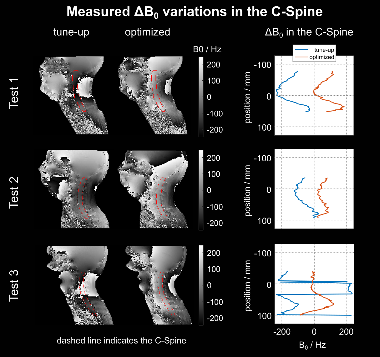
Fig.4 displays the measured ΔB0 variations in the C-Spine using the vendor-supplied tune-up and optimized B0 shims. A notable reduction in RMSE (296Hz vs. 102Hz) and standard deviation (117Hz vs. 102Hz) is observed. Tailored B0 shimming in the T/L-Spine region was not possible due to substantial motion artifacts in the Cartesian ΔB0 maps, necessitating further work to develop a motion-robust approach.
Fig. 5 presents a sagittal slice of the 3D GRE images acquired during free-breathing using US in the C-Spine and customized B0 shims. Additionally, a 2D GRE image was acquired during a breath-hold using US in the T/L-Spine for all three test subjects. Qualitatively, good image quality is evident, even at longer echo times, confirming the feasibility of calibration-free pTx across the entire SC.
Conclusion
This in-vivo study highlights the efficacy of US for calibration-free SC imaging at 7T for different SC regions and RF coils. The application of US enables seamless pTx in the SC, eliminating the requirement for time-consuming calibration procedures. This approach, in combination with motion-robust non-Cartesian encoding, holds promise for advancing SC-imaging capabilities at 7T and higher field strengths.Acknowledgements
We gratefully acknowledge funding from the German Research Foundation SCHM 2677/2-1, SCHM 2677/4-1 and GRK2260, BIOQIC.References
[1] Ladd, E, Bachert, P, Meyerspeer, M, et al. Pros and cons of ultra-high-field MRI/MRS for human application, Prog. Nuc. Magn. Reson. Spec. 2018; 109:1-50. doi: 10.1016/j.pnmrs.2018.06.001
[2] Barry, RL, Vannesjo, SJ, By, S, Gore, JC, Smith, SA. Spinal cord MRI at 7T. Neuroimage 168, 437–451. doi: 10.1016/j.neuroimage.2017.07.003.
[3] Padormo, F., Beqiri, A., Hajnal, J. V., and Malik, S. J. (2016), Parallel transmission for ultrahigh‐field imaging. NMR Biomed., 29: 1145– 1161. doi: 10.1002/nbm.3313
[4] Gras, V., Vignaud, A., Amadon, A., Le Bihan, D. and Boulant, N. (2017), Universal pulses: A new concept for calibration‐free parallel transmission. Magn. Reson. Med., 77: 635-643. doi:10.1002/mrm.26148
[5] Aigner, CS, Dietrich, S, Schaeffter, T, Schmitter, S. Calibration-free pTx of the human heart at 7T via 3D universal pulses. Magn Reson Med. 2021; 87: 70– 84. doi: 10.1002/mrm.28952
[6] Papp, D, Boulant, N, Massire, A, Mauconduit, F, Gras, V, Cohen-Adad, J. Universal pulses for the cervical spinal cord at 7T: a feasibility study, ISMRM 2023, 0199.
[7] Kazemivalipour, E, May, MW, Rangaprakash, D, Bilgic, B, Stockman, JP, Barry, RL, Keil, B, Wald, LL, Guerin, B. Design of a universal RF-shimming drive mode for head and neck imaging at 7 Tesla using a 16-channel pTx array, ISMRM 2023, 4406
[8] Schmitter, S., DelaBarre, L., Wu, X., Greiser, A., Wang, D., Auerbach, E.J., Vaughan, J.T., Uğurbil, K. and Van de Moortele, P.-F. (2013), Cardiac imaging at 7 tesla: Single- and two-spoke radiofrequency pulse design with 16-channel parallel excitation. Magn. Reson. Med., 70: 1210-1219. https://doi.org/10.1002/mrm.24935
[9] Dietrich, S, Aigner, CS, Kolbitsch, C, et al. 3D Free-breathing multichannel absolute B1+ Mapping in the human body at 7T. Magn Reson Med. 2021; 85: 2552– 2567. doi: 10.1002/mrm.28602
[10] Fautz HP, Vogel M, Gross P, Kerr A, Zhu Y. B1 mapping of coil arrays for parallel transmission. In Proceedings of the 16th Annual Meeting of ISMRM, Toronto, Canada, 2008. Abstract 1247.
[11] D'Astous A, Cereza G, Papp D, Gilbert KM, Stockmann JP, Alonso-Ortiz E, Cohen-Adad J. Shimming toolbox: An open-source software toolbox for B0 and B1 shimming in MRI. Magn Reson Med. 2022; 1-17. doi:10.1002/mrm.29528
Figures