1410
Intraoral Coax-based Dipole Antenna for Improved Dental Magnetic Resonance Imaging1School of Electrical and Biomedical Engineering, Hanyang University, seoul, Korea, Republic of, 2Lauterbur Imaging Research Center, Shenzhen Institutes of Advanced Technology, Chinese Academy of Sciences, Shenzhen, China, 3Key Laboratory for Magnetic Resonance and Multimodality Imaging of Guangdong Province, Shenzhen, China, 4Department of Biomedical Engineering and Department of Electronic Engineering, Hanyang University, seoul, Korea, Republic of
Synopsis
Keywords: Non-Array RF Coils, Antennas & Waveguides, High-Field MRI
Motivation: In this work, we investigated the performance of a coaxial dipole antenna compared with a single wire for dental MRI at 3 T.
Goal(s): A fully flexible coaxial dipole antenna was designed and optimized with an aim to improve the signal-to-noise ratio, image quality, and sensitivity while maintaining safety performances.
Approach: Based on electromagnetic simulations and MRI results it is demonstrated that in single conductor has a current distribution, which is strongly inhomogeneous leading to inhomogeneous B1+ field distribution.
Results: In contrast, coaxial cable with multiple gaps offers homogenous current distribution yielded to optimum B1+ field distribution.
Impact: The development of a fully flexible coaxial intraoral antenna with lightweight, capable of overcoming impedance and homogeneity challenges, has the potential to revolutionize dental MRI. Enhanced patient comfort and adaptability improve image quality, advancing dental MRI's utility in medical imaging.
Introduction
Magnetic Resonance Imaging (MRI) can be used in dentistry to obtain detailed, three-dimensional images of teeth, the jawbone, and surrounding tissues without emitting ionizing radiation. Various RF coils such as surface coils (intraoral and extraoral coils) are commonly used to improve the signal-to-noise ratio and sensitivity in the region of interest, which are typically teeth and the surrounding tissues [1], [2]. However, the aforementioned surface coils have limited sensitivity, and increase signal from the buccal fat and cheeks. Currently, the main challenge in dental MRI technology is designing a dipole antenna that can be seamlessly inserted into the mouth of the patient. This antenna should be lightweight, deliver peak performance, and effectively address impedance and homogeneity challenges. For this reason, we developed a fully flexible dipole antenna constructed from coaxial cable, which can be used by a wide range of patients.Methodology
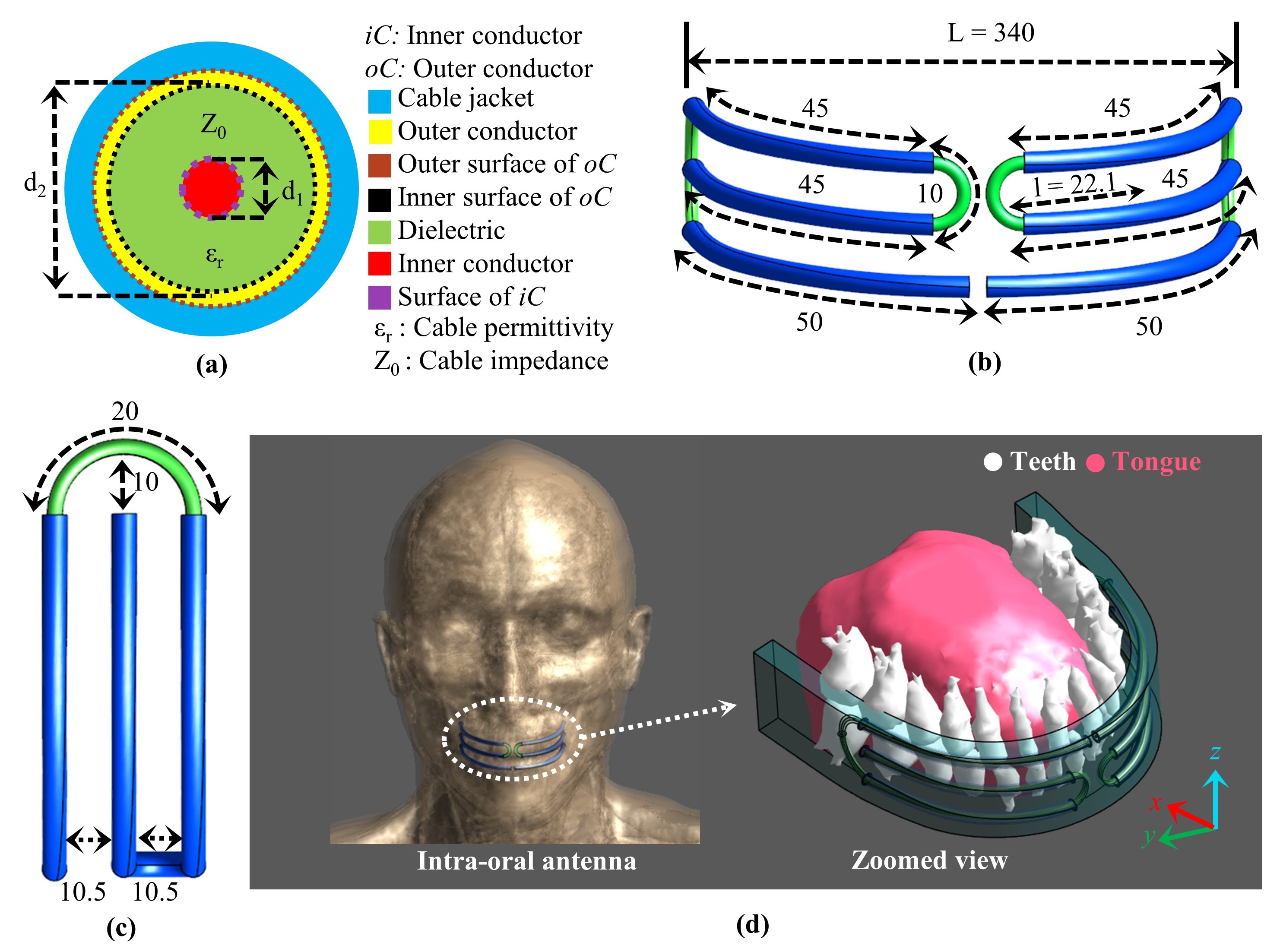
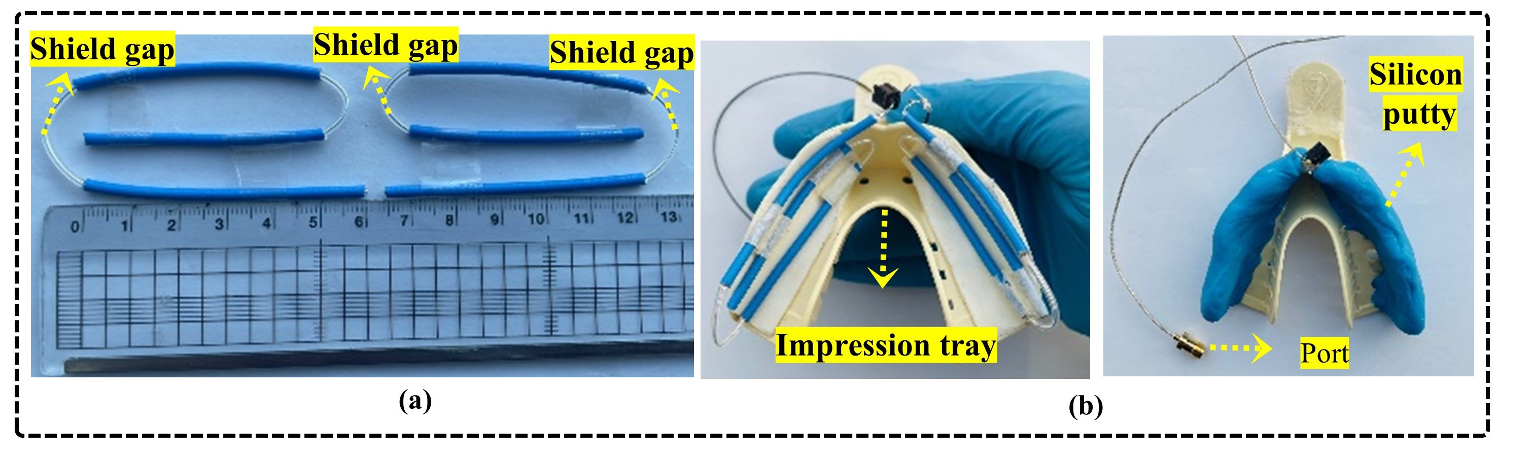
Finite-difference time-domain (FDTD) electromagnetic simulations were utilized to analyze the B1+ field distribution using an anatomically Duke model [3]. Figs. 1a, b, and c show the parameters of a coaxial cable with three distinct conductors, as well as the design of the intraoral dipole antenna. For accurate simulations, the intraoral antenna was designed according to the dimensions of the impression tray typically used in dental procedures for adults, as shown in Fig. 1 c. The fully flexible antenna was constructed using two sections of cable (Molex 086SC-2401, with a characteristic impedance of 50 ohms), as depicted in Fig. 2a. Fig. 2b shows how the intraoral antenna is placed within the M-impression tray, which can comfortably fit within an adult's mouth.Results
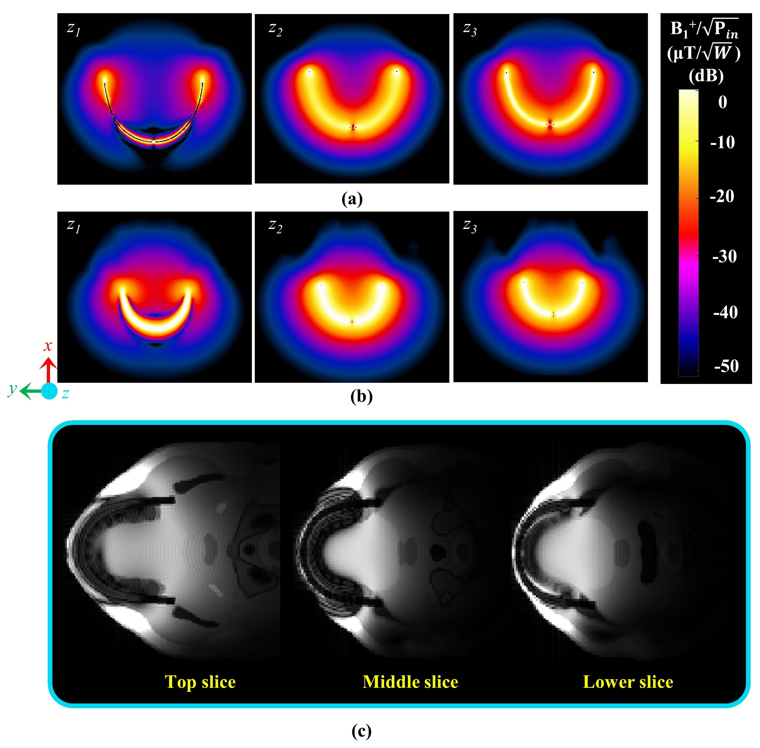
This research develops an intraoral antenna design optimized for MRI applications in dentistry, based on recent developments in loop coil technology [4], [5]. The design uses a coaxial cable to produce a perpendicular B1+ field orientation that effectively covers the roots of the molars. Additionally, a simulation was conducted using a single-core wire with the same dimensions as d1. Notably, the single-core wire exhibited non-uniformity within the z1 slice, as shown in Fig. 3a. Magnetic field intensities varied significantly near both ends and the center of the single-core wire, which resulted in irregular distributions. On the other hand, the distribution of B1+ in slices z2 and z3 was comparatively low. In contrast, the proposed coaxial antenna demonstrated an even distribution of B1+ throughout its structure, as shown in Fig. 3b. This uniformity is attributed to the balanced current flow between the inner and outer conductors of the coaxial antenna. Furthermore, the acquired images demonstrated a relatively homogeneous sensitivity across all the slices as shown in Fig. 3c. Therefore, the proposed coaxial antenna is superior to the single-core wire in terms of producing consistent and uniform magnetic fields.Conclusion
In this study, we proposed a new fully flexible dipole antenna inspired by recent advances in loop coils for intraoral MRI at 3 T. The RF fields were generated using coaxial cables, where the core conductor was driven and the shield was gap-filled, resulting in a relatively flat current profile outside the antenna. Furthermore, we demonstrated that intraoral MRI can be performed using a dipole antenna, improving transmit efficiency and homogeneity as compared to single-core wire.Acknowledgements
This work was supported by the National Research Foundation of Korea (NRF) Grant by the Korean Government through the Ministry of Science and ICT (MSIT) under Grant 2022R1A2C2003726.References
1. M. Prager, S. Heiland, D. Gareis, T. Hilgenfeld, M. Bendszus, and C. Gaudino, “Dental mri using a dedicated rf-coil at 3 tesla,” Journal of Cranio-Maxillofacial Surgery, vol. 43, no. 10, pp. 2175–2182, 2015.
2. J. Gradl, M. Horeth, T. Pfefferle, M. Prager, T. Hilgenfeld, D. Gareis, ¨ P. Baumer, S. Heiland, M. Bendszus, and S. H ¨ ahnel, “Application of a ¨ dedicated surface coil in dental mri provides superior image quality in comparison with a standard coil,” Clinical neuroradiology, vol. 27, pp. 371–378, 2017. 3.
3. Sim4Life by ZMT, https://www.zmt.swiss
4. T. Ruytenberg, A. Webb, and I. Zivkovic, “Shielded-coaxial-cable coils as receive and transceive array elements for 7t human mri,” Magnetic resonance in medicine, vol. 83, no. 3, pp. 1135–1146, 2020.
5. C. C. van Leeuwen, B. R. Steensma, D. W. Klomp, C. A. van den Berg, and A. J. Raaijmakers, “The coax dipole: A fully flexible coaxial cable dipole antenna with flattened current distribution for body imaging at 7 tesla,” Magnetic Resonance in Medicine, vol. 87, no. 1, pp. 528–540, 2022.
Figures