1408
A stitching method for dynamic field monitoring using NMR probes1State Key Laboratory of Brain and Cognitive Science, Institute of Biophysics, Chinese Academy of Sciences, Beijing, China, 2Institute of Artificial Intelligence, Hefei Comprehensive National Science Center, Hefei, China, 3University of Chinese Academy of Sciences, Beijing, China, 4CMRR, Radiology, University of Minnesota, Minneapolis, MN, United States
Synopsis
Keywords: System Imperfections, System Imperfections: Measurement & Correction
Motivation: NMR field probe methods for field monitoring are limited by probe signal loss due to relaxation, dephasing, or both.
Goal(s): To develop a method for characterizing sequences with higher resolution or readout length than allowed by current field monitoring approaches.
Approach: Long-duration 2D spiral readout gradients were characterized by acquiring and stitching multiple segment-specific dynamic field measurements per segmentation of the readout gradient determined by a signal loss model.
Results: For both long-duration and ultrahigh-resolution readouts, our method resulted in plausible 0th-2nd order dynamic field measurements throughout the entire spiral readout, correcting erroneous k-space traversal observed with a traditional approach.
Impact: The proposed stitching method provides an effective means to characterize challenging imaging gradients using commercially available hardware and without assuming a linear gradient system, thereby having utility for dynamic field measurements in ultrahigh-resolution MRI using a standard field monitoring system.
INTRODUCTION
Dynamic field monitoring1 using NMR probes2 has shown utility for various MRI applications3-5 owing to its ability to measure higher-order systematic and physiologically induced field fluctuations. However, measurement duration is limited by T2* decay within the probe, dephasing by strong gradients, or both, indicating that long-duration or ultrahigh-resolution readout gradients cannot be characterized. Here, we propose a new, stitching method and demonstrate its effectiveness for acquiring long-duration and ultrahigh-resolution readout gradients. Our results, obtained at 10.5 Tesla (10.5 T), of a 2D spiral readout show that our proposed method can be used to characterize a readout as lengthy as 88 ms or a readout targeting a resolution as high as 0.3 mm while using standard fluorine-19 probes (of 0.4 mm in radius and ~30 ms in T2*).METHODS
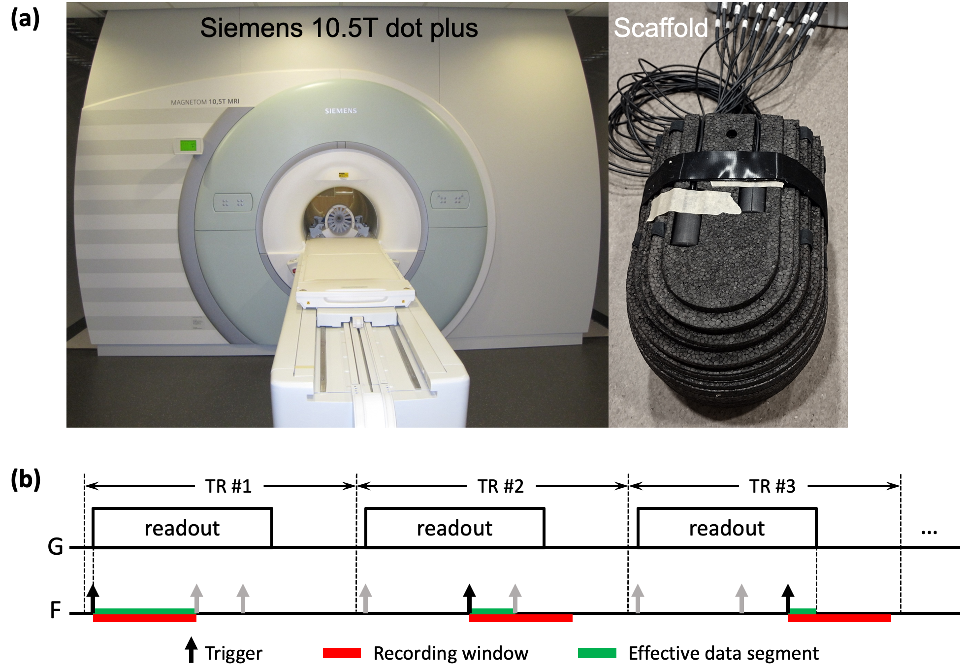
All experiments were performed on a Siemens 10.5 T Dotplus MR scanner (Siemens, Erlangen, Germany) (Fig. 1a) equipped with a whole-body gradient (80 mT/m @ 200 T/m/s). Dynamic field monitoring was conducted using a Clip-on Camera (Skope MRT, Zurich, Switzerland), placed in a scaffold that optimally places the 16 fluorine-19 probes for dynamic field measurement.To demonstrate the utility of our proposed method, we considered two extreme readout scenarios: 1) a long-duration readout and 2) a short readout that targets ultrahigh resolutions. For both, a Skope-compatible single-shot 2D spiral gradient-echo pulse sequence incorporating time-optimal readout waveforms6 was developed using the pulseq framework7.
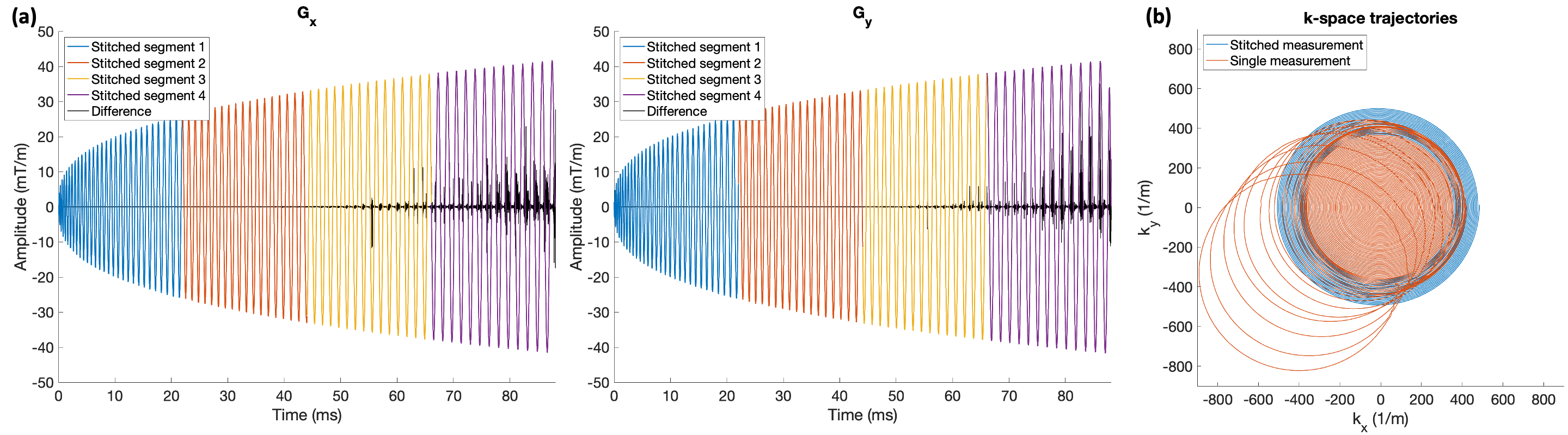
In scenario 1, the readout gradient was designed to acquire 1.0-mm isotropic resolutions with full k-space sampling, giving rise to a long readout of 88 ms. In scenario 2, the readout gradient was designed to achieve 0.3-mm isotropic resolutions with 30-fold under-sampling, leading to a short readout of 21 ms but a maximum gradient moment of 1.024 mT·s/m.
In either case, the entire readout gradient was characterized by stitching multiple segment-specific dynamic field measurements obtained across a matched number of consecutive TRs corresponding to a certain segmentation of the readout gradient (Fig. 1b).
For scenario 1 with T2* decay being a significant limiting factor, the segmentation was determined such that each segment would not exceed 22 ms in length (1/4 the total readout and within one T2* decay time of the probe), resulting in a total of four segments of constant durations.
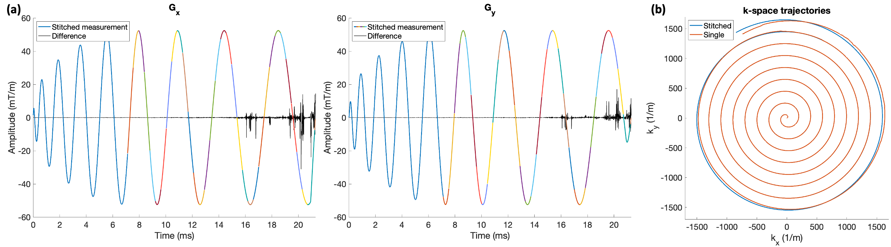
For scenario 2 with dephasing being a significant limiting factor, the segmentation was determined such that each segment would maintain at least 35% of the initial probe signal (assuming no T2* decay) per an intra-voxel signal dephasing model with the voxel size being the probe diameter, leading to a total of 36 segments with variable durations.
For comparison, dynamic field measurements were also obtained in both scenarios using the traditional single measurement (i.e., characterizing the entire readout gradient in a single TR).
RESULTS
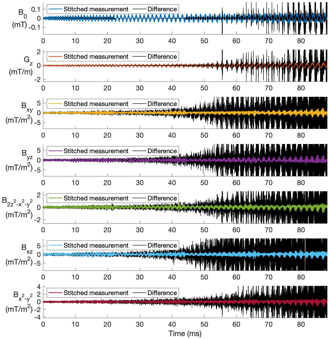
When characterizing the long-duration readout gradient, the use of our proposed method eliminated discontinuities associated with T2* signal loss, which generated a plausible gradient waveform and a sensible trajectory across the entire k-space (Fig. 2).Likewise, when comparing dynamic field measurements for other spherical harmonic terms, the use of our proposed method resulted in more sensible time courses than using the single measurement (Fig. 3).
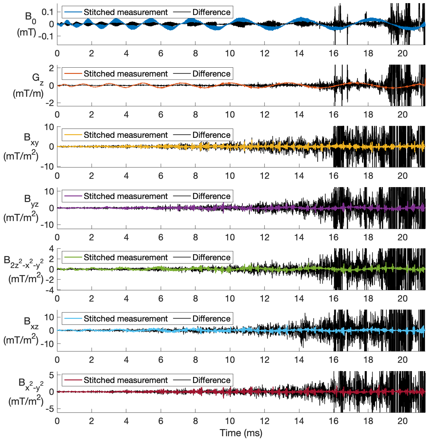
Similar results (Figs. 4 and 5) were observed when characterizing the short readout gradient but targeting ultrahigh resolutions, demonstrating efficacy for eliminating discontinuities associated with gradient-induced probe dephasing.
DISCUSSION
We have demonstrated the utility of a new method that can be used to measure dynamic field changes during long-duration or ultrahigh-resolution readout gradients, which would otherwise be difficult to measure using a standard field monitoring system. Our proposed method is compatible with both concurrent8 and post-monitored9 field monitoring schemes.Compared to existing approaches, our proposed method has the advantages of utilizing currently available field monitoring hardware10 and relaxes the assumption that the gradient system be linear11, but at the cost of a decrease in time efficiency.
Part of our future work is to validate our method by comprehensive image reconstruction12 incorporating high-order dynamic field measurements13.
CONCLUSION
Our proposed stitching method provides an effective means to extend traditional field monitoring approaches and is shown capable of making quality high-order dynamic field measurements for challenging readout gradients, thereby holding a promise to many imaging applications especially those at ultrahigh field in pursuit of ultrahigh resolutions.Acknowledgements
The authors are deeply indebted to Cameron Cushing and Paul Weavers from Skope MRT for their indispensable contribution to this work. ZZ was supported by Youth Innovation Promotion Association CAS (2022093). EA, AB, AG, KU, XW and all work conducted at the University of Minnesota were supported in part by USA NIH grants (NIBIB P41 EB027061, U01 EB025144, and S10 RR029672). ZZ, YZ, SH were supported in part by China Major S&T Projects (2022ZD0211901), NSFC of China (82271985, 31730039), and MOST of China (2019YFA0707103).References
1. Barmet, C., N. De Zanche and K. P. Pruessmann (2008). "Spatiotemporal magnetic field monitoring for MR." Magnetic Resonance in Medicine 60(1): 187-197.
2. De Zanche, N., C. Barmet, J. A. Nordmeyer-Massner and K. P. Pruessmann (2008). "NMR probes for measuring magnetic fields and field dynamics in MR systems." Magn Reson Med 60(1): 176-186.
3. Kasper, L., S. Bollmann, S. J. Vannesjo, S. Gross, M. Haeberlin, B. E. Dietrich and K. P. Pruessmann (2015). "Monitoring, analysis, and correction of magnetic field fluctuations in echo planar imaging time series." Magnetic Resonance in Medicine 74(2): 396-409.
4. Bollmann, S., L. Kasper, S. J. Vannesjo, A. O. Diaconescu, B. E. Dietrich, S. Gross, K. E. Stephan and K. P. Pruessmann (2017). "Analysis and correction of field fluctuations in fMRI data using field monitoring." Neuroimage 154: 92-105.
5. Engel, M., L. Kasper, C. Barmet, T. Schmid, L. Vionnet, B. Wilm and K. P. Pruessmann (2018). "Single-shot spiral imaging at 7T." Magnetic Resonance in Medicine 80(5): 1836-1846.
6. Lustig, M., S. J. Kim and J. M. Pauly (2008). "A fast method for designing time-optimal gradient waveforms for arbitrary k-space trajectories." IEEE Transactions on Medical Imaging 27(6): 866-873.
7. Layton, K. J., S. Kroboth, F. Jia, S. Littin, H. Yu, J. Leupold, J. F. Nielsen, T. Stöcker and M. Zaitsev (2017). "Pulseq: A rapid and hardware-independent pulse sequence prototyping framework." Magnetic Resonance in Medicine 77(4): 1544-1552.
8. Vannesjo, S. J., B. J. Wilm, Y. Duerst, S. Gross, D. O. Brunner, B. E. Dietrich, T. Schmid, C. Barmet and K. P. Pruessmann (2015). "Retrospective correction of physiological field fluctuations in high-field brain MRI using concurrent field monitoring." Magnetic Resonance in Medicine 73(5): 1833-1843.
9. Ma, R. Y., M. Akcakaya, S. Moeller, E. Auerbach, K. Ugurbil and P. F. Van De Moortele (2020). "A field -monitoring -based approach for correcting eddy -current -induced artifacts of up to the 2 nd spatial order in human-connectome-project-style multiband diffusion MRI experiment at 7T: A pilot study." Neuroimage 216.
10. Dietrich, B. E., D. O. Brunner, B. J. Wilm, C. Barmet and K. P. Pruessmann (2016). "Continuous Magnetic Field Monitoring Using Rapid Re-Excitation of NMR Probe Sets." Ieee Transactions on Medical Imaging 35(6): 1452-1462.
11. Wilm, B. J., B. E. Dietrich, J. Reber, S. J. Vannesjo and K. P. Pruessmann (2020). "Gradient Response Harvesting for Continuous System Characterization During MR Sequences." IEEE Trans Med Imaging 39(3): 806-815.
12. Wilm, B. J., C. Barmet, S. Gross, L. Kasper, S. J. Vannesjo, M. Haeberlin, B. E. Dietrich, D. O. Brunner, T. Schmid and K. P. Pruessmann (2017). "Single-shot spiral imaging enabled by an expanded encoding model: Demonstration in diffusion MRI." Magnetic Resonance in Medicine 77(1): 83-91.
13. Wilm, B. J., C. Barmet, M. Pavan and K. P. Pruessmann (2011). "Higher Order Reconstruction for MRI in the Presence of Spatiotemporal Field Perturbations." Magnetic Resonance in Medicine 65(6): 1690-1701.
Figures