1406
Reducing motion artefact in high resolution 7T scans using a new head stabilisation device1Biomedical Engineering Department, School of Biomedical Engineering and Imaging Sciences, King's College London, London, United Kingdom, 2London Collaborative Ultra high field System (LoCUS), London, United Kingdom, 3Experimental Psychology, University College London, London, United Kingdom, 4Imaging Neuroscience, University College London, London, United Kingdom
Synopsis
Keywords: High-Field MRI, Motion Correction
Motivation: Long scan durations and ultra-high field both facilitate the acquisition of high-resolution quantitative brain MRI, but they increase motion sensitivity.
Goal(s): We aimed to limit the occurrence of deliberate motion by testing a device (‘MinMo’) designed to increase head stability at 7T.
Approach: Using two k-space phase encoding orders with different motion sensitivity profiles we obtained data with and without the MinMo device.
Results: This showed that the MinMo increased image quality visually and as measured quantitatively via reduced gradient entropy in scans of ~10 and ~20 minute duration.
Impact: Reducing head motion would have a significant impact on image quality in high-resolution long duration research scans and clinical imaging. A preliminary investigation of a prototype device aiming to stabilize the head showed efficacy in most subjects, warranting further investigation.
Introduction
High-resolution quantitative brain MRI is important for neuroanatomical research and its alteration in pathology[1]. Both long scan durations and ultra-high field facilitate its acquisition, but they increase motion sensitivity. At high resolution even small undeliberate motion can cause visible artefacts. Many methods to reduce the impact of motion have been proposed[2], however, this remains a challenging problem, particularly at ultra-high field where motion interacts strongly with the B0 field[3]. We therefore aimed to limit the occurrence of motion during long-duration, high resolution scans using a head stabilisation device called the MinMo[4] (Fig 1), which might reduce motion artefacts. This was tested using long (10min) and very long (20min) scan durations. In addition to increased sensitivity to small undeliberate motion in healthy volunteers (HVs), we used both linear and random-checkered DISORDER sampling patterns[5] the latter to enhance motion sensitivity. Our main aim was to evaluate the effect of the MinMo on image quality visually and quantitatively using gradient entropy and normalised gradient squared (NGS)[6] within the brain.Methods
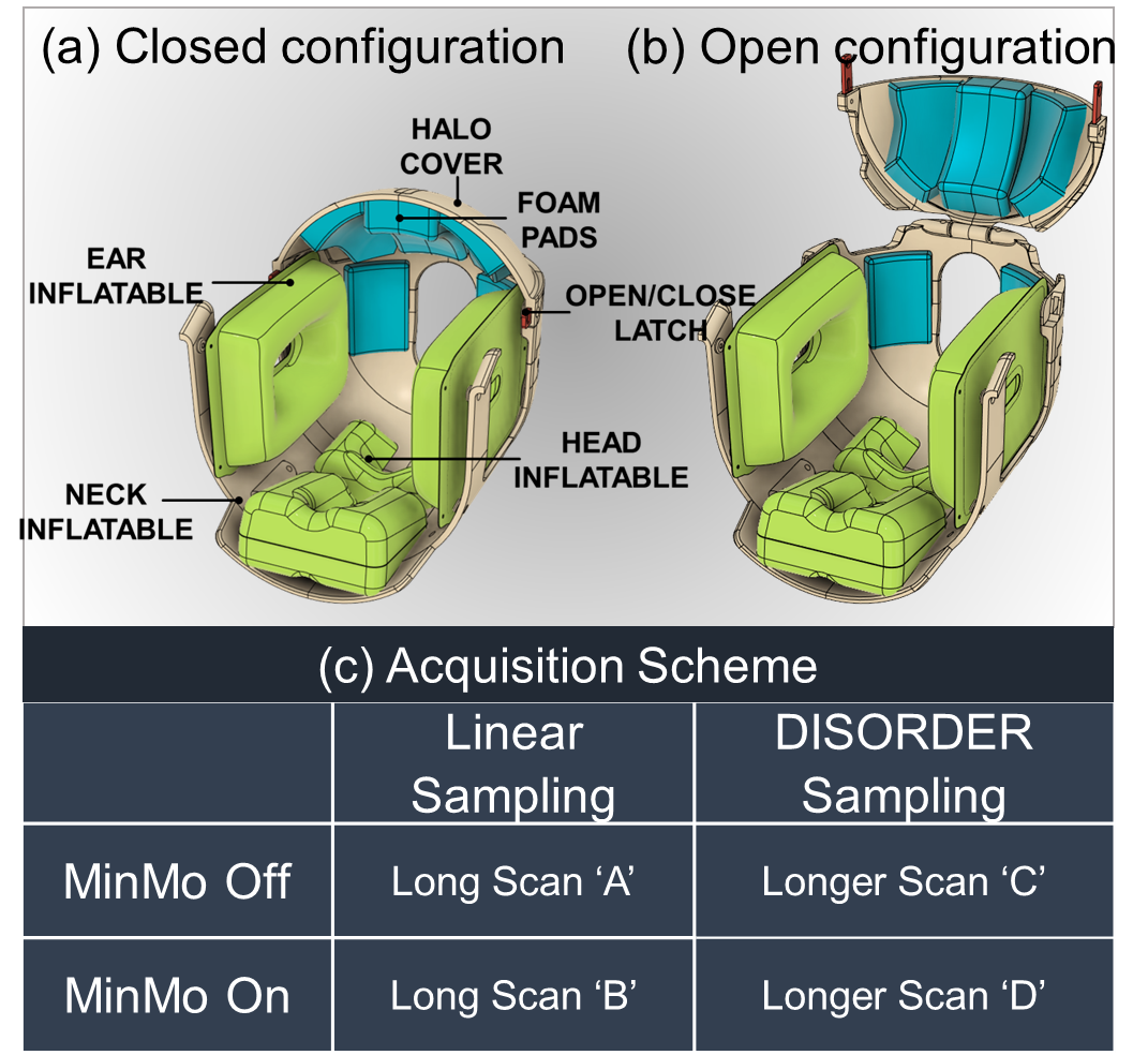
Fig. 1 shows the schematic diagram of the unloaded MinMo device with components of the head frame labelled on it for the closed and open configuration in Fig.1(a) and (b) respectively.Acquisition: To determine whether the MinMo effectively reduced motion artefact in long duration scans, we obtained whole-brain data using an optimized multi-echo GRE (MEGRE) protocol with flip angle=36°, TR/TE1,...TE10=30/2.3,4.9,..26.4ms for receiver bandwidth=420Hz/Px. Data was acquired at 0.6mm3 resolution (FOV=265x228x172mm3 (HF/AP/LR) with elliptical shutter) in 5 HVs.
For each volunteer subject, 4 MEGRE scans were obtained 2x10 min (long) and 2x20 min (very long). The scan acquisition order is described in Fig.1(c). The long scans [A&B] were acquired with linear cartesian sampling IPAT=2x2, Ref lines=2 and time of acquisition Tacq=10:40[min:s]. The very long scans [C&D] were acquired with the random-checkered DISORDER sampling pattern in the phase-encoding direction, IPAT=2x2, phase/slice oversampling=0.44/0.41 and Tacq=21:30[min:s]. The overall undersampling factor was limited to ~1.44 due to reconstruction limitations.
For scans A&C head stabilization was achieved using conventional foam pads following local standard radiographer practice. For scans B&D the participant was loaded in the MinMo with the inflatable pads inflated to provide comfortable stabilization as reported by each subject. The HVs were asked to lie still and were all accustomed to volunteer MRI research scans
To randomize the acquisition, the chronological order of scans for the 5 HVs was followed as (1) DB | AC, (2) BD | CA, (3) CA | DB, (4) BD | CA and (5) DB | AC respectively, where ‘|’ represents that the HV was repositioned into the MinMo or standard foam pads. Reference low-resolution (6mm3 iso) fully sampled scans were acquired before and after . All acquisitions were done on the 7T scanner (MAGNETOM Terra, Siemens Healthcare) 32ch receiver, 8ch PTx
Reconstruction: The coil sensitivity profiles used in the reconstruction were computed from the reference scans using ESPIRiT[7]. Reconstructions were done on down-sampled data at 1mm3 resolution for all 5 HVs using the conjugate-gradient SENSE method[
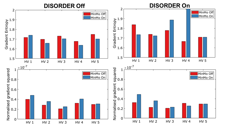
Data analysis: Gradient entropy and normalized gradient squared, two of the methods with the closest correspondence to visual image quality assessment, within whole brain extracted volumes[9] were calculated. Image quality was compared between the MinMo Off and MinMo On cases for both the long scans. MATLAB2023b was used for all reconstructions and analysis
Results
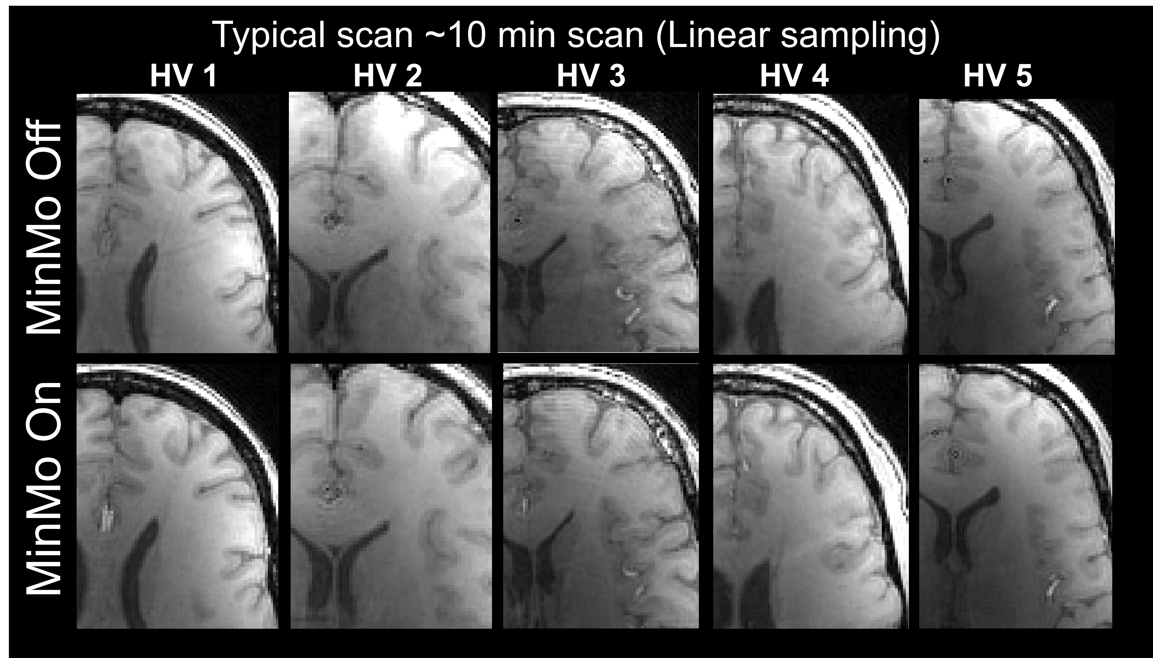
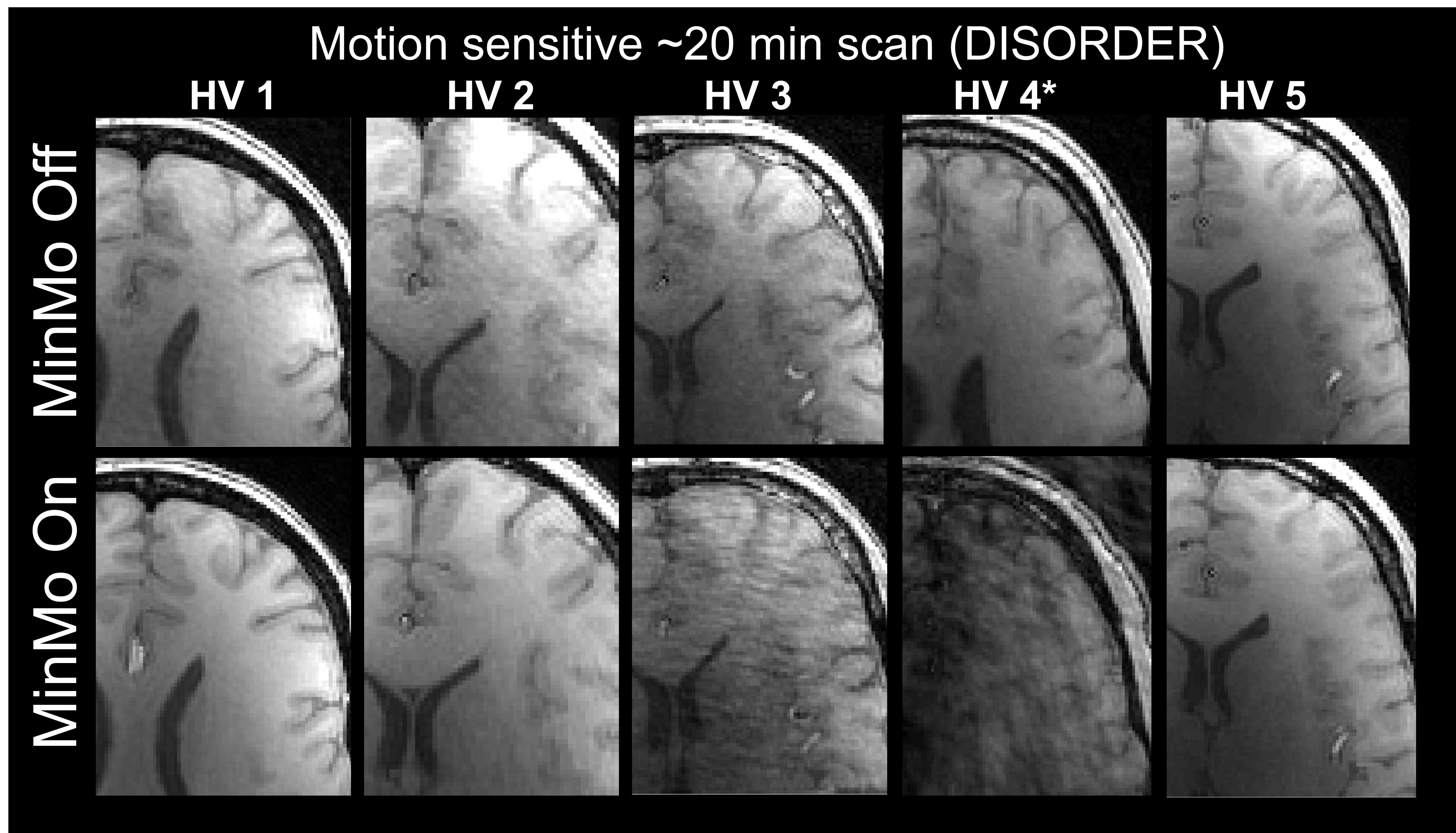
Fig. 2(a) shows representative zoomed-in images for the 5 HVs reconstructed for the long scan with MinMo Off and On. Fig. 2(b) shows the images for the same HVs reconstructed corresponding to the very long scan. Fig. 3(a) and Fig(b) shows the bar plots representing the gradient entropy values calculated on the brain volumes shown in Fig. 2 for the long and very long scans. For most cases, using MinMo improved the image quality (gradient entropy reduced, NGS increased). In one case (HV4) with worse performance with the MinMo the subject was too large to fit in the closed device. P value for normalised gradient square excluding the HV4 was reported to be 0.006, and for gradient entropy was reported 0.42Discussion
In our experiment, a 10-minute scan duration was used as it is a typical duration for an MPM study. The ~20min scan was obtained aiming to maximise motion sensitive owing to its long duration and motion sensitised phase encoding ordering scheme. Motion correction techniques such as aligned-SENSE can be used to aid motion correction but were not yet unexplored.Conclusion
A preliminary investigation suggested the MinMo device improved image quality in compliant subjects during most of the long duration (10-20 minutes) scans obtained.Acknowledgements
This work was supported by EPSRC CDT PhD studentship (JM). This work was supported also by the Wellcome/EPSRC Centre for Medical Engineering [WT203148/Z/16/Z] and by the National Institute for Health Research (NIHR) Biomedical Research Centre based at Guy’s and St Thomas’ NHS Foundation Trust and King’s College London and/or the NIHR Clinical Research Facility. The views expressed are those of the author(s) and not necessarily those of the NHS, the NIHR or the Department of Health and Social Care. This research was also supported by GOSHCC Sparks Grant V4419 (DC)References
[1] Carey, D., Caprini, F., Allen, M., Lutti, A., Weiskopf, N., Rees, G., Callaghan, M. F., & Dick, F. (2018). Quantitative MRI provides markers of intra-, inter-regional, and age-related differences in young adult cortical microstructure. Neuroimage, 182, 429.
[2] Maclaren J, Herbst M, Speck O, Zaitsev M. Prospective motion correction in brain imaging: a review. Magn Reson Med. 2013 Mar 1;69(3):621-36.
[3] Brackenier, Y, Cordero-Grande, L, Tomi-Tricot, R, et al. Data-driven motion-corrected brain MRI incorporating pose-dependent B0 fields. Magn Reson Med. 2022; 88: 817-831
[4] Richardson S., Dick F., Carmichael D., Callaghan M., European Patent No. GB 2205139.5, Head Immobilisation in MRI Head Coils, King's College London
[5] Cordero-Grande L, Ferrazzi G, Teixeira RPAG, O'Muircheartaigh J, Price AN, Hajnal JV. Motion-corrected MRI with DISORDER: Distributed and incoherent sample orders for reconstruction deblurring using encoding redundancy. Magn Reson Med. 2020 Aug;84(2):713–26
[6] McGee, K.P., Manduca, A., Felmlee, J.P., Riederer, S.J. and Ehman, R.L. (2000), Image metric-based correction (Autocorrection) of motion effects: Analysis of image metrics. J. Magn. Reson. Imaging, 11: 174-181
[7] Uecker M, Lai P, Murphy MJ, Virtue P, Elad M, Pauly JM, Vasanawala SS, Lustig M. ESPIRiT--an eigenvalue approach to autocalibrating parallel MRI: where SENSE meets GRAPPA. Magn Reson Med. 2014 Mar;71(3):990-1001
[8] Pruessmann KP, Weiger M, Scheidegger MB, Boesiger P. SENSE: sensitivity encoding for fast MRI. Magn Reson Med. 1999 Nov;42(5):952-62. PMID: 10542355
[9] Penny, W. D., Friston, K. J., Ashburner, J. T., Kiebel, S. J., & Nichols, T. E. (Eds.). (2006). Statistical Parametric Mapping: The Analysis of Functional Brain Images (1st ed.). Academic Press.
Figures