1401
MRI-based Biological Age Estimation for Multiple Organs in the UK Biobank Cohort1University Hospital of Tübingen, Tübingen, Germany, 2University of Stuttgart, Stuttgart, Germany
Synopsis
Keywords: Analysis/Processing, Aging, Biological Age
Motivation: MRI is a valuable tool for providing health-related information, including visualizing age-associated changes. Aging is influenced by chronic diseases, and assessing the true organ-specific biological age is essential for accurate diagnosis.
Goal(s): Development of an organ-specific age estimation for investigation in large imaging cohort with associated non-imaging information.
Approach: While prior studies focused on age estimation from non-imaging data or single organs, we propose a multi-organ age estimation framework, operating on brain, cardiac, and abdominal MRIs, and OCT scans.
Results: Our results prove the feasibility of imaging-based organ age estimation and initiate further investigations to identify risk factors for accelerated aging.
Impact: Reliable imaging-based estimation of biological age in multiple organ systems facilitates research efforts to identify risk factors of accelerated aging, advancing the goal of age-related phenotyping.
Introduction
MRI is an imaging technique which provides valuable information for supporting diagnosis and monitoring of diseases, including the visualization of age-related changes in the human body. Age plays an important role in shaping medical decisions due to its link to chronic diseases1-4. However, the physiological changes associated with aging can vary significantly among individuals and even between different organ systems of the same person. The concept of biological age was introduced to better reflect an individual’s true aging process5,7. With the increased availability of large medical population studies, such as the UK Biobank, multi-organ assessments of biological aging offer significant promise in investigating factors that contribute to accelerated aging. These findings support efforts to age-related phenotyping and treatment. So far, research exploring biological age in multiple organ systems has focused on single organs8-12 or non-imaging data13-16, failing to consider the potential of leveraging multi-organ aging information encoded in MR images. In our previous work, we showed the feasibility of using deep learning to extract age-related information in whole-body MR images17 and brain MRIs18,19. We propose a deep-learning-based multi-organ age estimation framework that is based on brain, cardiac, and abdominal MRIs as well as Optical-Coherence Tomography (OCT) scans to extract organ-specific age information in the image content.Methods
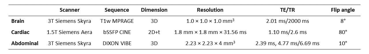
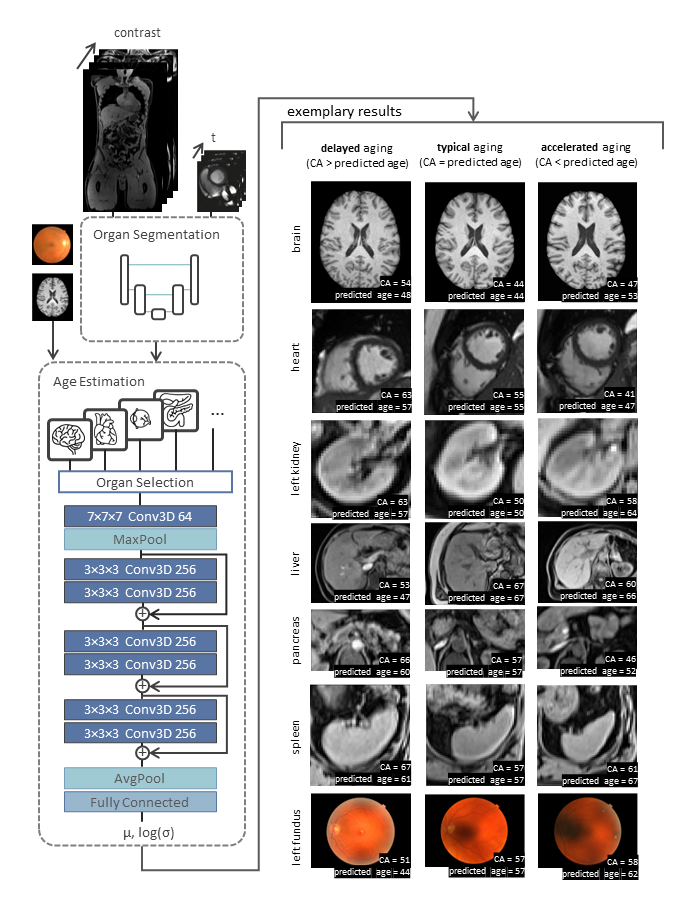
Data: Data was provided by the UK Biobank20, including MRI data from ~40,000 participants for brain, cardiac, and abdominal imaging, as well as OCT scans from ~80,000 participants. MRI parameters are listed in Fig. 1. Retinal images were obtained by a Topcon 3D OCT1000 Mark II device. The participants are 53.53% female and 46.46% male between 39-73 years with a mean age of 56.29±7.95 years. Artifact-corrupted organ images were excluded through manual quality control. For brain imaging, the brain MRIs after skull stripping serve as input. For cardiac imaging, the motion-resolved mid-ventricular short-axis Cine slices after heart segmentation are used. For abdominal imaging, all four Dixon contrasts are fed to the network with kidney (left/right), liver, spleen and pancreas segmented images being obtained from a semantic organ segmentation21.Network: The network adopts a modified ResNet-based structure18 (Fig. 2). It takes 3D images of the brain, heart (2D+t), and the abdominal organs or 2D fundus images as input. The images are passed through three sequential building blocks, consisting of convolutional and instance normalization layers. The network predicts the mean μ(x) and the logarithm of the standard deviation log(σ(x)) of a heteroscedastic Gaussian predictive distribution of the estimated age for the input image x. σ(x) indicates the level of uncertainty while μ(x) represents the age estimate.
Experiments: Age estimation was conducted for brain, heart, left/right kidney, liver, spleen, pancreas, and left/right fundus. Training was performed on eight Nvidia RTX A6000 GPUs using Adam optimizer. Mean absolute errors (MAE) and Pearson correlation coefficients r were calculated between predicted and chronological age. As a starting point for further correlation analyses of atypical aging, we conducted Kaplan-Meier survival curve analysis and regression analysis using lifestyle and health-related non-imaging information.
Results and Discussion
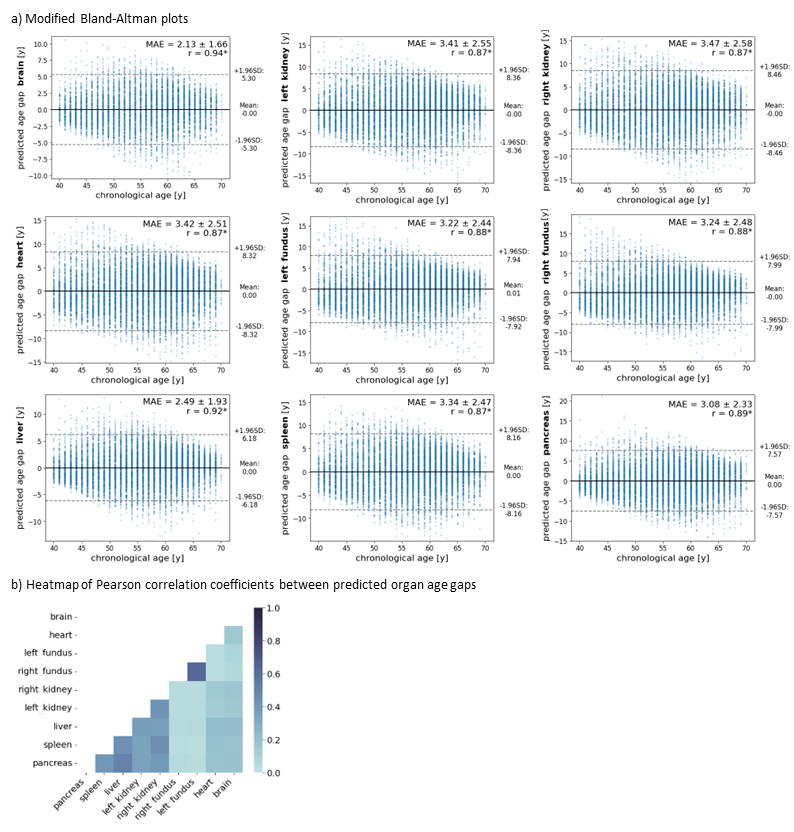
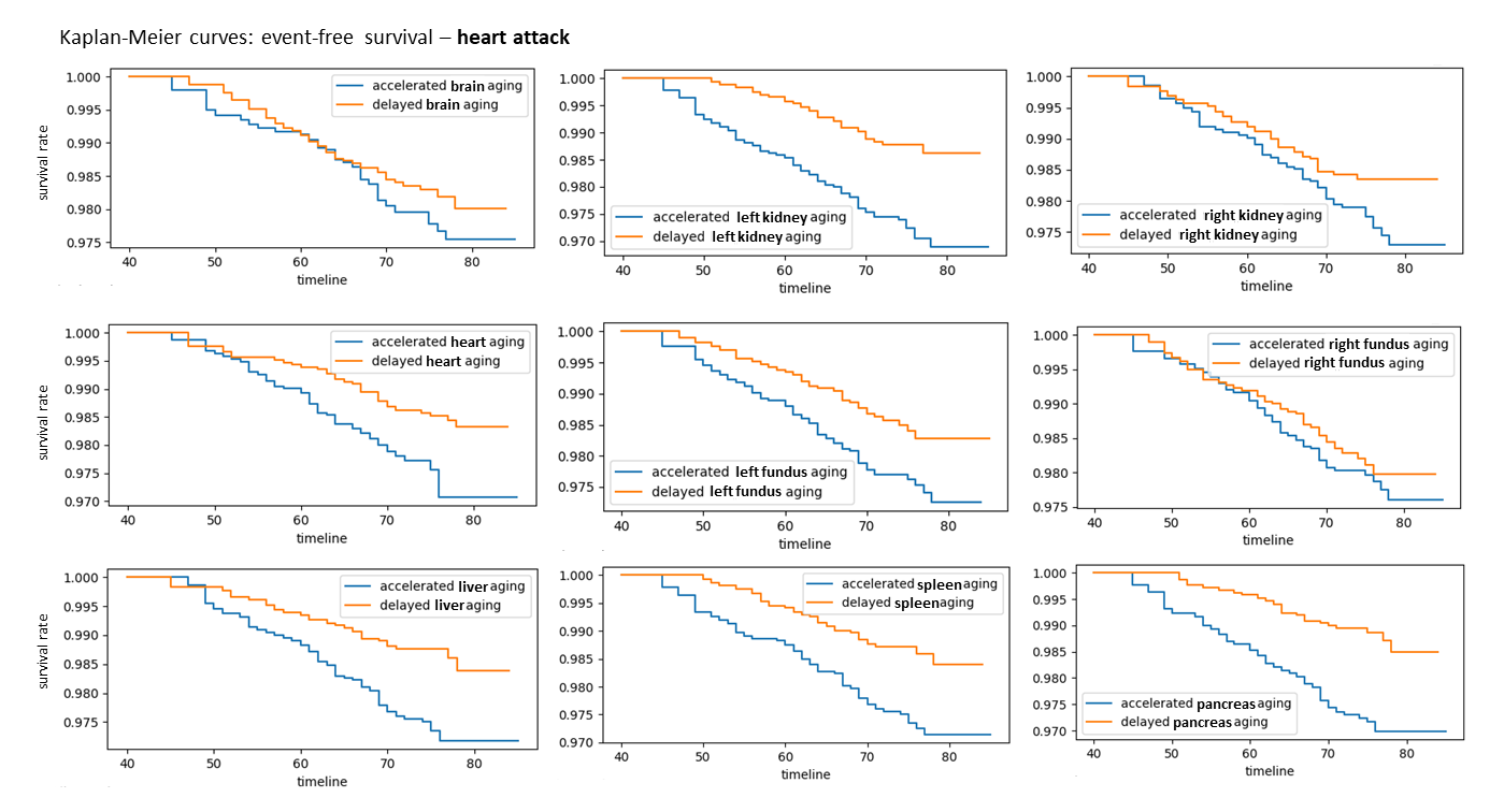
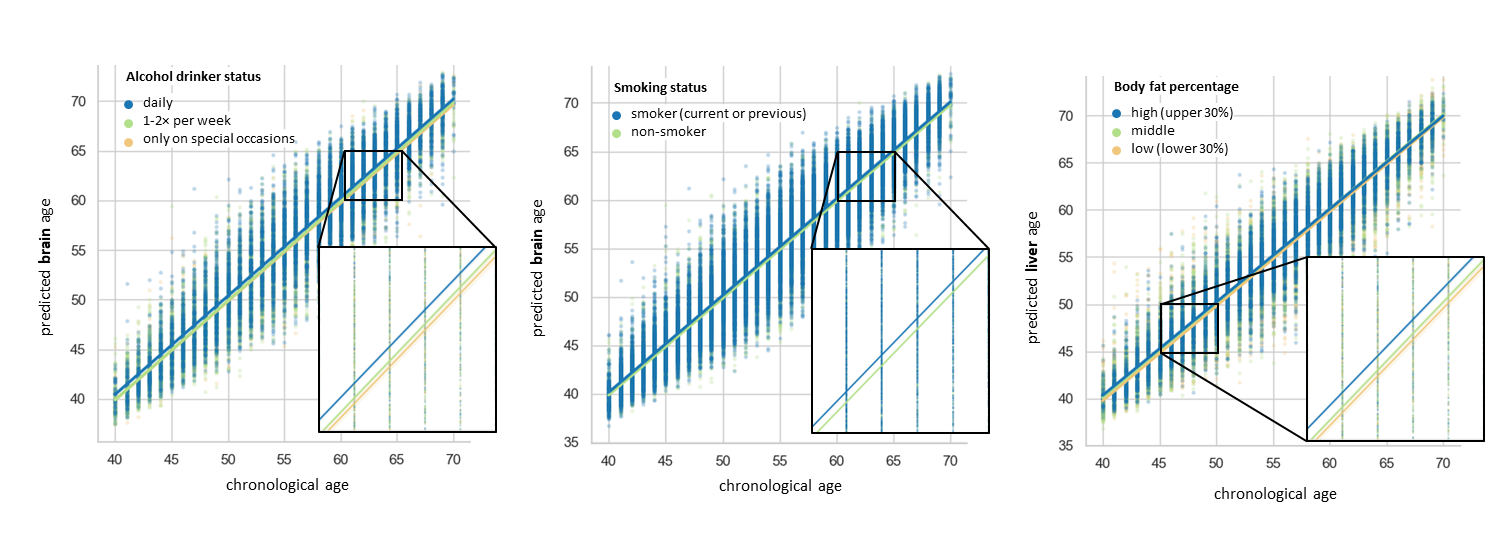
We observe reasonable quantitative results for all organs, proving the feasibility of age estimation for all selected organs (Fig. 3a). As anticipated, there is a strong correlation in the rate of accelerated or delayed aging between abdominal organs, and left and right fundus (Fig. 3b). The Kaplan-Meier-curves for heart attack in Fig. 4 show the likelihood that, at a certain age, the event has not yet occurred. The probability of the event is elevated in the group exhibiting accelerated organ aging not only for the primarily affected heart, but in all organs, highlighting the significance of a comprehensive multi-organ analysis. Additionally, we performed a regression analysis taking into consideration the alcohol drinker status and smoking status for brain aging, and the body fat percentage for liver aging. The results in Fig. 5 suggest that increased alcohol consumption and smoking are associated with slightly faster brain aging, while higher body fat percentages, especially among younger participants, are linked to accelerated liver aging. We acknowledge several limitations. Further examinations of the estimation framework are required to ensure that factors like image artifacts do not influence age estimates. Given the large volume of data, an automatic quality control should be considered. For a more robust performance, it may also be beneficial to employ decision-visualization techniques and to build upon existing non-imaging-based approaches and leverage both image-related and non-imaging information for age estimation.Conclusion
Our proposed multi-organ age estimation framework demonstrates the feasibility of organ-specific age estimation based on MR scans and serves as a starting point to investigate factors that contribute to atypical aging.Acknowledgements
This work was funded by the Deutsche Forschungsgemeinschaft (DFG,German Research Foundation) project #428219130 and supported under Germany’s Excellence Strategy EXC 2064/1 #390727645. This work was carried out under UK Biobank Application 60520. We thank all participants who took part in the UKBB study and the staff in this research program.
References
1. Kaeberlein et al. Healthy aging: The ultimate preventative medicine. Science. 2015 Dec 4;350(6265):1191-3.
2. North et al. The intersection between aging and cardiovascular disease. 110(8):1097–1108. Publisher: American Heart Association.
3. Azbel’. Phenomenological theory of mortality and aging. 249(1):472–481
4. Budovsky et al. Molecular links between cellular senescence, longevity and age-related diseases - a systems biology perspective. Aging (Albany NY). 2011 Dec;3(12):1178-91.
5. Baker et al. Biomarkers of aging. vol. 23, no. 4, pp. 223–239, 1988.
6. Lowsky et al. Heterogeneity in healthy aging. J Gerontol A Biol Sci Med Sci. 2014 Jun;69(6):640-9.
7. Hayflick L. Biological aging is no longer an unsolved problem. Ann N Y Acad Sci. 2007 Apr;1100:1-13.
8. Jonsson et al. Brain age prediction using deep learning uncovers associated sequence variants. vol. 10, no. 1, p. 5409, 2019. Number: 1 Publisher: Nature Publishing Group.
9. Mehta et al. Deep learning framework for automatic bone age assessment. in 2021 43rd Annual International Conference of the IEEE Engineering in Medicine and Biology Society (EMBC), pp. 3093–3096, 2021. ISSN: 2694-0604.
10. Lindow et al. Heart age gap estimated by explainable advanced electrocardiography is associated with cardiovascular risk factors and survival. p. ztad045, 2023.
11. Goallec et al. Using deep learning to predict abdominal age from liver and pancreas magnetic resonance images. vol. 13, no. 1, p. 1979, 2022. Number: 1 Publisher: Nature Publishing Group.
12. Raghu et al. Deep learning to estimate biological age from chest radiographs. vol. 14, no. 11, pp. 2226– 2236, 2021. Publisher: American College of Cardiology Foundation.
13. Tian et al. Heterogeneous aging across multiple organ systems and prediction of chronic disease and mortality. vol. 29, no. 5, pp. 1221–1231, 2023. Number: 5 Publisher: Nature Publishing Group.
14. Nie et al. Distinct biological ages of organs and systems identified from a multi-omics study. 38(10). Publisher: Elsevier.
15. Rahman et al. Deep learning using convolutional LSTM estimates biological age from physical activity. vol. 9, no. 1, p. 11425, 2019. Number: 1 Publisher: Nature Publishing Group.
16. Belsky et al. Impact of early personal-history characteristics on the pace of aging: implications for clinical trials of therapies to slow aging and extend healthspan. vol. 16, no. 4, pp. 644–651, 2017.
17. Küstner et al., Age estimation from whole-body MR images: A proof-of-principle study. ISMRM 2020.
18. Hepp et al. Uncertainty estimation and explainability in deep learning-based age estimation of the human brain: Results from the German national cohort MRI study. vol. 92, p. 101967, 2021.
19. K. Armanious et al. Age-Net: An MRI-Based Iterative Framework for Brain Biological Age Estimation. in IEEE Transactions on Medical Imaging, vol. 40, no. 7, pp. 1778-1791, July 2021.
20. Sudlow et al. UK biobank: An open access resource for identifying the causes of a wide range of complex diseases of middle and old age. vol. 12, no. 3, p. e1001779, 2015. Publisher: Public Library of Science.
21. Kart et al. Deep learning-based automated abdominal organ segmentation in the UK biobank and german national cohort magnetic resonance imaging studies. vol. 56, no. 6, pp. 401–408, 2021.
Figures