1396
Detection and Quantification of Acute Ischemic Lesions using Deep Learning-Based Super-resolution Portable Low-Field-Strength MRI1Department of Radiology, Beijing Chaoyang Hospital, Beijing, China, 2Subtle Medical, Shanghai, China, 3MR Research Collaboration, Siemens Healthineers, Beijing, China, 4Department of Radiology, Beijing Chaoyang Hospital, Shanghai, China, 5Laboratory for Clinical Medicine, Capital Medical University, Beijing, China
Synopsis
Keywords: AI/ML Image Reconstruction, Ischemia
Motivation: The diagnostic performance of portable low-field-strength MRI (LF-MRI) is constrained by low spatial-resolution and signal-to-noise ratio.
Goal(s): To evaluate the performance in detecting and quantifying ischemic lesions among SynthMRI, LF-MRI and real high-field-strength MRI (HF-MRI).
Approach: We created a deep learning-based model to generate the synthetic super-resolution (3T) MRI (SynthMRI) based on LF-MRI (0.23T). We evaluated the performance in detecting and quantifying ischemic lesions among SynthMRI, LF-MRI and HF-MRI.
Results: SynthMRI demonstrated high sensitivity in detecting the number and locations of ischemic lesions. Moreover, SynthMRI exhibited strong correlations with HF-MRI in the quantitative assessment of ischemic lesions, and significantly higher than portable LF-MRI.
Impact: Synthetic super-resolution MRI images overcome the limitations of low spatial resolution and signal-to-noise ratio in portable low-field-strength MRI. It has the potential to replace high-field-strength MRI images in the neuroimaging of AIS, enabling portable low-field-strength MRI examinations with comparable performance.
Introduction
Portable low-field-strength MRI (LF-MRI) has been integrated into the MRI-based stroke care pathway for participants with acute ischemic stroke (AIS). However, its diagnostic performance is constrained by low spatial-resolution and signal-to-noise ratio. The deep learning-based super-resolution portable LF-MRI has a potential to address this limitation by generating synthetic super-resolution MRI (SynthMRI). Deep learning-based super-resolution methods have shown promising performance in generating a synthetic high-spatial-resolution (3-7T) output from a low-spatial-resolution (1.5-3T) input 1–3. It is suggested that deep learning-based super-resolution methods have the potential to enhance the imaging performance of LF-MRI (≤0.3T). This study investigates the potential of deep learning-based super-resolution MRI in AIS assessment by comparing the performance in detecting ischemic lesions and quantifying their volumes and intensities among synthetic super-resolution MRI, low-field-strength MRI and real high-field-strength MRI.Methods
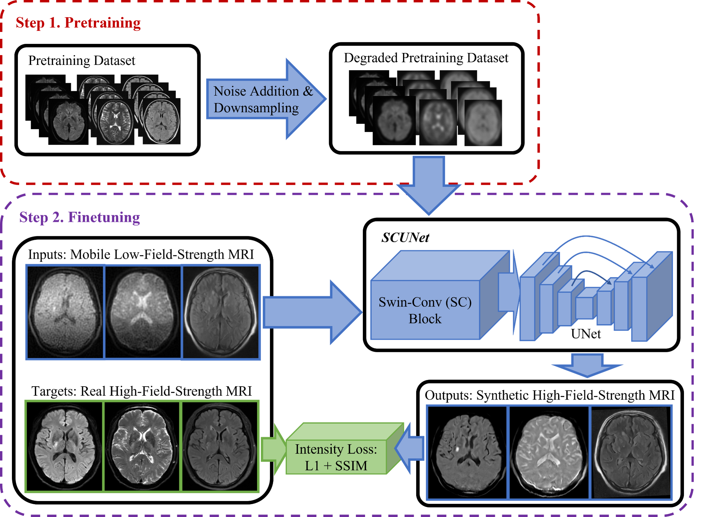
We retrospectively enrolled participants with AIS symptoms, who underwent both LF-MRI (0.23T, ACUTA Elfin, Rayplus) and HF-MRI (3.0T, Prisma, SIMENSE) examinations. The deep learning model (Figure 1) used to generate SynthMRI by inputting LF-MRI, was pretrained using 966 opensource MRI datasets. It was then finetuned and tested using small-scale datasets. We detected the number and locations of ischemic lesions for each participant on LF-MRI, SynthMRI and HF-MRI, respectively, and quantified their volumes and intensities with the commercial software (imSTROKE v1.0, YueXi). With HF-MRI as the reference standard, the performance of SynthMRI and LF-MRI in AIS assessment were evaluated using diagnostic values and Pearson’s correlation coefficients. We compared them using McNemar and Steiger Z test.Results
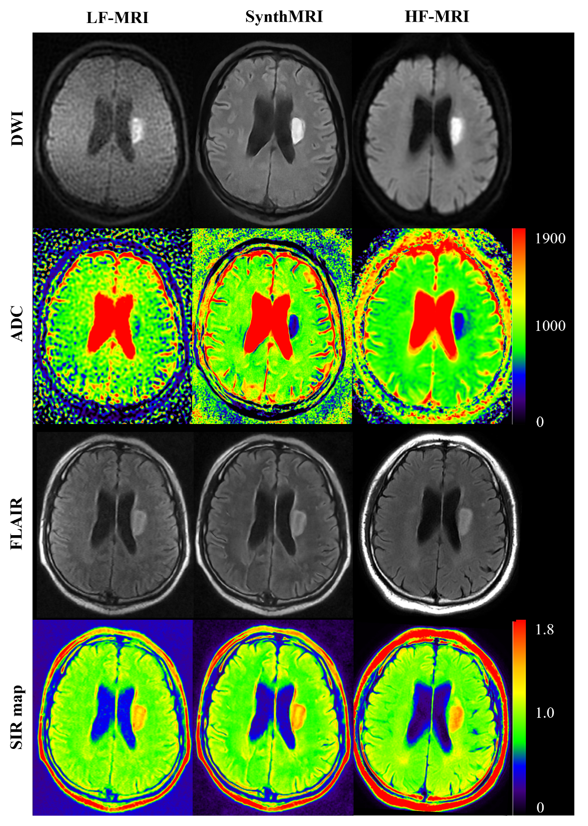
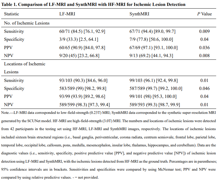
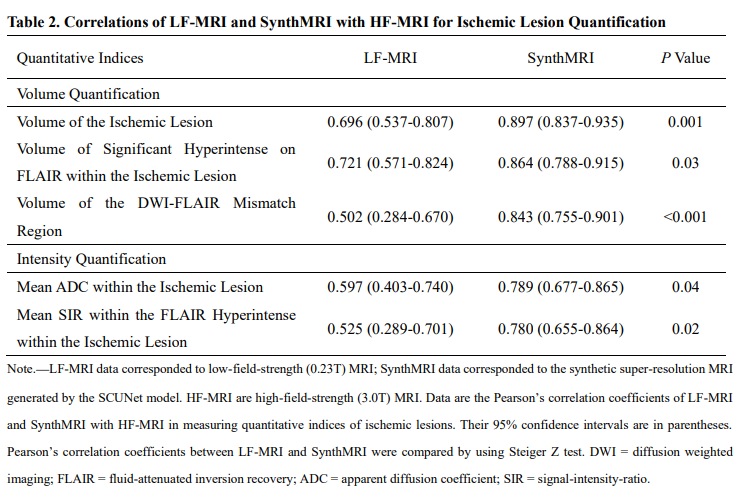
We included 101 participants (68 male; mean age, 61.8±14.7 years; 19 with unknown onset time), yielding 59 and 42 paired LF-MRI with HF-MRI images for finetuning and testing, respectively. SynthMRI demonstrated high sensitivity in detecting the number and locations of ischemic lesions (94.4% [95% CI: 89.0, 99.7] and 96.1% [95% CI: 92.4, 99.8], respectively) (Table 1). Furthermore, SynthMRI exhibited strong correlations (Pearson’s correlation coefficients > 0.75, p < 0.001) with HF-MRI in both volume and intensity quantification of ischemic lesions. SynthMRI provided a significant improvement (p < 0.05) in both detection and quantification of ischemic lesions compared to LF-MRI (Table 2). Figure 2 illustrates an example of qualitative and quantitative comparison of LF-MRI, SynthMRI and HF-MRI images on DWI, FLAIR, ADC and SIR map for one participant with a periventricular ischemic lesion in the testing set.Discussion and Conclusion
MRI-based pretreatment neuroimaging is mainly used to evaluate DWI Alberta Stroke program early CT score (DWI-ASPECTS), ischemic lesions and the DWI-FLAIR mismatch, all of which can guide clinical decision-making 4–6. To accurately measure these imaging indices, MRI images should exhibit highly qualitative and quantitative capabilities for ischemic lesions. In the qualitative analysis, we found that both SynthMRI and LF-MRI have high sensitivities (sensitivity > 80%) for ischemic lesions, but the sensitivity of SynthMRI was significantly higher than that of LF-MRI (84.5% vs 94.4%, respectively, p = 0.009). It means that SynthMRI can meet the requirement of estimating DWI-ASPECTS, which assesses ischemic changes in ten brain structural regions within the territory of the middle cerebral artery. The results of quantitative analysis indicated that SynthMRI exhibited a strong correlation (mean ADC value within the ischemic lesion: 0.789 [95% CI: 0.677, 0.865], p < 0.001; volume of ischemic lesions: 0.780 [95% CI: 0.655, 0.864], p < 0.001) with HF-MRI in evaluating mean ADC value within the ischemic lesion and volume of ischemic lesions. It suggests that SynthMRI enables to be utilized to quantify ischemic lesions like HF-MRI. Therefore, synthetic super-resolution MRI overcomes the limitations of low spatial resolution and signal-to-noise in portable low-field-strength MRI, and demonstrates the comparable performance with real high-field-strength MRI in AIS assessment.Acknowledgements
This work was supported by grants from the National Natural Science Foundation of China (82025018 and 92249301), Capital’s Funds for Health Improvement and Research (No. 2022-1-2031), Beijing Hospitals Authority’s Ascent Plan (No. DFL20220303), Beijing Key Specialists in Major Epidemic Prevention and Control.References
1. Iglesias JE, Schleicher R, Laguna S, et al. Quantitative Brain Morphometry of Portable Low-Field-Strength MRI Using Super-Resolution Machine Learning. Radiology. 2023;306(3). doi:10.1148/radiol.220522
2. Benzakoun J, Deslys MA, Legrand L, et al. Synthetic FLAIR as a Substitute for FLAIR Sequence in Acute Ischemic Stroke. Radiology. 2022;303(1):153-159. doi:10.1148/RADIOL.211394
3. Masutani EM, Bahrami N, Hsiao A. Deep learning single-frame and multiframe super-resolution for cardiac MRI. Radiology. 2020;295(3):552-561. doi:10.1148/radiol.2020192173
4. Powers WJ, Rabinstein AA, Ackerson T, et al. Guidelines for the Early Management of Patients with Acute Ischemic Stroke: 2019 Update to the 2018 Guidelines for the Early Management of Acute Ischemic Stroke a Guideline for Healthcare Professionals from the American Heart Association/American Stroke A. Vol 50.; 2019. doi:10.1161/STR.0000000000000211
5. Jovin TG, Willinsky RA, Sapkota BL, et al. Randomized Assessment of Rapid Endovascular Treatment of Ischemic Stroke. Published online 2015:1019-1030. doi:10.1056/NEJMoa1414905
6. A., Shafiq CWNSSJRB. A multicenter randomized controlled trial of endovascular therapy following imaging evaluation for ischemic stroke (DEFUSE 3). Physiol Behav. 2017;176(3):139-148. doi:10.1177/1747493017701147.A
Figures