1388
Whole Heart, Whole Cardiac Cycle, Motion-Resolved, 3D T2*-Mapping Reveals Myocardial T2* Differences Between Wild-Type and HCM Mice1Berlin Ultrahigh Field Facility (B.U.F.F.), Max Delbrueck Center for Molecular Medicine in the Helmholtz Association, Berlin, Germany, 2Biomedical Imaging Research Institute, Cedars-Sinai Medical Center, Los Angeles, CA, United States, 3School of Medicine, Indiana University, Indianapolis, IN, United States, 4Department of Bioengineering, University of California- Los Angeles (UCLA), Los Angeles, CA, United States, 5DZHK (German Centre for Cardiovascular Research), Berlin, Germany, 6Krannert Cardiovascular Research Center, Indiana University, Indianapolis, IN, United States, 7Experimental and Clinical Research Center, Charité—Universitätsmedizin Berlin, Berlin, Germany
Synopsis
Keywords: Myocardium, Animals, Relaxometry, T2*, Motion-resolved, Low-rank-tensor, Multi-echo
Motivation: To explore various stages of myocardial pathophysiology and improve prediction and interception of disease progression by capturing myocardial T2* variations across cardiac cycle.
Goal(s): To establish a framework that operates independently of ECG- and respiratory-gating, enabling cinematic, whole-heart myocardial T2*-mapping in mouse models.
Approach: A tailored framework was developed using modified Multi-Gradient-Echo (MGE) sequence and Low-Rank-Tensor (LRT) reconstruction technique. In vivo study was performed.
Results: Our preliminary findings demonstrate the feasibility of flow-compensated, free breathing, fully ungated, cardiac motion-resolved, whole heart and whole cardiac cycle coverage CINE imaging and T2* mapping in healthy and diseased mice heart.
Impact: The differences in myocardial T2* obtained for wild-type mice and HCM model provide a new metric ΔT2*,rel for myocardial tissue characterization, and springboard to inform on the different stages of myocardial pathophysiology and improve prediction and interception of disease progression.
Introduction
Myocardial tissue changes, such as hemorrhage, oxygenation and fibrosis can be reflected in alterations of myocardial T2*. This tissue parameter provides valuable insights into pathophysiology of cardiomyopathies or heart failure1. Cinematic T2*-mapping is required to detail transient changes in T2* across the cardiac cycle. Standard gated Multi-Gradient-Echo (MGE) T2* imaging is prone to motion artifacts due to disturbance of the ECG signal and inaccurately monitored respiratory motion. This challenge is more pronounced in mouse models, with heart rates ranging between 400-600 bpm. Recognizing this constraint and opportunity, we developed a novel imaging and reconstruction framework tailored for mouse hearts. We employed an MGE sequence with a random Gaussian trajectory combined with a Low-Rank-Tensor (LRT) formulation to facilitate high-resolution, motion-resolved 3D T2* mapping of the whole mouse heart in approximately 15 minutes. We evaluated the feasibility of our approach in mouse HCM models representing different disease states and examined T2* changes throughout the cardiac cycle.Method
The animal study was conducted in accordance with local animal welfare guidelines. This study included female mice with 2 genotypes in C57BL/6J background representing different disease status: wild-type (WT;11 weeks old) and mybpc3 knock-in (KI;22 weeks old) represent human HCM2. To address cardiac and respiratory motion in T2* mapping, we adopted a randomized Gaussian trajectory3 to densely sample the center of k-space for spatial basis estimation (Figure1). To determine the temporal dynamics, we used a zero-encoded line (k-space center line) as a navigator in an interleaved manner (indicated by blue line in middle illustration of Figure1). Additionally, we introduced a first-order Gradient-Moment-Nulling (GMN) into the MGE sequence to mitigate flow artifacts in the heart's blood pools. An ICA-based retrospective gating technique was adopted to provide a reliable respiratory and cardiac phase estimation3. Sequence development and data acquisition were carried out on a preclinical 9.4T Bruker scanner (Biospec 94/20, Bruker, Germany). MGE images were acquired with the parameters summarized in Table1 without cardiac and respiratory gating. Subsequently, 3D, motion-resolved MGE images were reconstructed using an LRT formulation modeling a 6D tensor with spatial (Ux) and temporal dimensions for Cardiac (Uc), Respiratory (Ur), and T2* (Ut) evolution (Figure 1)4. T2* was calculated using mono-exponential fitting utilizing 6 MGE images (TE = 2.6, 4.02, 5.44, 6.86, 8.28, 9.7) reconstructed by LRT reconstruction.Result
Figure 2 shows exemplary whole heart coverage, short-axis view T2*-maps (apex, mid-slice, and base) of the heart obtained from 3D whole heart coverage acquisitions of two mouse genotypes. Mean myocardial T2* was calculated for each slice across all cardiac phases. Myocardial T2* was significantly reduced in the HCM model versus wild-type mice (apex = 7.0 ± 0.4 vs 12.8 ± 1.1 (p<0.0001), mid-slice = 9.2 ± 0.3 vs 12.6 ± 0.9 (p<0.0001), base = 6.2 ± 0.4 vs 11.0 ± 1.7 (p<0.0001)). Unlike the HCM model the wild-type mouse showed T2* alterations across the cardiac cycle as illustrated in Figure 2. This difference between the diseased and the normal model offers the opportunity to establish a marker ΔT2*,rel for myocardial tissue characterization, which is represented by the averaged T2* across all cardiac phases and normalized to T2* obtained for a mid-systolic phase. For the HCM model a ΔT2*,rel=13% was observed. Wild-type mice showed ΔT2*,rel=27%.Discussion
Our preliminary findings demonstrate the feasibility of flow-compensated, free breathing, fully ungated, cardiac motion-resolved, whole heart and whole cardiac cycle coverage CINE imaging and T2* mapping in healthy and diseased mice heart. The differences in myocardial T2* obtained for wild-type mice and a HCM model provide a new metric ΔT2*,rel for myocardial tissue characterization, and springboard to inform on the different stages of myocardial pathophysiology and improve prediction and interception of disease progression.Conclusion
Flow-compensated, free breathing, fully ungated, cardiac motion-resolved, whole heart and whole cardiac cycle coverage T2* mapping facilitates detection of differences in myocardial T2* across genotypes and phenotypes. These T2* differences provide a potential metric ΔT2*,rel and springboard to inform on the different stages of myocardial pathophysiology and improve prediction and interception of disease progression.Acknowledgements
No acknowledgement found.References
1. M. Gastl, A. Gotschy, J. von Spiczak, M. Polacin, F. Bönner, C. Gruner, M. Kelm, F. Ruschitzka, H. Alkadhi, and S. Kozerke, "Cardiovascular magnetic resonance T2* mapping for structural alterations in hypertrophic cardiomyopathy," European journal of radiology open, vol. 6, pp. 78-84, 2019.
2. Vignier N, Schlossarek S, Fraysse B, Mearini G, Krämer E, Pointu H, Mougenot N, Guiard J, Reimer R, Hohenberg H, Schwartz K, Vernet M, Eschenhagen T, Carrier L. "Nonsense-mediated mRNA decay and ubiquitin-proteasome system regulate cardiac myosin-binding protein C mutant levels in cardiomyopathic mice," Circ Res. 2009 Jul 31;105(3):239-48.
3. X. Guan, H.-J. Yang, Z. Hu, N. Wang, A. Christodoulou, B. Sharif, D. Li, and R. Dharmakumar, "Free-breathing Fully Ungated 3D Cardiac T2* MR Mapping using a Low-Rank Tensor Framework."
4. A. G. Christodoulou, J. L. Shaw, C. Nguyen, Q. Yang, Y. Xie, N. Wang, and D. Li, "Magnetic resonance multitasking for motion-resolved quantitative cardiovascular imaging," Nature biomedical engineering, vol. 2, pp. 215-226, 2018.
Figures
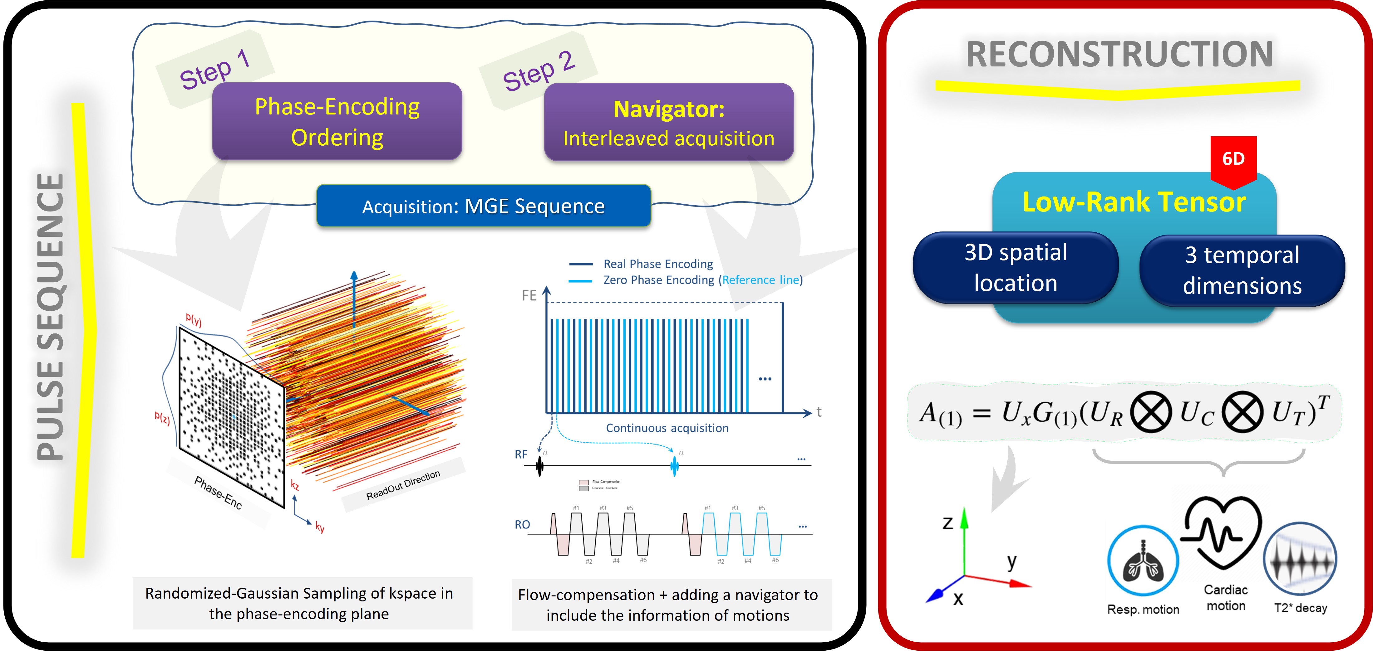
Figure 1: Overview of proposed fully ungated T2*-mapping framework. We utilized 3D MGE for data collection. A first-moment GMN applied to MGE to mitigate flow ghosting artifacts. Phase-Encoding (PE) ordering modified to a randomized Gaussian pattern to densely sample the center of k-space. A zero-PE reference line was acquired after each real PE to capture motion info for reconstruction. The LRT reconstruction framework models the data as a 6D tensor, incorporating 3 spatial, and 3 temporal dimensions for T2* decay, Cardiac and Respiratory motion.
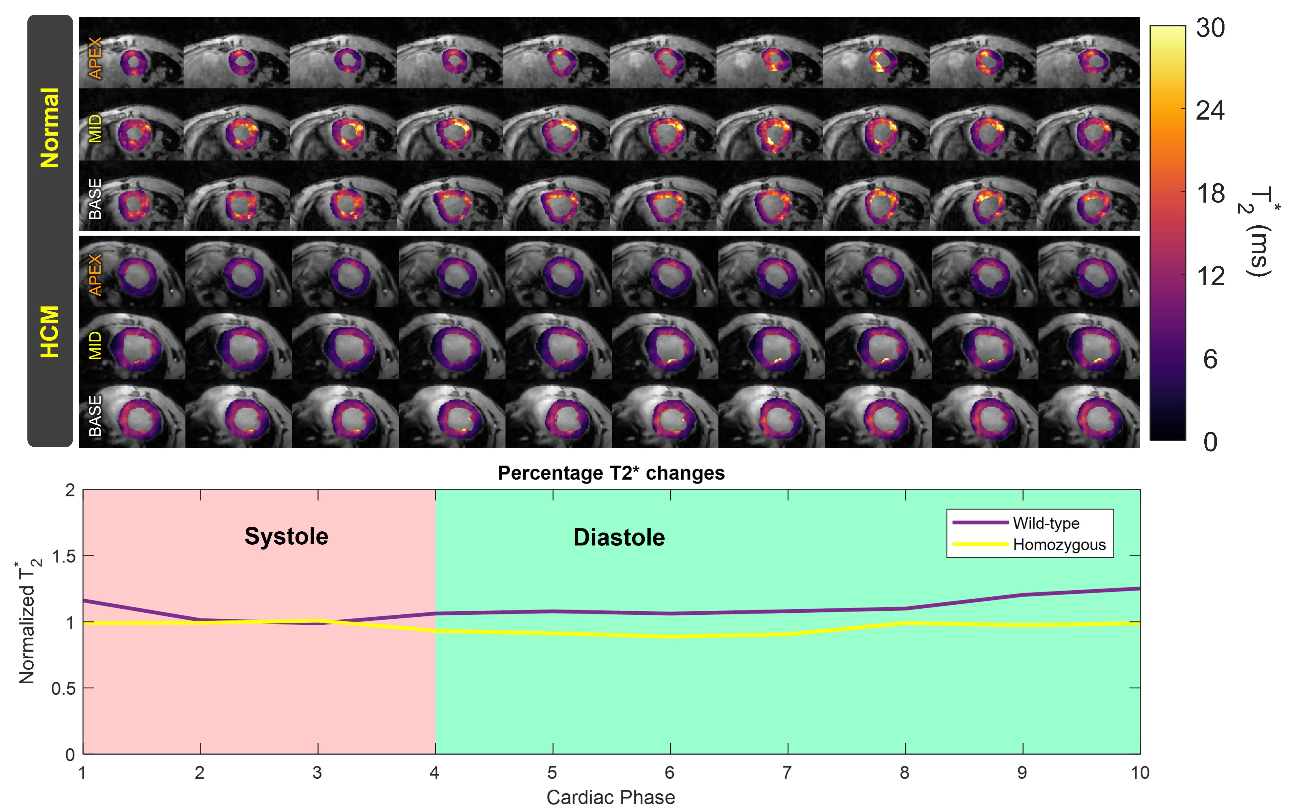
Figure 2. Top: Exemplary T2*-maps derived from ungated, motion resolved whole heart coverage 3D MGE of HCM and wild-type mice heart at apex-, mid-slice and base-SAX views reconstructed using the LRT framework for 10 cardiac phases. Bottom: These curves depict cardiac phase-resolved mean myocardial T2* values across all imaging planes, normalized relative to the mid-systolic phase. These findings illustrate higher T2* variations throughout the cardiac cycle in the wild-type mouse compared to the diseased mouse.
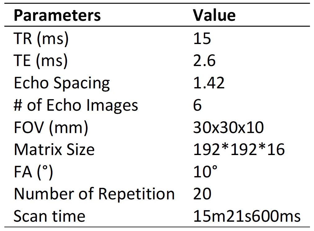
Table 1: Imaging parameters used for in vivo scanning in mice heart.