1381
Accelerating whole brain vessel wall imaging of isotropic 0.4 mm3 on 5T by 10-fold using deep learning reconstruction1Paul C. Lauterbur Research Center for Biomedical lmaging, Shenzhen Institute of Advanced Technology, Shenzhen, China, 2Research Center for Medical AI, Shenzhen Institute of Advanced Technology, Shenzhen, China, 3University of Chinese Academy of Sciences, Beijing, China
Synopsis
Keywords: Vessel Wall, Atherosclerosis
Motivation: Whole brain vessel wall imaging (VWI) of isotropic 0.4 mm3 on 3T can’t utilize higher than 5-fold acceleration to reduce the scan time due to insufficient signal-to-noise.
Goal(s): To achieve 10-fold accelerated whole brain VWI of isotropic 0.4 mm3 on the 5T scanner with a 48-channel transmit receive head coil.
Approach: Deep learning (DL) reconstruction equipped with 3D convolution neural network was developed to alleviate the nonuniform noise amplified by SPIRiT reconstruction and the B1 inhomogeneity of 5T scanner.
Results: The proposed DL SPIRiT reconstruction achieves 10-fold accelerated intracranial VWI scan on 5T in 6 minutes and give better VWI quality than 3T.
Impact: This work develops a 10-fold accelerated whole brain vessel wall imaging of isotropic 0.4 mm3 in 6 minutes using deep learning (DL) unrolled SPIRiT reconstruction on the 5T scanner equipped with a 48-channel transmit receive head coil.
Introduction
Three-dimensional (3D) MR vessel wall imaging (VWI) with resolution higher than isotropic 0.5 mm3 is highly demanded in detecting and characterizing intracranial wall diseases such as atherosclerosis plaque, arterial dissection, and aneurysms. Currently, intracranial VWI performed on the clinical 3T scanner often utilize resolution lower than 0.5 mm3 to reduce the clinical scan time2. Conventional acceleration schemes such as parallel imaging (PI) and compressed sensing (CS) are quite challenging to achieve acceleration factors higher than 5-fold for intracranial VWI due to the insufficient signal-to-noise (SNR) from the small voxel size 3,4. This work develops a 10-fold accelerated whole brain VWI of isotropic 0.4 mm3 in 6 minutes using deep learning (DL) unrolled SPIRiT reconstruction5,6 on the 5T scanner equipped with a 48-channel transmit receive head coil.Methods
The 5T scanner could promote the baseline SNR for intracranial VWI, and benefits the undersampling based acceleration, especially with a 48-channel parallel receiving coil system. However, the increased B0 and B1 inhomogeneity on 5T and higher than 9-fold underdsampling would lead to heavy and nonuniform noise amplification during reconstruction.This work proposed a deep learning-based parallel image reconstruction method for highly accelerated VWI scan on 5T. SPIRiT model is utilized due to its robustness and fast coil sensitivity calibration. The 3D reconstruction is decomposed to separate 2D reconstruction tasks after coil compression from 48 channels to 24 channels and 1D inverse Fourier Transform (FT) along the fully sampled readout. The iterative algorithm solving sparsity regularized SPIRiT reconstruction was unrolled to a multi-layer CNN. Each layer utilized a 3D CNN to replace the original 2D wavelet sparsity regularization for effective noise suppression. The 3D CNN could exploit both the inter- and intra-slice correlation, leading to high fidelity and continuous depiction of vessel walls. Inputs to the proposed DL-SPIRiT network were the initial noisy GRAPPA reconstruction results of the CAIPIRINHA undersampled k-space of multiple adjacent slices and corresponding SPIRiT kernels, and outputs were reconstructed multi-slice magnitude images (coil combined by sum-of-squares). The network was trained using a database containing 900 training samples and was deployed for inline reconstruction through the open-source Gadgetron platform with a Nvidia RTX 8000 GPU (48 GB GPU memory).
Institutional review board approved in-vivo experiments were performed on six healthy volunteers (4 males and 2 females) with informed consent obtained. All scans were performed on a 5T scanner (uMR Jupiter, United Imaging Healthcare, Shanghai, China) with a 48-channel transmit and receive head coil. VWI scan parameters included: T1 weighted MATRIX sequence with non-selective excitation and sagittal imaging orientation, whole brain coverage with FOV = 170 (RO) x 192 (PE1) x 156 (PE2) $$$mm^3$$$, imaging resolution = 0.4$$$mm^3$$$, matrix size = 416x480x388, TE/TR = 15.6/1000 ms, echo train length (ETL) = 46, bandwidth = 500 Hz/pixel, prescribed T1/T2 = 1000/200 ms with minimal flip angle (FA) = 48°. 10-fold CAIPIRINHA undersampling with elliptical scanning being enabled lead to a scan duration of 6 minutes. Reconstruction parameters included: 1.5x interpolation, and image inhomogeneity correction using combined prospective B1 map and retrospective N4 algorithm.
Result
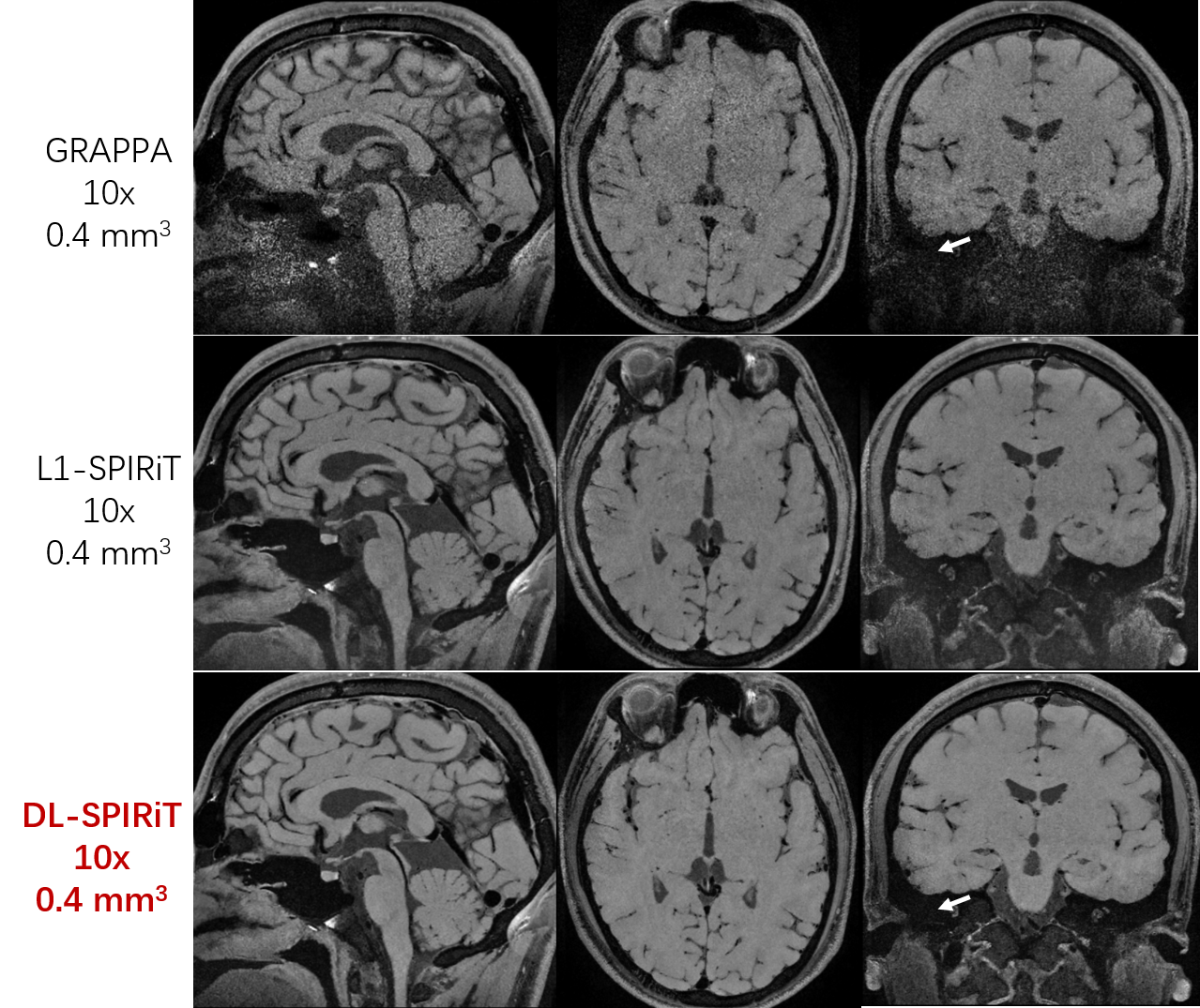
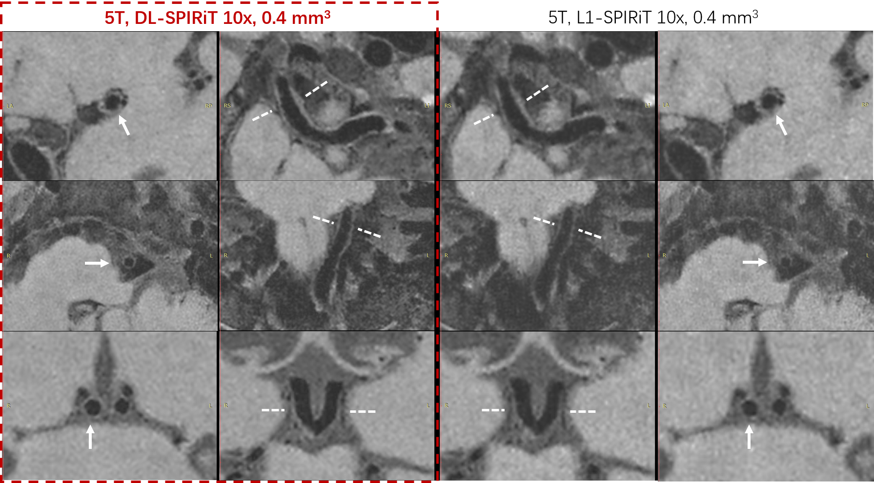
Figure 1 compares the reconstruction results for 10-fold accelerated VWI scan of 0.4 mm3 on a 24-year-old volunteer by GRAPPA, iterative wavelet sparsity regularized SPIRiT (L1-SPIRiT) and the proposed DL-SPIRiT methods. Both DL-SPIRiT and L1-SPIRiT reconstruction could effectively suppress the reconstruction noise which varies spatially and is heavier in the region where the B1 field is inhomogeneity.Figure 2 shows that DL-SPIRiT reconstruction of 10-fold accelerated VWI data acquired from a 71-ear-old volunteer gives more clear and continuous depiction of intracranial vessel walls than L1-SPIRiT. Moreover, DL-SPIRiT is free of parameter adjusting, while L1-SPIRiT needs manually adjusting the L1 regularization weights.
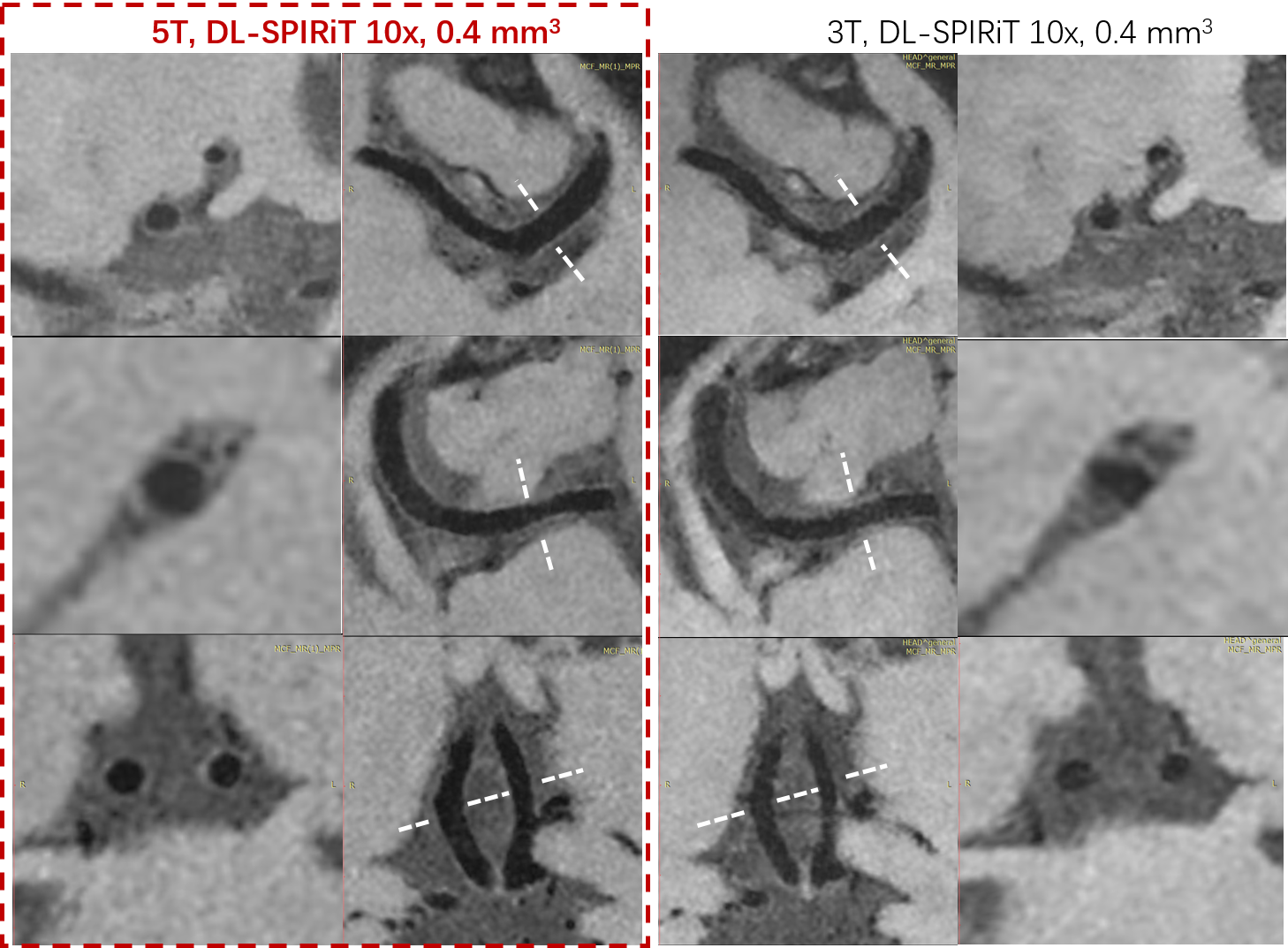
Figure 3 compares the DL-SPIRiT reconstruction results of two intracranial VWI scans with identical sequence parameters, performed separately on the 5T and 3T (uMR 790, UIH, Shanghai, China, with 32 channel head coil) scanners. The higher SNR on 5T dramatically improve the VWI quality.
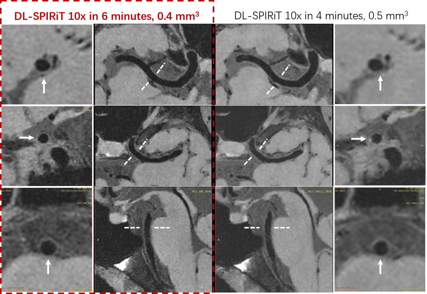
Figure 4 demonstrates that increasing the imaging resolution of VWI on a 22-years-old volunteer from 0.5 mm3 to 0.4 mm3 could depict the intracranial vessel walls more sharply and alleviate the partial volume effects.
Discussion
Based on the increased SNR and acceleration capability from the 5T scanner and the 48-channel head coil, this work demonstrates the great potential of achieving 10-fold accelerated whole brain vessel wall imaging of isotropic 0.4 mm3 in 6 minutes using deep learning (DL) unrolled SPIRiT reconstruction method. Further work will focus on optimizing the VWI contrast and investigating the feasibility of clinical deployment.Acknowledgements
This work is supported by the State Key Program of National Natural Science Foundation of China (Grant No. 81830056, 2021YFF0501503 and 2022YFA1004203) and the National Natural Science Foundation of China (Grant No. 81801691, 62125111).References
- Murphy M, Alley M, Demmel J, et al. Fast l1-SPIRiT Compressed Sensing Parallel Imaging MRI: Scalable Parallel Implementation and Clinically Feasible Runtime. IEEE Trans. Medical Imaging 2012, 31:1250-1262.
- Fan Z, Yang Q, Deng Z et al. Whole-brain intracranial vessel wall imaging at 3 Tesla using cerebrospinal fluid-attenuated T1-weighted 3D Turbo Spin Echo. Magn Reson Med 2017; 77:1142-1150.
- Jia S, Zhang L, Ren L, et al. Joint Intracranial and Carotid Vessel Wall Imaging in 5 minutes using Compressed Sensing accelerated DANTE-SPACE. Eur Radiol 2020, 30:119-127.
- Zhu C, Tian B, Chen L, et al. Accelerated whole-brain intracranial vessel wall imaging using black-blood fast spin-echo with compressed sensing (CS-SPACE). Magn Reson Mater Phy 2018; 31, 457–467.
- Monga V, Li YL, Eldar YC. Algorithm Unrolling: Interpretable, efficient deep learning for signal and image processing. IEEE Signal Processing Magazine 2021; 38:18-44.
- Hammernik K, Schlemper J, Qin C, et al. Systematic evaluation of iterative deep neural networks for fast parallel MRI reconstruction with sensitivity-weighted coil combination. Magn Reson Med 2021; 86:1859-1872.
Figures