1378
Intracranial aneurysm wall enhancement predicts aneurysm growth and rupture: a large-scale multi-center longitudinal study1Radiology, University of Washington, Seattle, WA, United States, 2Neurosurgery, Tiantan Hosptial, Beijing, China, 3Neurosurgery, University of Washington, Seattle, WA, United States
Synopsis
Keywords: Vessel Wall, Stroke, Aneurysm, vessel wall MRI
Motivation: Unruptured intracranial aneurysm (UIA) with wall enhancement identified on vessel wall MRI was considered at high risk of rupture and growth. But previous longitudinal studies were limited by small sample size (n<130).
Goal(s): To evaluate whether wall enhancement can predict UIA growth or rupture in a large-scale multi-center longitudinal study.
Approach: 709 UIA patients were followed by 2 years. Growth or rupture was recorded as primary outcome.
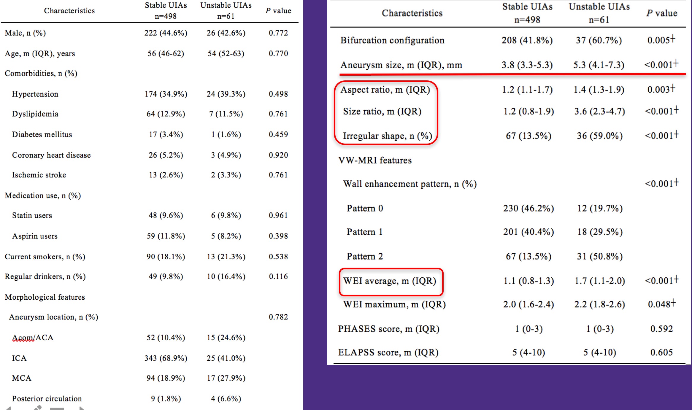
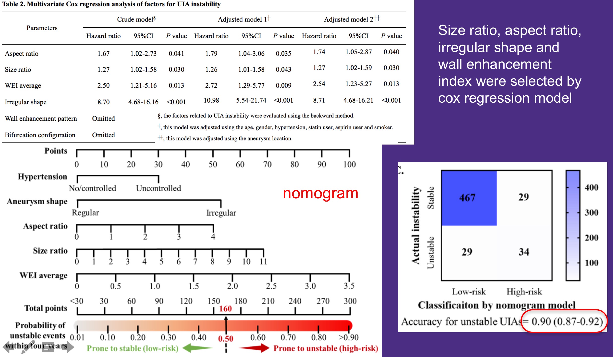
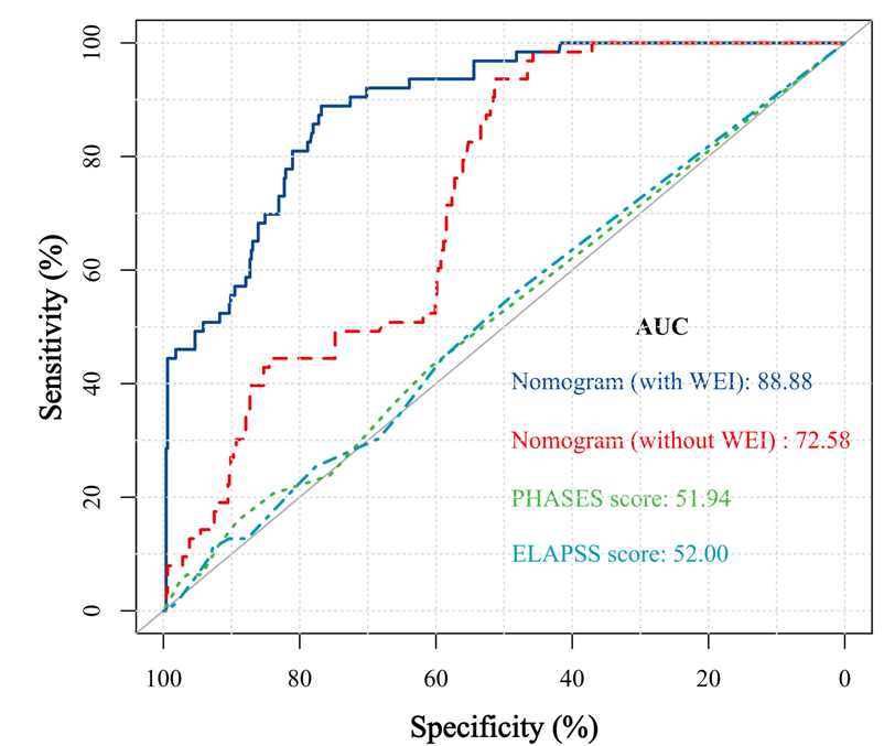
Results: Size ratio, aspect ratio, irregular shape and wall enhancement index were identified as factors of UIA instability. The final model has an AUC of 0.89, which was superior to traditional risk models (AUC 0.67-0.70, p<0.001).
Impact: To our best knowledge, this is the largest longitudinal study using vessel wall MRI to predict the natural risk of UIA rupture and growth. The model can potentially help select small but high-risk UIAs for early intervention.
Background
Unruptured intracranial aneurysm (UIA) with wall enhancement identified on vessel wall MRI was considered at high risk of rupture and growth. But previous longitudinal studies were limited by small sample size (n<130) and single center cohorts. This study aimed to evaluate whether wall enhancement and morphological features can predict UIA growth or rupture in a large scale multi-center longitudinal study.Materials and Methods
Single-UIA patients were enrolled from two prospective, multicenter studies, and set as the derivation cohort and validation cohort. All patients were scanned by vessel wall MRI on 3T Siemens scanners both pre- and post-contrast. Quantitative wall enhancement index (WEI) was measured. The primary endpoint was aneurysm rupture, growth, or morphology change, during 2-year follow-up. Based on the result of Cox analysis, a model was established within the derivation cohort, and then validated within the validation cohort. The performance of model was evaluated using the area under curve (AUC).Results
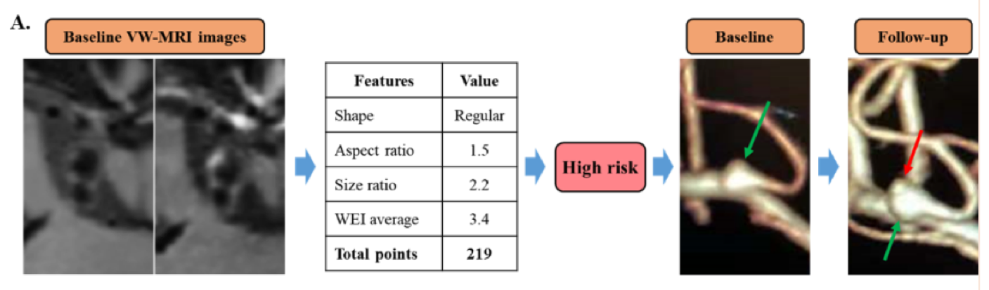
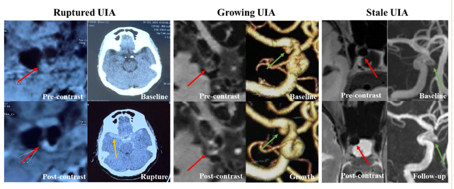
Derivation cohort included 559 single-UIA patients (248 male, median age 55 years). 61 (10.9%) patients had events during follow up (16 rupture and 45 growth). Size ratio, aspect ratio, irregular shape and wall enhancement index were identified as factors of UIA instability. A model including these features predicted UIA instability within the derivation cohort (AUC, 0.89). In the validation cohort of 146 patients (28 unstable and 118 stable), the AUC was 0.84, which was superior to traditional risk models (AUC: PHASES score 0.67 and ELAPSS score 0.70, p<0.001).Conclusion and Discussion
This study provided a radiological model incorporating wall enhancement and morphological features to predict UIA growth and rupture, which may help guide treatment decision-making.To our best knowledge, this is the largest longitudinal study using vessel wall MRI to predict the natural risk of UIA rupture and growth. Our sample size (n=709) was more than 5 times larger the previous largest study (n=129), providing the strongest evidence so far in the field.
Wall enhancement is a marker of inflammation and wall remodeling, which might weaken the wall and leads to growth or rupture. The use quantitative wall enhancement characters also helped the model to achieve high accuracy, which was much better than the traditional risk scores and provide additional value in the outcome prediction. The utility of our model needs to be validated in other international cohorts and also in randomized controlled clinical trials (RCTs).
Acknowledgements
No acknowledgement found.References
No reference found.Figures