1375
Achieving Robust Labeling Above the Circle of Willis with Vessel-Encoded Arterial Spin Labeling1Institute of Science and Technology for Brain-inspired Intelligence, Fudan University, Shanghai, China, 2University of Oxford, Wellcome Centre for Integrative Neuroimaging, FMRIB Division, Nuffield Department of Clinical Neurosciences, Oxford, United Kingdom, 3Key Laboratory of Computational Neuroscience and Brain-Inspired Intelligence (Fudan University), Ministry of Education, Shanghai, China
Synopsis
Keywords: Blood Vessels, Perfusion, ASL
Motivation: The ability to distinguish arterial blood supply above CoW is crucial in the study of collateral circulation, but it is challenging due to the complex positioning of arteries around the CoW.
Goal(s): To achieve a robust vascular territories separation above CoW.
Approach: We propose improvements to the original OES method, optimizing its SNR efficiency while minimizing high spatial frequencies to avoid mislabeling. We have also selected a set of PCASL parameters that facilitate thin-slice labeling and ensure high labeling efficiency to overcome issues related to vascular tortuosity.
Results: Combining optimized parameters and improved OES, the vascular territories separation has significantly improved.
Impact: Our study optimized the fully automatic encoding pattern design above the circle of Willis, and achieved a robust vascular territories separation, combining with optimized PCASL parameters.
Introduction
The ability to distinguish arterial blood supply to specific brain regions is crucial in the study of collateral blood flow. Vessel-encoded arterial spin labeling (VEASL) can potentially achieve this non-invasively. However, an appropriate encoding scheme is required to distinguish many arterial territories above the circle of Willis (CoW). An optimal encoding scheme (OES) automated approach has been suggested1, but the high tortuosity of these arteries makes it challenging to ensure that multiple arterial branches are perpendicular to the labeling plane, resulting in reduced labeling efficiency and errors in vessel-decoding. To enhance the stability of labeling above the CoW, we propose improvements to the original OES method, optimizing its SNR efficiency while minimizing excessively high spatial frequencies to avoid mislabeling due to subject motion. We have also selected a set of PCASL optimal parameters that facilitate thin-slice labeling and ensure high labeling efficiency to overcome issues related to vascular tortuosity. We assessed the efficacy of territorial separation above the CoW by comparing our "optimized" parameters combined with the improved OES ("IOES") approach, to the conventional "default" parameters, in combination with the original "OES".Materials and methods
SimulationThe IOES method, built upon the originally proposed OES, involved the random permutation of Hadamard matrix columns, which considered real ASL acquisition parameters to simulate the final magnetization at each vessel location. The condition number of the encoding matrix and the shortest spatial wavelength ($$$\lambda $$$) were calculated and plugged into Equation 1, which encourages high condition numbers and long wavelength encodings. The IOES process iterated 100 times to obtain the final encoding design with the minimum cost function.$$\mathrm{cost} = \frac{\left ( 1-\frac{1}{\mathrm{condition} } \right ) ^{2} }{2} +\frac{\left ( \frac{1}{\frac{\mathrm{min\left ( \lambda \right ) } }{\mathrm{motion} } } -1 \right ) ^{2} }{2}\qquad\qquad\qquad(1) $$To achieve thin-slice labeling, a long RF duration is required. We fixed the RF interval at 1560 μs, the maximum value that can be set on the scanner, trying to stick to around a 50% duty cycle. We explored a range of Gmax, Gmean, RF duration and flip angle. Finally, a parameter set was chosen to ensure that the thickness of the labeling plane z, as defined in Equation 22, was less than 6 mm, whilst maintaining high labeling efficiency.$$z = \frac{2}{\frac{\gamma }{2\pi }\cdot \mathrm{G_{max}\cdot \mathrm{RF\_duration} } } \qquad\qquad\qquad(2) $$
Image acquisition
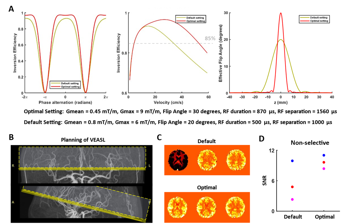
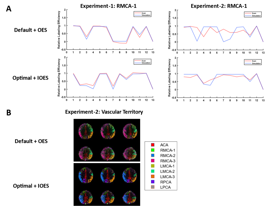
The optimized parameters were listed in Figure 2, and for default parameters, we used settings similar to previous work3. We performed scans on three healthy volunteers at a 3T Siemens Prisma scanner. Each subject underwent two VEASL scans, one with default parameters using OES, and the other with optimal parameters using IOES. Experiment-1: for two of the subjects, we carefully selected optimal labeling planes, maximizing perpendicularity and spacing between vessels. Experiment-2: for the third subject, we intentionally chose suboptimal labeling planes, resulting in some vessel segments running parallel to the labeling plane or located at bifurcation sites. Maximum a posteriori solution to the Bayesian framework was used for VEASL analysis4.
Results
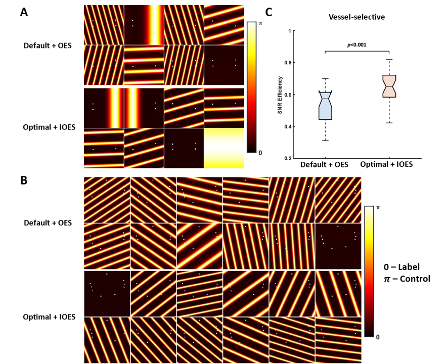
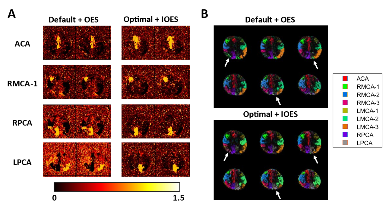
Figure 1 illustrates the comparison between OES and IOES. Clearly, IOES eliminated the high spatial frequency encoding patterns that might occur with OES and significantly enhanced the encoding's SNR efficiency. Figure 2, we observed that the optimal parameters could achieve a thin-slice labeling while maintaining high labeling efficiency across a broad range of blood flow velocities and exhibited a favorable inversion profile. The SNR under optimal parameters showed an improvement in non-selective images, albeit not significantly. Due to the bottom imaging slice being very close to the labeling plane, reduced static tissue saturation effects when using the optimized settings could also be observed. Figure 3A illustrates the posterior probability maps obtained through the Bayesian analysis in one subject, revealing that the combination of optimal parameters with IOES effectively led to more robust vascular territory separation. In Experiment 1, the perfusion signals within the specific territories were in good agreement with the simulated signal (Figure 4). However, in Experiment 2, the combination of default settings and OES clearly did not match the simulated signals as effectively as the optimal settings with IOES for the RMCA-1 territory due to vessel tortuosity within the thicker-labeling region. Consequently, this territory was not well separated in the analysis.Discussion and conclusions
Using optimized parameters and the IOES allowed thin-slab labeling, improved labeling efficiency and vessel-encoding, enabling more stable vascular territory separation, even for more complex vascular structures above the CoW. However, the selection of the labeling plane remains crucial. If we cannot accurately position it in relation to the vessel’s location, vascular territory imaging remains challenging. AI-assisted methods for automatically finding the optimal labeling plane are worth considering in future work.Acknowledgements
This work was enabled by a Sir Henry Dale Fellowship jointly funded by the Wellcome Trust and the Royal Society (220204/Z/20/Z). The Wellcome Centre for Integrative Neuroimaging is supported by core funding from the Wellcome Trust (203139/Z/16/Z).References
1. Berry, E.S.K., Jezzard, P. & Okell, T.W. An Optimized Encoding Scheme for Planning Vessel-Encoded Pseudocontinuous Arterial Spin Labeling. Magnetic Resonance in Medicine 74, 1248-1256 (2015).
2. Dai, W., Garcia, D., de Bazelaire, C. & Alsop, D.C. Continuous flow-driven inversion for arterial spin labeling using pulsed radio frequency and gradient fields. Magnetic Resonance in Medicine 60, 1488-1497 (2008).
3. Okell, T.W., et al. Vessel-encoded dynamic magnetic resonance angiography using arterial spin labeling. Magnetic Resonance in Medicine 64, 698-706 (2010).
4. Chappell, M.A., Okell, T.W., Payne, S.J., Jezzard, P. & Woolrich, M.W. A fast analysis method for non-invasive imaging of blood flow in individual cerebral arteries using vessel-encoded arterial spin labelling angiography. Medical Image Analysis 16, 831-839 (2012).
Figures