1374
Using water beads as static tissue in a Circle of Willis flow phantom in 4D flow MRI1Department of Medical Physics, University of Greifswald, Greifswald, Germany, 2Department of Radiology, Northwestern University Feinberg School of Medicine, Chicago, IL, United States, 3Institute of Diagnostic Radiology and Neuroradiology, University Medicine Greifswald, Greifswald, Germany
Synopsis
Keywords: Blood Vessels, Blood vessels
Motivation: While previous research has focused on realistic vessel and flow replication, the background tissue mimicking in 4D flow MRI remains unexplored despite its significant impact on phase correction accuracy.
Goal(s): Aims to identify a suitable material for a Circle of Willis flow phantom that can mimic background static tissue, ensure transparency, and constrain vessel motion.
Approach: Transparent water beads soaked in a Gd-doped solution were used as background static tissue in a flow phantom. A MATLAB tool was used for the post-processing of the 4D-flow data.
Results: Water beads effectively minimized motion artifacts, and increased the number of time-averaged streamlines and their quality.
Impact: This study introduces a practical solution, to enhance the accuracy of in-vitro 4D Flow MRI of complex vessel phantoms by mitigating motion artifacts. The transparent water beads offer a cost-effective and easily exchangeable alternative for mimicking background static tissue.
Motivation:
Phase contrast MRI is a commonly employed method for evaluating the blood flow and tissues movement. This approach depends on identifying variations in the signal phase caused by motion or flow in the presence of recognized linear magnetic gradient fields1. Realistic flow phantoms are of the essence to validate new flow MRI sequences and algorithms for the quantitation of flow parameters. However, most works concentrate on mimicking realistic vessels and flow. The material to use in a flow phantom to mimic background static tissue hasn’t been investigated even though its properties affect 4D-flow MRI quality2. Moreover, this material should be able to fully embed the complicated vessel structure (e.g., a circle of Willis replicate) without changing its diameter and ensure transparency to allow visibility and access. This would allow air bubble detection and removal or visual guidance for placing catheters. Additionally, it should mimic the property of static in vivo tissue by constraining vessel motion due to pulsatile blood flow while retaining its original geometry. Alternatives such as agar3 and ballistic gel4 were suggested but are impractical due to their complicated handling when using complex vessel architectures, their limited opacity, the inevitable air gaps between the phantom and the surrounding material, and the need for toxic chemicals to prevent mold growth. To circumvent these issues, we propose a new solution that is easily exchangeable, prevents vessel motion, and mimics the brain static tissue T1 contrast.Methods:
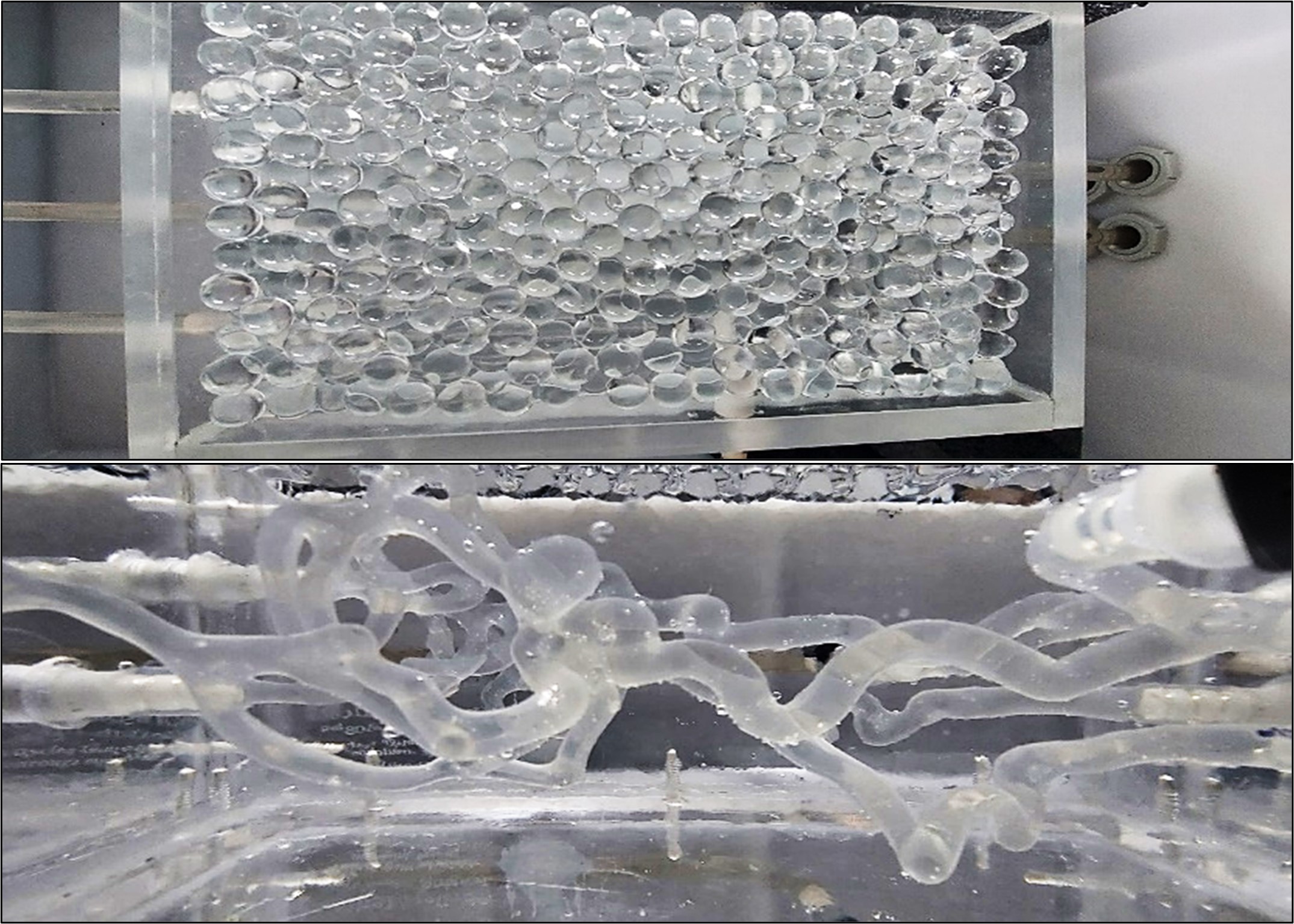
The phantom used is the “HN-S-A-010” complete intracranial model with an anterior cerebral artery aneurysm made from silicone (ELASTRAT, Geneva, Switzerland). 30g of transparent water beads were soaked in 5 liters of water mixed with 0.5ml of Gadovist 0.1mol/ml (Bayer, Leverkusen, Germany) for 48 hours (Fig.1). T1 of the solution was around 770ms mimicking the values of gray matter (768ms), measured ex-vivo in brain tissue of pigs.The phantom was first scanned with Gd-doped water-embedded beads. In a second step, the beads were removed, and the phantom box was filled with Gd-doped water for comparison (Fig. 1). All scans were performed on a 3T VIDA MRI (Siemens, Erlangen, Germany) using a Small Ultra Flex 18-channel coil, a dual-venc 4D flow MRI sequence5 (Venc1=50cm/s, Venc2=100cm/s, TE=3.8ms, TR=5.7ms, temporal resolution =79.8ms, spatial resolution=1mm isotropic, FA=15°), and a standard 3D time-of-flight (TOF) GRE sequence. A CompuFlow 1000-MR pump (Shelley Medical Imaging Technologies, London, Canada) provided a pulsatile flow of 0.55L/min with 72 strokes/min. The circuit was filled with a blood-mimicking solution (60% water and 40% glycerol, viscosity of 0.003kg/(m·s), density of 1100kg/m3). Gadovist 0.1mol/ml (Bayer, Leverkusen, Germany) was added to this solution (≈0.2% by volume) to increase SNR1.
An in-house build semi-automatic MATLAB tool was used for eddy current correction, noise masking, dual-venc reconstruction, and phase-contrast MR angiogram (MRA) calculation5,6. Subsequently, volumetric flow quantification was performed by combining the velocity data from 4D flow MRI with the anatomical information obtained from the automatic segmentation of 3D TOF MRA7. additionally, TOF vessels were segmented with 3DSlicer for further comparison.
Results:
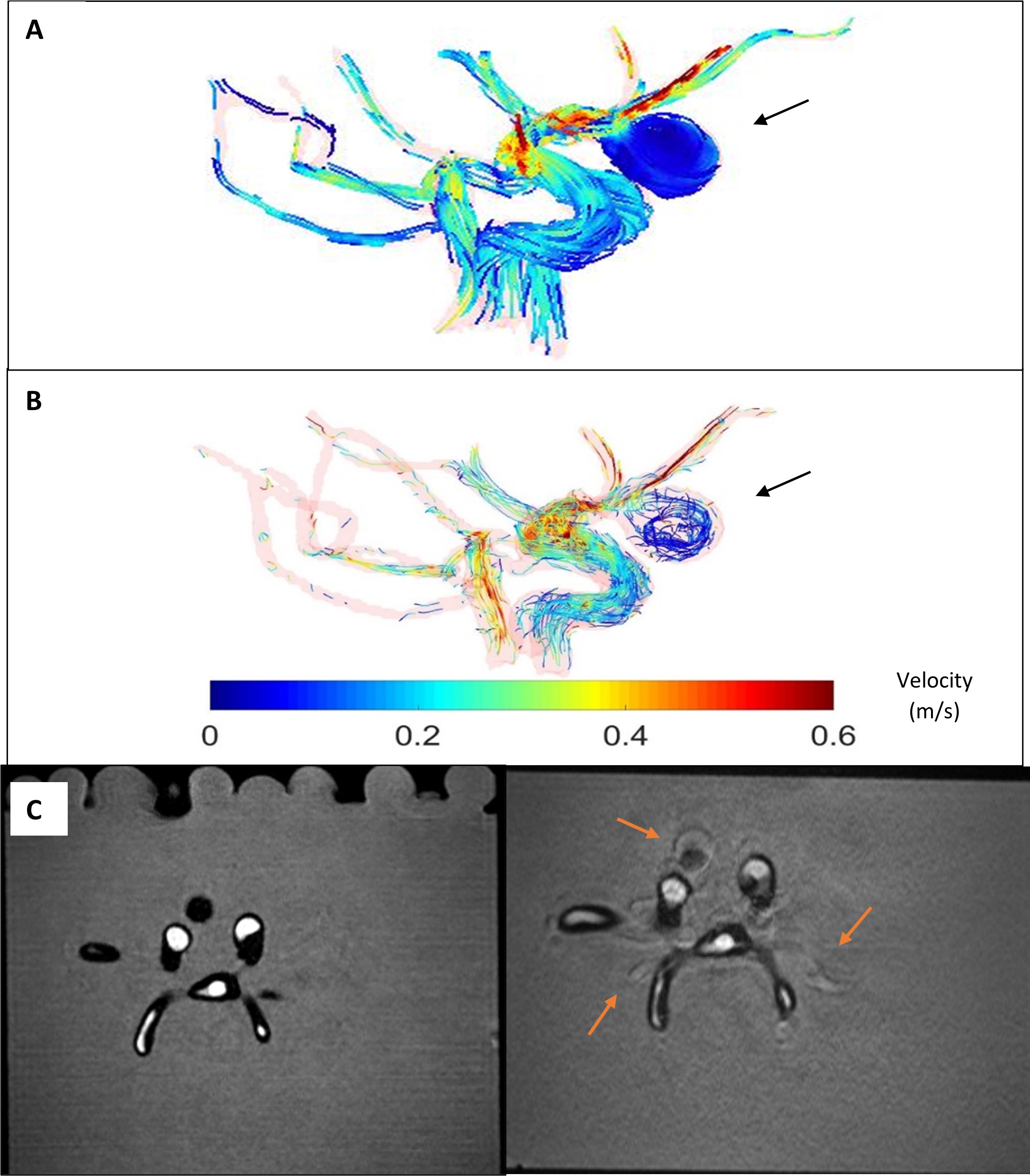
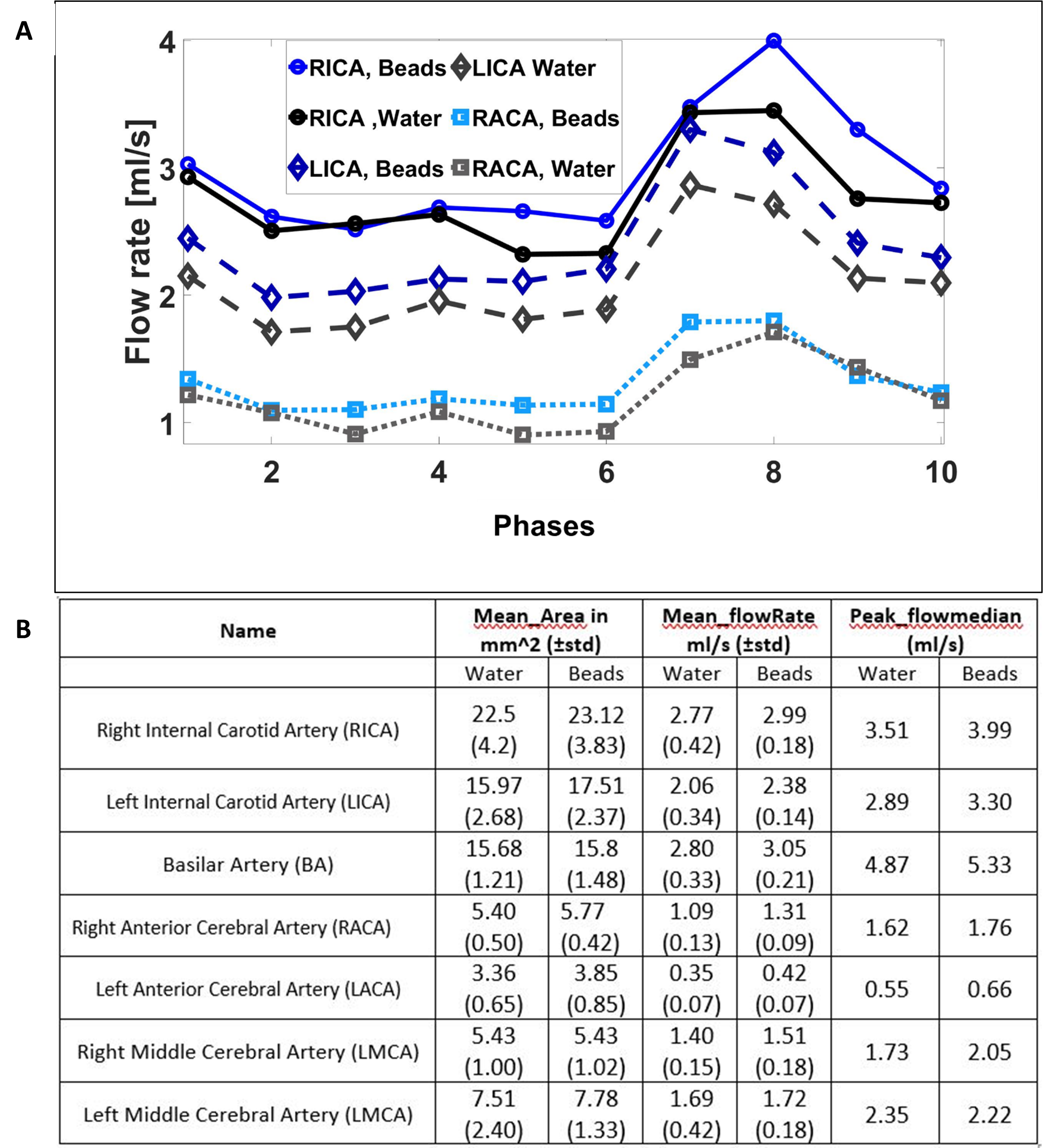
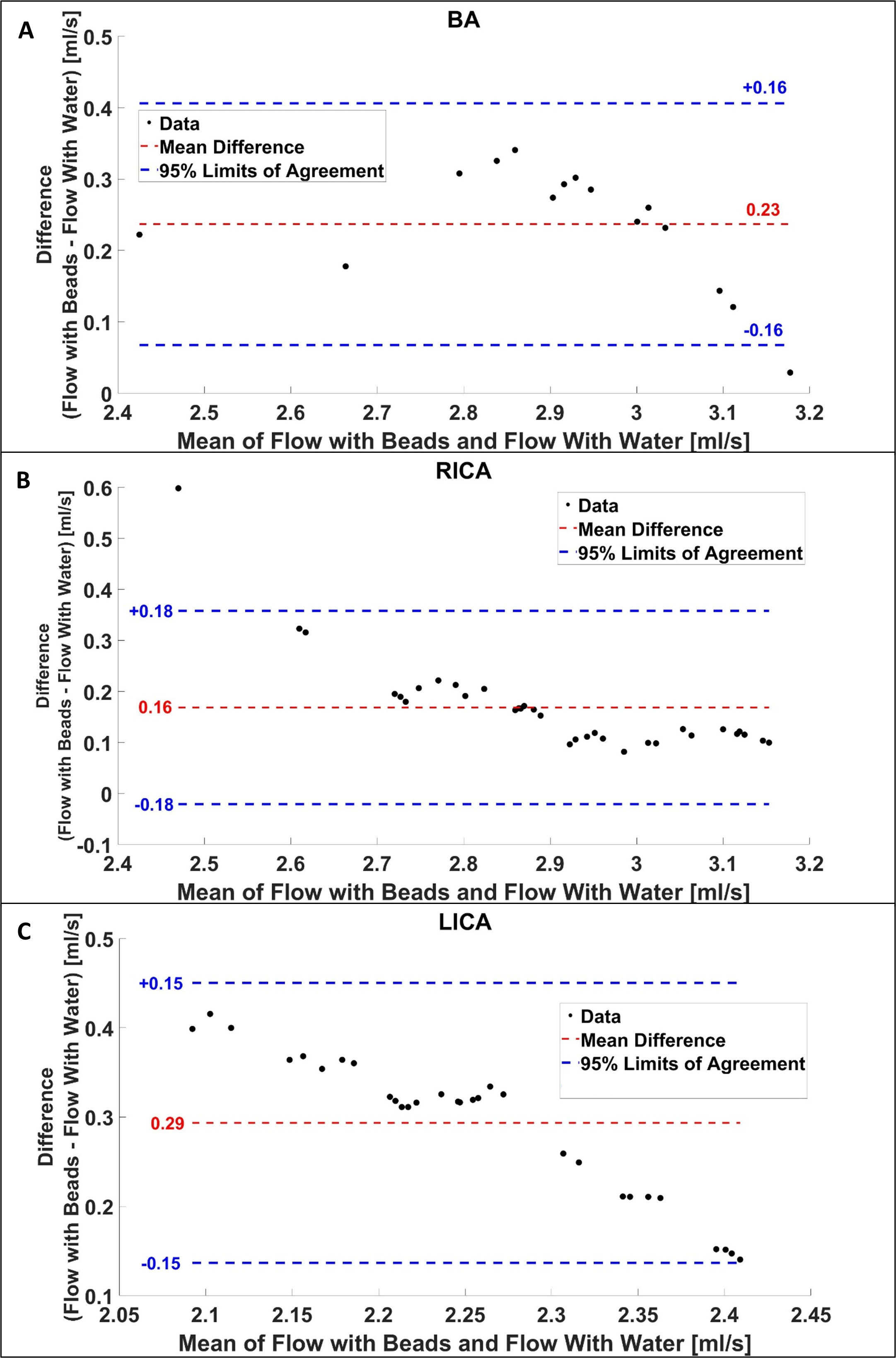
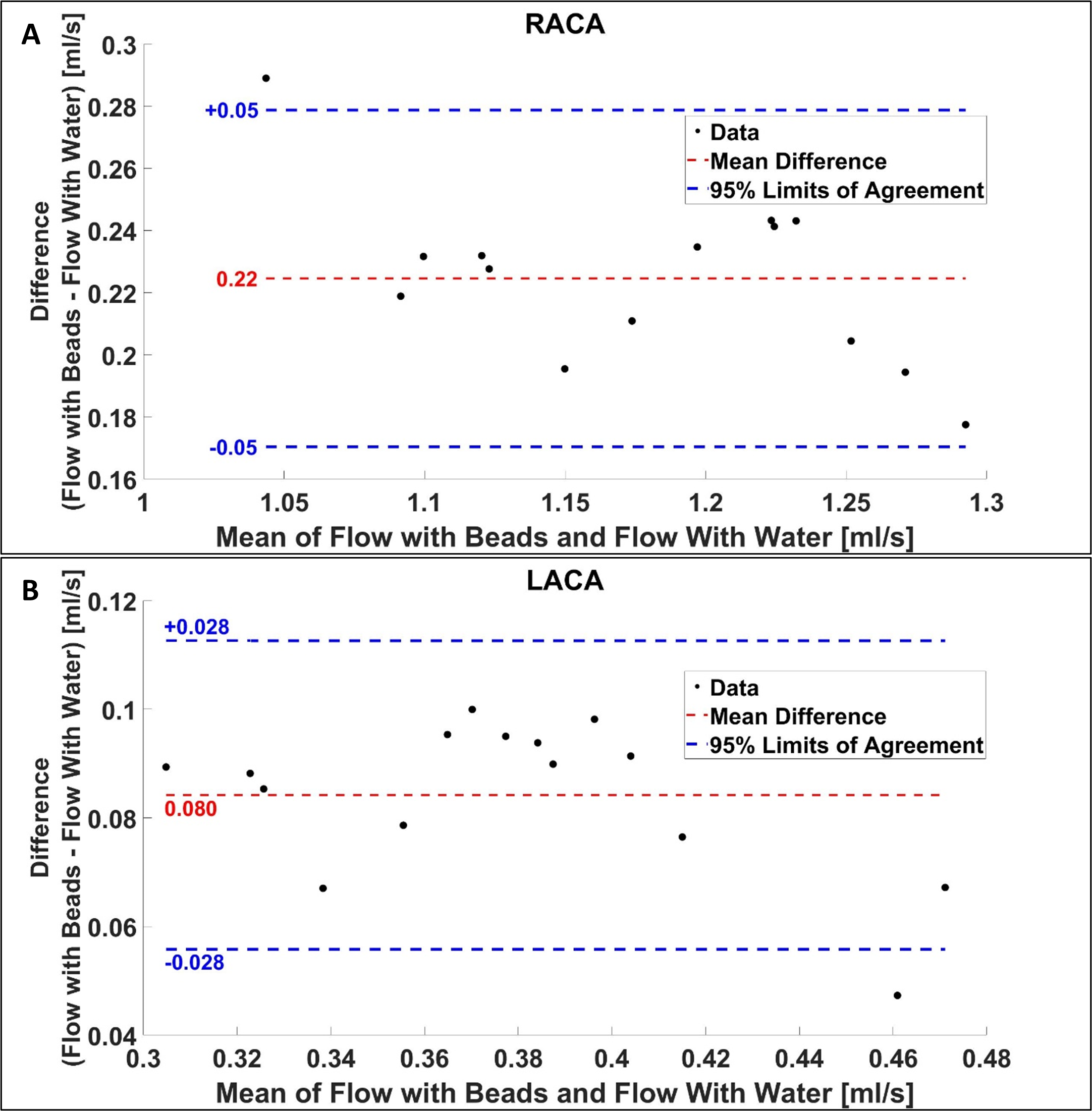
The time-averaged streamline visualization in Fig.2 indicates the presence of helical recirculating flow with slower velocities in the aneurysm. Qualitatively, the use of water beads showed noticeable improvement in streamline length and increased number of streamlines compared to water (Fig.2). In Fig.2.C, increased motion artifacts can be seen in the phantom with water compared to in water-embedded beads (see arrows). Flow evaluation showed a lower median transient flow rate and mostly a higher standard deviation when water was used compared to water beads (Fig.3), whereas the Bland Altman assessment yields a higher mean flow for beads in comparison to water (Fig.4 and Fig.5). Moreover, the phantom position in the container shifted higher in water due to buoyancy. Taking the upper tip of the aneurysm as a reference, the difference between the water and beads position was around 9.5 mm.Discussion and conclusion:
Water beads successfully minimized motion artifacts by holding the vasculature in place. They also allowed for a noticeable improvement in overall streamline quality, length, and an increased number of streamlines compared to water. The simplicity of handling and working with water beads, their low price, their good exchangeability, and their ability to absorb and retain water alone made them well-suitable. In addition, they reduced vessel motion, which quantitatively resulted in larger vessel areas as well as higher flow rates compared to the setup of only water. Thus, using water beads as background static tissue for complex vascular phantoms was found to be very well-suited. In the future, we aim to further quantify the performance of phase offset correction with beads.Acknowledgements
This work was funded by the German Research Foundation (DFG INST 292/155-1 FUGG) and the National Institutes of Health (NIH 1R01HL149787, 5R21NS122511).References
1. Markl, M., Bammer, R., Alley, M. T., Elkins, C., Draney, M. T., Barnett, A., Moseley, M. E., Glover, G. H., & Pelc, N. J. (2003). Generalized reconstruction of phase contrast MRI: Analysis and correction of the effect of gradient field distortions. Magnetic Resonance in Medicine, 50(4), 791–801.
2. Busch, J., Giese, D., & Kozerke, S. (2017). Image-based background phase error correction in 4D flow MRI revisited. Journal of Magnetic Resonance Imaging, 46(5), 1516–1525.
3. Aristova, M., Pang, J., Ma, Y., Ma, L., Berhane, H., Rayz, V. L., Markl, M., & Schnell, S. (2022). Accelerated dual‐venc 4D flow MRI with variable high‐venc spatial resolution for neurovascular applications. Magnetic Resonance in Medicine, 88(4), 1643–1658.
4. Zimmermann, J., Loecher, M., Kolawole, F. O., Bäumler, K., Gifford, K., Dual, S. A., Levenston, M. E., Marsden, A. L., & Ennis, D. B. (2021b). On the impact of vessel wall stiffness on quantitative flow dynamics in a synthetic model of the thoracic aorta. Scientific Reports, 11(1).
5. Schnell, S., Ansari, S. A., Wu, C., García, J., Murphy, I., Rahman, O., Rahsepar, A. A., Aristova, M., Collins, J. D., Carr, J. C., & Markl, M. (2017). Accelerated dual-venc 4D flow MRI for neurovascular applications. Journal of Magnetic Resonance Imaging, 46(1), 102–114.
6. Bock, J., Kreher, B.W., Hennig, J., & Markl, M. (2007). Optimized pre-processing of time-resolved 2 D and 3 D Phase Contrast MRI data. Proceedings of the 15th Annual Meeting of ISMRM.
7. Vali, A., Aristova, M., Vakil, P., Abdalla, R., Prabhakaran, S., Markl, M., Ansari, S. A., & Schnell, S. (2019). Semi‐automated analysis of 4D flow MRI to assess the hemodynamic impact of intracranial atherosclerotic disease. Magnetic Resonance in Medicine, 82(2), 749–762.
Figures